RESEARCH ARTICLE
Two Half-Sandwiched Ruthenium (II)
Compounds Containing 5-Fluorouracil
Derivatives: Synthesis and Study of DNA
Intercalation
Zhao-Jun Li1, Yong Hou2, Da-An Qin3, Zhi-Min Jin4*, Mao-Lin Hu3*
1Institute of Agricultural Resources and Regional Planning, Chinese Academy of Agricultural Sciences, Key Laboratory of Plant Nutrition and Fertilizer, Ministry of Agriculture, Beijing, China,2Institute of Biotechnology and Nucleic Technology, Sichuan Academy of Agricultural Sciences, Chengdu, China,3College of Chemistry and Materials Engineering, Wenzhou University, Wenzhou, China,4College of Pharmaceutical Sciences, Zhejiang University of Technology, Hangzhou, China
*zimichem@sina.com(ZMJ);maolin@wzu.edu.cn(MLH)
Abstract
Two novel coordination compounds of half-sandwiched ruthenium(II) containing
2-(5-fluorouracil)-yl-N-(pyridyl)-acetamide were synthesized, and their intercalation bind-ing modes with calf thymus DNA were revealed by hyperchromism of ultraviolet-visible spectroscopy; the binding constants were determined according to aLangmuiradsorption equation that was deduced on the base of careful cyclic voltammetry measurements. The two compounds exhibited DNA intercalation binding activities with the binding constants of 1.13×10⁶M-1and 5.35 ×10⁵M-1, respectively.
Introduction
Cisplatin shows a strong ability of binding to and causing crosslinking of DNA invivo, which ultimately triggers cell apoptosis, and is used successfully to treat some special types of cancers. The discovery of cisplatin has aroused great effort toward the design of metal-based anticancer drugs [1]. Particular interest is attracted toward ruthenium element, a member of platinum-group, due to its low toxity and its ability to mimic iron in binding with proteins in the plasma [2]. In the recent decades, as a potential anticancer medicine, organometallic ruthenium com-pounds of the type [Ru(η6-arene)] are under intensive investigation, owing to their promising activity invitroand invivotoward cancer cell, including cisplatin-resistant cells [3].
Combining two or more multifunctionalities into one is a popular strategy in design new therapeutic agents. 5-Fluorouracil (5-FU) has been used extensively in the treatment of solid tumors for years, and in the late, its strong toxicities to the gastric system, intestinal mucosa and bone marrow is evidenced eventually. Therefore, attempts are made to improve its antican-cer activity and to minimize its side effects by exploiting several prodrug, such as amines, alco-hols, and peptides [4]. In the literature, covalent compound containing platinum(II) and 5-FU
OPEN ACCESS
Citation:Li Z-J, Hou Y, Qin D-A, Jin Z-M, Hu M-L (2015) Two Half-Sandwiched Ruthenium (II) Compounds Containing 5-Fluorouracil Derivatives: Synthesis and Study of DNA Intercalation. PLoS ONE 10(3): e0120211. doi:10.1371/journal.pone.0120211
Academic Editor:Heidar-Ali Tajmir-Riahi, University of Quebect at Trois-Rivieres, CANADA
Received:November 4, 2014
Accepted:January 20, 2015
Published:March 19, 2015
Copyright:© 2015 Li et al. This is an open access article distributed under the terms of theCreative Commons Attribution License, which permits unrestricted use, distribution, and reproduction in any medium, provided the original author and source are credited.
Data Availability Statement:All relevant data are within the paper and its Supporting Information files.
fragment has been reported [5], but covalent compound containing ruthenium and 5-FU frag-ment is not available.
The interaction of DNA with small molecule had been a hot subject of research [6–11] for long time, due to the interaction modes and kinetic mechanism reflected various senses for de-veloping molecular drugs and probers to monitor DNA structure [12–15]. In this respect, we synthesized two novel half-sandwiched ruthenium(II) compounds from dichloro(p-cymene) ruthenium(II) dimer and 2-[(5-fluorouracil)-yl]-N-pyridyl-acetamide for the first time, and studied the interaction of calf thymus DNA (CT-DNA) with them by cyclic voltammetry and UV-vis spectrometry.
Experimental
1. Apparatus and reagents
CT-DNA with high molecular weight was extracted and purified by a method described elsewhere [16]. All other chemicals, including dichloro(p-cymene)ruthenium(II) dimer {[(η6-p-cymene)RuCl2]2} and 5-FU, were of analytical reagent grade, and were purchased from sigma-aldrich Co. (USA).
The CT-DNA solution was prepared with 0.1 M KCl / 0.05 M Tris—HCl (pH 7.16) and stored at 4°C. The concentration of DNA was determined by UV absorbance at 260 nm, with the extinction coefficient (ε260) taken as 6600 M–1 cm–1.
The electrochemical measurements were performed on a CHI1030b electrochemical work-station (CH Instrumental, Chenhua Corp., Shanghai, China). Supporting electrolyte for all experiments was a 0.1 M KCl / 0.05 M Tris—HCl buffer solution, which was adjusted to pH 7.16 by HCl.
IR spectra were recorded on an EQUINOX-55 instrument (Bruker Optics, Germany). Ele-mental analysis was performed on a Perkin-Elmer 2400 CHNS/O eleEle-mental analyzer (USA). The UV-vis spectra were recorded at room temperature on a U-3010 spectrophotometer (Hita-chi, Japan) equipped with 1.0 cm quartz cells.1HNMR and13CNMR spectra were recorded on a AVANCE-500 instrument using tetramethylsilane (TMS) as an internal standard at room temperature, and the chemical shifts were given in relative to TMS.
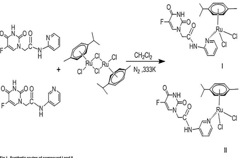
2. Synthesis of Ruthenium compounds I and II
Two isomeric 5-FU derivatives, N- (pyridin-2-yl)-acetamide (L1) and 2-[5-fluoro-2,4-dioxo-3,4-dihydropyrimidin-1(2H)-yl]-N-(pyridin- 3-yl)-acetamide (L2), were synthesized (Fig. 1) in accordance with a procedure described previously [17]. The two titled compounds were synthesized from the 5-FU deriva-tive and dichloro(p-cymene)ruthenium(II) dimer. Typically, 2-[(5-fluorouracil)-yl]-N-pyridyl-acetamide (66 mg, 0.25 mmol) was added dropwise to a suspension of [(η6-cymene)RuCl2]2 (76.5 mg, 0.125 mmol) in freshly distilled anhydrous methanol (25 mL). The color of the result-ing mixture changed immediately with stirrresult-ing at 25°C under argon, and precipitate was formed after storage in a freezer at -18°C for 24 h. The fine yellow solid was collected by filtra-tion, recrystallized from methanol/ether, washed with methanol followed by ether, and dried overnight in vacuo.
½ðZ6 p cymeneÞRuðL1ÞCl2 ðIÞ
Yield based on [(η6-cymene)RuCl2]2: 54.4%. IR: 3390 (υN-H), 3291, 3044 (υC-H), 2843
(υ-CH2-), 1705 (υC = O), 1479 (υC-N), 1430, 1375, 1234 (υC-F), 879, 763, 686 cm-1(S1 Fig.).
Anal. calc. for C20H23Cl2FN4O3Ru (558.37), C 43.02% N 10.03% H 4.15%; found: C42.98% data collection and analysis, decision to publish, or
preparation of the manuscript.
N 10.11% H 4.10%.1HNMR (500 MHz, DMSO-d6)δ: 11.96 (1H, s), 8.11 (1H, d,J= 6.8 Hz),
7.90 (1H, dd,J= 5.1, 1.0 Hz), 7.43 (1H, ddd,J= 8.8, 7.2, 1.9 Hz), 6.51 (1H, d,J= 3.5 Hz), 6.49 (1H, d,J= 3.5 Hz), 5.84 (2H, d,J= 6.3 Hz), 5.80 (2H, d,J= 6.3 Hz), 4.36 (2H, s), 2.84 (1H, dt,
J= 13.8, 6.9 Hz), 2.10 (3H, s), 1.20 (6H, d,J= 6.9 Hz) ppm (S2 Fig.). 13C NMR (126 MHz, DMSO-d6)δ: 169.49 (s), 159.10 (s), 157.47 (d,J= 25.8 Hz), 149.67 (s), 146.23 (s), 139.28
(d,J= 228.6 Hz), 137.78 (s), 111.77 (s), 108.62 (s), 106.37 (s), 100.09 (s), 85.95 (d,J= 107.1 Hz), 48.78 (s), 29.98 (s), 21.51 (s), 17.88 (s) ppm (S3 Fig.).
½ðZ6 p cymeneÞRuðL2ÞCl2 ðIIÞ
Yield based on [(η6-cymene)RuCl2]2: 61.3%. IR: 3520 (υN-H), 3256, 3050 (υC-H), 2926
(υ-CH2-), 1704 (υC = O), 1485 (υC-N), 1430, 1381, 1220 (υC-F), 809, 694 cm-1(S4 Fig.).
Anal. calc. for C20H23Cl2FN4O3Ru (558.37), C 43.04% N 10.04% H 4.18%; found: C 43.02% N 10.03% H 4.15%.1HNMR (500 MHz, DMSO-d6)δ: 11.95 (1H, d,J= 5.0 Hz), 10.54 (1H, s),
8.72 (1H, d,J= 2.2 Hz), 8.29 (1H, d,J= 3.6 Hz), 8.11 (1H, d,J= 6.7 Hz), 8.06–7.97 (1H, m), 7.37 (1H, dd,J= 8.3, 4.7 Hz), 5.82 (2H, d,J= 6.3 Hz), 5,78 (2H, d,J= 6.3 Hz), 4.53 (2H, s), 2.83 (1H, dt,J= 13.7, 6.9 Hz), 2.08 (3H, s), 1.19 (6H, d,J= 6.9 Hz) ppm (S5 Fig.). 13C NMR (126 MHz, DMSO-d6)δ:165.98 (s), 157.50 (d,J= 25.6 Hz), 149.77 (s), 144.60 (s), 140.43 (d,J= 64.6
Fig 1. Synthetic routes of compound I and II.
doi:10.1371/journal.pone.0120211.g001
Hz), 138.36 (s), 135.16 (s), 131.01 (d,J= 34.0 Hz), 126.14 (s), 123.77 (s), 106.38 (s), 100.08 (s), 85.92 (d,J= 106.7 Hz), 50.14 (s), 29.95 (s), 21.48 (s), 17.84 (s) ppm (S6 Fig.).
3. Preparation of (CT-DNA)-modified electrode
The electrodes were modified with CT-DNA in a procedure reported previously [18,19]. Gold disk electrodes were polished with a series of alumina powder (1.0, 0.5 and 0.05μm), then was
purified and placed in fresh piranha solution (30% H2O2and 70% H2SO4) to remove adsorbed organic impurities and sonicated in highly purified water for 5 min. Prior to the modification, the electrode surface was electrochemically activated by sweeping from -0.3 to +1.5 V in 0.1 M H2SO4solution until a stable cyclic voltammogram characteristic of a clean gold electrode was obtained. After being washed with twice-distilled water, the freshly polished gold electrode was immediately modified by transferring a droplet of 10μL of 1.0μgμL-1CT-DNA solution onto
its surface, followed by air-drying overnight. The CT-DNA-modified electrode (CT-DNA/Au) was then soaked in sterile water for about 4 h and rinsed with water to remove unadsorbed CT-DNA.
4. Cyclic voltammetry
Voltammetric measurements were carried out in a conventional cell consisting of three-electrode, namely a bare gold (or CT-DNA/Au) as the working three-electrode, a saturated calomel electrode, and a platinum wire auxiliary electrode. A CT-DNA/Au electrode was soaked in buffer solution with different concentrations of the compound for voltammetric test designed to investigate the interaction of CT-DNA. Typical cyclic voltammetry experiment was carried out in a 5 mM solution of K3[Fe(CN)6] / K4[Fe(CN)6] (1:1) in the supporting electrolyte at room temperature (25°C). All solutions were deaerated with highly pure nitrogen, and the elec-trochemical experiments were performed at a scan rate of 0.1 Vs-1.
5. UV-visible spectra
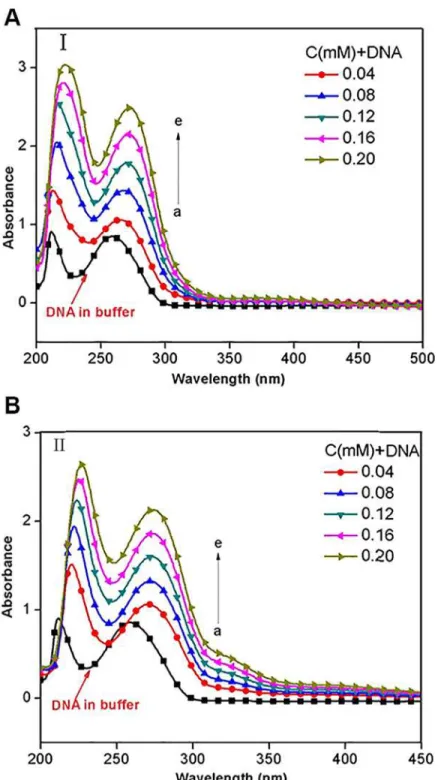
The UV-vis spectrum was performed in 0.1 M KCl / 0.05 M Tris—HCl buffer at pH 7.16 and a temperature of 25°C, in the wavelength range of 200–600 nm. The concentration of CT-DNA was kept constant and the concentrations of two compounds were ranged from 0.04 mM to 0.20 mM.
Results and Discussion
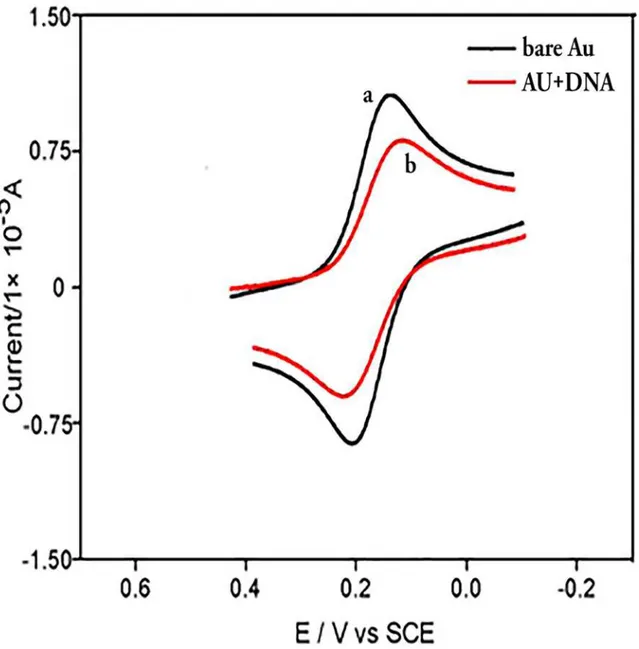
1. Electrochemical characterization of CT-DNA-modified electrode
2. Interaction of the compounds with CT-DNA-modified gold electrode
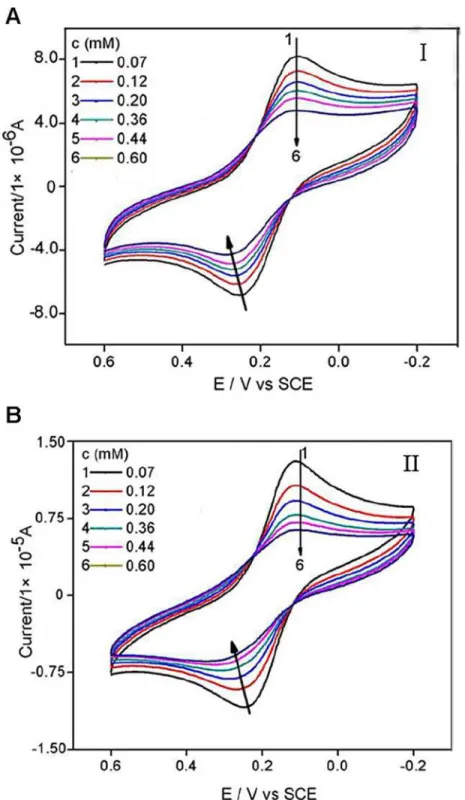
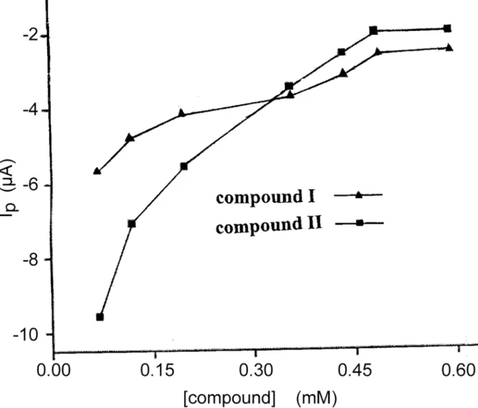
As was shown inFig. 3, the peak current of Fe(CN)63-/4-decreased as compounds (I or II) were added into the test solution. The more compound was added, the more the peak current of probe molecule decreased. As shown inFig. 4, both peak currents of the cyclic voltammograms decreased with the concentrations of compounds increasing and tended to achieve a saturation value, i.e. at about 0.60 mM, as expected according toLangmuiradsorption behaviour. The rea-son might be attributed to that CT-DNA films made the redox process of Fe(CN)63-/4-at the gold electrode more difficult due to the physical blockage as well as possible electrostatic
Fig 2. Cyclic voltammograms of Fe(CN)63-/4-in pH 7.16 Tris
—HCl buffer solution at a bare Au electrode (a) and CT-DNA/Au (b), respectively.The scan rate was 0.1 V s-1and the concentrations of Fe(CN)
63-/4-and KCl were 5 mM. doi:10.1371/journal.pone.0120211.g002
Fig 3. Cyclic voltammograms of Fe(CN)63-/4-in Tris
—HCl buffer solution (pH 7.16) containing different
concentrations of compound I and II.The scan rate was 0.1 V s-1 and the concentrations of Fe(CN)6 3-/4-and KCl were 5 mM.
repulsion, and that the addition of the two compounds to the solution caused the CT-DNA film denser due to molecular interaction, therefore harder for Fe(CN)63-/4-ions to
migrate through.
According to a previously reported method [22,23], it was assumed that CT-DNA and DRUG produced a single complex of DNADRUGmonly.
DNA þm DRUG$DNADRUGm ð1Þ
The constant (K) was descripted as follows:
K ¼ ½DNADRUGm
½DNA ½DRUGm ð1:1Þ
Fig 4. The dependence of decrease value of the peak current on the concentration of compound I and II.
doi:10.1371/journal.pone.0120211.g004
And the following equations could be deduced, where I indicated cyclic voltammetry current.
DIMAX¼k 0C
DNA ð1:2Þ
andDI¼k0 ½DNADRUG
m ð1:3Þ
½DNA þ ½DNADRUGm ¼CDNA ð1:4Þ
DImax DI¼k 0ðC
DNA ½DNADRUGmÞ ð1:5Þ
DImax DI¼k
0 ½DNA ð1
:6Þ
As equations (1.3) and (1.6) were put into (1.1), it yielded: log DI
DImax DI
¼log Kþm log½DRUG ð1:7Þ
1
DI¼
1
DImax
þ 1
DImaxK
1
½DRUGm ð1:8Þ
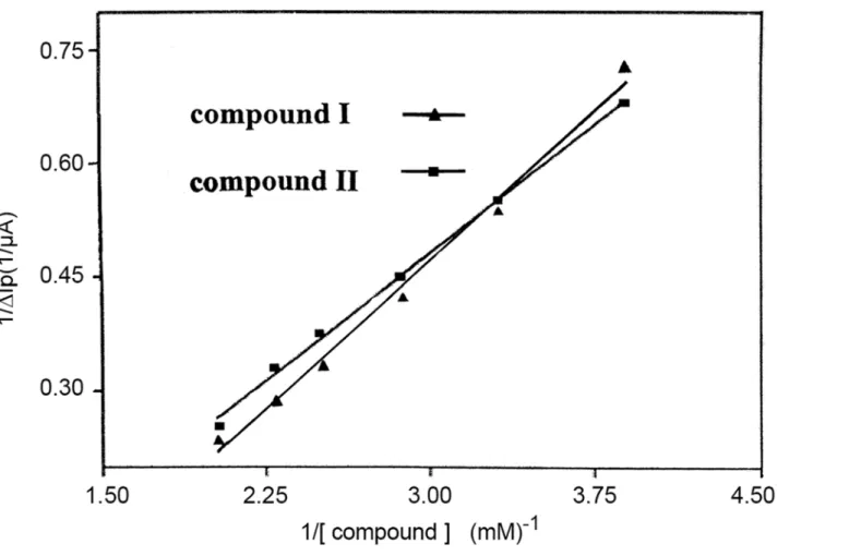
As forEquation (1.8), we assumedm= 1, usingΔIPto representΔI, andΔIp, maxto represent ΔImax. As shown inFig. 5, 1/[DRUG] demonstrated a good linear relationship with 1/ΔIp,
indi-cating reasonable assumptive value ofmfor compounds I and II. Thus, theequation (1.9) was deduced [24], whereΔIp= Ip-Ipo, Ipand Iporepresent the oxidation peak current of Fe
(CN)63-/4-in the presence and absence of the drugs,respectively;ΔIp,maxwas the maximum
dif-ference of the oxidation peak current; and [DRUG] represented the concentration of the drug.
1
DIP
¼ 1
DIp;max
þ 1
DIp;maxK
1
½DRUG ð1:9Þ
The binding constant (K) between the compounds and CT-DNA, 1.13×10⁶M-1and 5.35 ×10⁵M-1for compound I and II respectively, was calculated according to theequation (1.9). These values were typical for metal compounds that bind to DNA via intercalation mode [25]. The binding constant of compound I was about 2.1 times larger than that of compound II, which indicated that the CT-DNA-binding strength of compound I was stronger than that of compound II. A stronger bonding indicated a more stable combination with CT-DNA and thus a better anticancer activity [26].
3. Ultraviolet-visible absorbance spectra
Generally, the DNA double-helix structural change, which is caused by its binding with other molecules, may be revealed in spectral features of hyperchromism (rise in absorption intensity) and hypochromism (fall in absorption intensity). The hyperchromism arises from disassembl-ing of DNA double strand [27–28] upon molecular contact, and has been observed frequently in the interaction of DNA with porphyrins, phenanthroliens and salophen complex [29]. In contrast, the hypochromism is resulted from the tightenning of DNA duplex assembly [30–32].
concentrations of compoundI. When the compoundIconcentration reached 0.08 mM, the hyperchromism was 49.4% and the red shift was 14–16 nm from band 260 nm. In contrast, as shown inFig. 6B, the hyperchromism of 53.5% and the red shift of about 13 nm from band 260 nm were observed for compoundII.
The hyperchromism appearing in absorbance around 258 nm without any hypsochromic effect, but with a bathochromic shift, can be attributed to the breaking of hydrogen bonds be-tween complementary DNA strands, which resulting from intercalations of DNA with small molecules that leads the opening of DNA double helix [33–34]. The present results indicated the existence of strong intercalation of CT-DNA with the two compounds, this was consistent with a typical berberine-DNA intercalation which caused a hyperchromism of 35% with a red shift about 15 nm [35]. Further, the red shift might be ascribed to the intercalation of CT-DNA base pairs with aromatic chromophores of cymene and pyridyl [36–37], due to the decrease in energy gap between the highest and the lowest molecular orbitals (HUMO and LUMO) after CT-DNA binding with the compounds [38].
Conclusions
Cyclic voltammetry showed that the two compounds, [(η6-p-cymene)Ru(L1)Cl2and
(η6-p-cymene)Ru(L1)Cl2], were adsorbed by CT-DNA which was coated on gold electrode sur-face, the adsorption behavior was fit forLangmuirequation. Further, the hyperchromism of ultraviolet-visible absorbance spectra revealed the nature of the adsorption was intercalation of
Fig 5. The relationship between 1/ΔIPand 1/C.
doi:10.1371/journal.pone.0120211.g005
the two half-sandwich ruthenium(II) compounds with CT-DNA. The binding constant of compound I was approximately 2.1 times larger than that of compound II, which indicated that the compound I showed stronger CT-DNA binding ability than compound II. It might be expected boldly that compound I was a better antitumor agent than compound II.
Fig 6. UV absorption spectra of CT-DNA (0.02mM) with various concentrations of compound I (A) and II (B).
Supporting Information
S1 Fig. The infrared spectrum of compound I.
(TIF)
S2 Fig. The1H-NMR spectrum of compound I.
(TIF)
S3 Fig. The13C-NMR spectrum of compound I.
(TIF)
S4 Fig. The infrared spectrum of compound II.
(TIF)
S5 Fig. The1H-NMR spectrum of compound II.
(TIF)
S6 Fig. The13C-NMR spectrum of compound II.
(TIF)
Acknowledgments
We thank Qian Miao for technical assistance, Shun Wang for many helpful discussions.
Author Contributions
Conceived and designed the experiments: ZMJ MLH ZJL YH DAQ. Performed the experiments: ZJL YH DAQ MLH ZMJ. Analyzed the data: ZMJ MLH ZJL YH DAQ. Contributed reagents/ materials/analysis tools: MLH ZJL YH DAQ ZMJ. Wrote the paper: ZJL ZMJ MLH YH DAQ.
References
1. Ronconi L, Sadler PJ (2007) Using coordination chemistry to design new medicines. Coord Chem Rev 251: 1633−1648.
2. Ruiz J, Rodríguez V, Cutillas N, Espinosa A, Hannon MJ (2011) A potential ruthenium (II) antitumor complex bearing a lipophilic levonorgestrel group. Inorg Chem 50: 9164−9171. doi:10.1021/ ic201388nPMID:21830785
3. Dougan SJ, Sadler PJ, Chimia (2007) The design of organometallic ruthenium arene anticancer agents. Int J Chem 61: 704–715.
4. Conejo-Garcia A, Schofield CJ (2005) A prodrug system for hydroxylamines based on esterase cataly-sis. Bioorg Med Chem Lett 15: 4004–4009. PMID:15990293
5. Liu KG, Cai XQ, Li XC, Qin DA, Hu ML (2012) Arene-ruthenium(II) complexes containing 5-fluorouracil-1-methyl isonicotinate: Synthesis and characterization of their anticancer activity. Inorg Chim Acta 388: 78−83.
6. Shi Y, Guo CL, Sun YJ, Liu ZL, Xu FG, et al. (2011) Interaction between DNA and Microcystin-LR stud-ied by Spectra Analysis and Atomic Force Microscopy. Biomacromol 12: 797−803.
7. Ding YH, Zhang L, Xie J, Guo R (2010) Binding characteristics and molecular mechanism of tnteraction between ionic liquid and DNA. J Phys Chem B 114: 2033−2043. doi:10.1021/jp9104757PMID: 20088558
8. Elder RM, Emrick T, Jayaraman A (2011) Understanding the effect of polylysine architecture on DNA binding using molecular dynamics simulations. Biomacromol 12: 3870−3879.
9. Campbell NH, Smith DL, Reszka AP, Neidle S, O'Hagan D (2011) Fluorine in medicinal chemistry:
β-fluorination of peripheral pyrrolidiness attached to acridine ligands affects their interactions with G-quadruplex DNA. Org Biomol Chem 9: 1328−1331. doi:10.1039/c0ob00886aPMID:21221451 10. Akiyama Y, Ma Q, Edgar E, Laikhter A, Hecht SM (2008) Identification of strong DNA binding motifs for
bleomycin. J Am Chem Soc 130: 9650–9651. doi:10.1021/ja802905gPMID:18597467
11. Lyles M. B, Cameron IL (2002) Interactions of the DNA intercalator acridine orange, with itself, with caf-feine, and with double stranded DNA. Biophys Chem 96: 53−76. PMID:11975993
12. Froehlich E, Mandeville JS, Kreplak L, Tajmir-Riahi HA (2011) Aggregation and particle formation of tRNA by dendrimers. Biomacromol 12: 2780−2787.
13. Liang MM, Liu XR, Liu GZ, Dou SP, Cheng DF, et al. (2010) Reducing the background fluorescence in mice receiving fluorophore/inhibitor DNA duplexes. Mol Pharm 8: 126−132. doi:10.1021/mp100229z PMID:21133414
14. Tong CL, Xiang GH, Bai Y (2010) Interaction of paraquat with calf thymus DNA: a terbium (III) fluores-cence probe and multi-spectral study. J Agric Food Chem 58: 5257−5262. doi:10.1021/jf1000748 PMID:20402507
15. Sinha R, Saha I, Kumar GS (2011) Protoberberine alkaloids berberine, palmatine, and coralyne binding to poly(dT).poly(dA)*poly (dT) triplex: comparative structural aspects and energetics profiles of the in-teraction. Chem Biodiv 8: 1512−1528.
16. Bathaie SZ, Movahedi AAM, Saboury AA (1999) Energetic and binding properties of DNA upon interac-tion with dodecyl trimethylammonium bromide. Nucleic Acids Res 27: 1001−1005. PMID:9927732 17. Yin P, Hu ML, Hu LC (2008) Synthesis, structural characterization and anticarcinogenic activity of a
new Gly-Gly dipeptide derivative: Methyl 2-(2-(5-fluoro-2,4-dioxo-3,4-dihydropyrimidin-1-(2H)-yl) ceta-mido) acetate. J Mol Struct 882: 75−79.
18. Pang DW, Abruna HD (1998) Micromethod for the investigation of the interactions between DNA and redox-active molecules. Anal Chem 70: 3162−3169. PMID:11013719
19. Jin BK, Ji XP, Nakamura T (2004) Voltammetric study of interaction of Co(phen)33+with DNA at gold nanoparticle self-assembly electrode. Electrochim Acta 50: 1049−1055.
20. Erkkila KE, Odom DT, Barton JK (1999) Recognition and reaction of metallointercalators with DNA. Chem Rev 99: 2777−2795. PMID:11749500
21. Carter MT, Rodriguez M, Bard AJ (1989) Voltammetric studies of the interaction of metal chelates with DNA. 2. Tris-chelated complexes of cobalt(III) and iron(II) with 1,10-phenanthroline and 2,2'-bipyridine. J Am Chem Soc 111: 8901−8911.
22. Xu ZQ, Zhou B, Jiang FL, Dai J, Liu Y (2013) Interaction between a cationic porphyrin and ctDNA inves-tigated by SPR, CV and UV—vis spectroscopy. Coll Surf B: Biointer 110: 321−326.
23. Kalanur SS, Seetharamappa J, Prashanth SN (2011) Interaction of an antidepressant buzepide methio-dide with DNA immobilized on the glassy carbon electrode. Coll Surf B: Biointer 82: 438−442. 24. Liu SQ, Xu JJ, Chen HY (2004) A reversible adsorption—desorption interface of DNA based on
nano-sized zirconia and its application. Coll Surf B: Biointer 36: 155−159.
25. Li LL, Cao WQ, Zheng WJ, Fan CD, Chen TF (2012) Ruthenium complexes containing 2,6-bis(benzimi-dazolyl)pyridine derivatives induce cancer cell apoptosis by triggering DNA damage-mediated p53 phosphorylation. Dalton Transact 41: 12766−12772. doi:10.1039/c2dt30665dPMID:22968364 26. Li XC, Liu KG, Qin DA, Cheng CC, Chen BX, et al. (2012) Influence of bromoethyl group on biological
activity of 5-fluorouracil prodrug: Insights from X-ray crystallography and molecular docking. J Mol Struct 1027: 104–110.
27. Hobro AJ, Rouhi M, Blanch EW, Conn GL (2007) Raman and raman optical activity (ROA) analysis of RNA structural motifs in Domain I of the EMCV IRES. Nucl Acids Res 35: 1169−1177. PMID:17264119 28. Li Q, Yang P, Wang H, Guo M (1996) Diorganotin (IV) antitumor agent. C2H5SnCl2(phen)/nucleotides
aquous and solid state coordination chemistry and its DNA binding studies J Inorg Biochem 64: 181−195. PMID:8893519
29. Kashanian S, Gholivand MB, Ahmadi F, Taravati A, Hosseinzadeh Colagar A. (2007) DNA interaction with Al—N,N0-bis(salicylidene) 2,20-phenylendiamine complex. Hosseinzadeh Colagar, Spectrochim.
Acta Part A Mol Biomol Spectrosc 67: 472−478. PMID:17011818
30. Rahban M, Divsalar A, Saboury AA, Golestani A (2010) Nanotoxicity and spectroscopy studies of silver nanoparticle: Calf thymus DNA and K562 as targets. J Phys Chem C 114: 5798–5803.
31. Wang BD, Yang ZY, Li TR (2006) Synthesis, characterization, and DNA-binding properties of the Ln(III) complexes with 6-hydroxy chromone-3-carbaldehyde-(2'-hydroxy) benzoyl hydrazine. Bioorg Med Chem 14: 6012–6021. PMID:16781158
32. Wang BD, Yang ZY, Crewdson P, Wang DQ (2007) Synthesis, crystal structure and DNA-binding stud-ies of the Ln(III) complex with 6-hydroxychromone-3-carbaldehyde benzoyl hydrazoneJ Inorg Biochem 101: 1492−1504. PMID:17692381
33. Kashanian S, Gholivand MB, Ahmadi F, Ravan H (2008) Interaction of diazinon with DNA and the protective role of selenium in DNA damage. DNA Cell Biol 27: 325−332. doi:10.1089/dna.2007.0718PMID:18447756 34. Arjmand F, Mohani B, Parveen S (2006) New dihydro O,O0-bis(salicylidene) 2,20aminobenzothiazolyl
35. Li XL, Hu YJ, Wang H, Yu BQ, Yue HL (2012) Molecular spectroscopy evidence of berberine binding to DNA: comparative binding and thermodynamic profile of intercalation. Biomacromol 13: 873–880. 36. Arjmand F, Mohani B, Ahmad S (2005) Synthesis, antibacterial, antifungal activity and interaction of
CT-DNA with a new benzimidazole derived Cu (II) complex. Eur J Med Chem 40: 1103–1110. PMID:
16006016
37. Baidini M, Ferrari MB, Bisceglie F, Pelosi G, Pinelli S, et al. (2003) Cu(II) complexes with heterocyclic substituted thiosemicarbazones: the case of 5-Formyluracil synthesis, characterization, X-ray struc-tures, DNA interaction studies, and biological activity. Inorg Chem 42: 2049–2055. PMID:12639140 38. Tan JH, Lu Y, Huang ZS, Gu LQ, Wu JY (2007) Spectroscopic studies of DNA binding modes of
cation-substituted anthrapyrazoles derived from emodin. Eur J Inorg Chem 42:1169–1175.