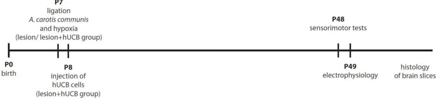
Human umbilical cord blood cells restore brain damage induced changes in rat somatosensory cortex.
Texto
Imagem
Documentos relacionados
Loss of cannabinoid CB1 receptor expression in the 6-hydroxydopamine- induced nigrostriatal terminal lesion model of Parkinson’s disease in the rat. Brain
Fig 4: Hypoxia and a microdosage stromal cell-derived factor-1α (SDF-1α) augmented the surface expression of cognate recep- tor CXC chemokine receptor 4 (CXCR4) on mesenchymal
Three weeks after cell therapy, electrocardiographic parameters in the groups treated with fresh and cryopre- served hUCB cells were typical of infarcted animals..
Effect of hyperbaric oxygenation on mitochondrial function of neuronal cells in the cortex of neonatal rats after hypoxic-ischemic brain damageL. Hu
The increase in sand content in aggregates in the clay plantation soil was not accompanied by any statistically significant changes in C contents or concentrations, but the decrease
In the group to which 0.5% levobupivacaine was administered (Group L4) staining was shown in the extracellular matrix structure in the calcified cartilage area ( Fig.. An increase
Regarding the secondary outcomes, brain relaxation was better achieved in the hypertonic saline group, since brain relax- ation in the mannitol group was considered inadequate in
However, we found that across the monetary and the physical condition subjects showed the same level of trust and reciprocity. Moreover, subjects’ behavior across
