Positive Selection of Natural Poly-Reactive B
Cells in the Periphery Occurs Independent of
Heavy Chain Allelic Inclusion
Ying Xing1,2,3, Qiuhe Ji2, Ying Lin4, Meng Fu1, Jixin Gao1, Ping Zhang4, Xingbin Hu3, Lei Feng3, Yufeng Liu1, Hua Han3*, Wei Li1*
1Department of Dermatology, Xijing Hospital, Fourth Military Medical University, Xi’an, China,2Department of Endocrinology and Metabolism Disease, Xijing Hospital, Fourth Military Medical University, Xi’an, China, 3State Key Laboratory of Cancer Biology, Department of Medical Genetics and Developmental Biology, Fourth Military Medical University, Xi’an, China,4Department of Otolaryngology Head and Neck surgery, Xijing Hospital, Fourth Military Medical University, Xi’an, China
*liwei1@fmmu.edu.cn(WL);huahan@fmmu.edu.cn(HH)
Abstract
Natural autoreactive B cells are important mediators of autoimmune diseases. Receptor ed-iting is known to play an important role in both central and peripheral B cell tolerance. How-ever, the role of allelic inclusion in the development of natural autoreactive B cells is not clear. Previously, we generatedμchain (TgVH3B4I) andμ/κchains (TgVH/L3B4) transgenic
mice using transgene derived from the 3B4 hybridoma, which produce poly-reactive natural autoantibodies. In this study, we demonstrate that a considerable population of B cells ed-ited their B cells receptors (BCRs) via light chain or heavy chain allelic inclusion during their development in TgVH3B4I mice. Additionally, allelic inclusion occurred more frequently in
the periphery and promoted the differentiation of B cells into marginal zone or B-1a cells in TgVH3B4I mice. B cells from TgVH/L3B4 mice expressing the intact transgenic 3B4 BCR
without receptor editing secreted poly-reactive 3B4 antibody. Interestingly, however, B cell that underwent allelic inclusion in TgVH3B4I mice also produced poly-reactive
autoantibod-ies in vivo and in vitro. Our findings suggest that receptor editing plays a minor role in the positive selection of B cells expressing natural poly-reactive BCRs, which can be positively selected through heavy chain allelic inclusion to retain their poly-reactivity in the periphery.
Introduction
The ability of B cells receptor (BCR) variable (V) region gene fragments to rearrange randomly during early B cell development is of great significance. It not only increases the diversity of BCR specificities [1], but also increases the possibility of autoantibody production. It has been suggested that the prevalence of poly-reactive B cells to various autoantigens is more than 50% in early B cells precursors [2]. However, this number is reduced to approximately 5% after B cell maturation. Many studies based on immunoglobulin (Ig) gene transgenic mice have shown that the deletion of autoreactive B cell clones is induced by central tolerance mechanisms,
OPEN ACCESS
Citation:Xing Y, Ji Q, Lin Y, Fu M, Gao J, Zhang P, et al. (2015) Positive Selection of Natural Poly-Reactive B Cells in the Periphery Occurs Independent of Heavy Chain Allelic Inclusion. PLoS ONE 10(5): e0125747. doi:10.1371/journal. pone.0125747
Academic Editor:Antonio A Freitas, Institut Pasteur, FRANCE
Received:November 29, 2014
Accepted:March 25, 2015
Published:May 19, 2015
Copyright:© 2015 Xing et al. This is an open access article distributed under the terms of theCreative Commons Attribution License, which permits unrestricted use, distribution, and reproduction in any medium, provided the original author and source are credited.
Data Availability Statement:All relevant data are within the paper and its Supporting Information files.
Funding:National Natural Science Foundation of China: 30901336 (YX), 31070793 (WL), 81070663 (QJ), 81273320 (WL), 81271750 (YL). The funders had no role in study design, data collection and analysis, decision to publish, or preparation of the manuscript.
including clonal deletion, anergy and receptor editing [3–7], during B cells development. Among these mechanisms, receptor editing is critical for central B cells tolerance [8], through which autoreactive B cells that are destined for clonal deletion or anergy can be rescued by suc-cessful secondary rearrangement of their BCR genes. Receptor editing plays important roles in both positive and negative selection of autoreactive B cells [9], suggesting a relationship be-tween receptor editing and autoimmune diseases [10,11]. Consistently, the persistence of path-ological autoantibodies has been associated with attenuated receptor editing in the bone marrow (BM) or periphery in autoimmune disease mouse models and patients [12–14]. Stud-ies with other models have suggested that significant receptor editing is elicited in the develop-ment of autoreactive B cells [15–17]. However, there is no direct evidence showing that defects in receptor editing enhance autoantibody production in autoimmune diseases.
Most of the naturally-occuring autoantibodies are poly-reactive and exist in healthy individ-uals [18,19]. Recent studies have suggested that 5~20% of long-lived B cells are autoreactive in humans [2]. However, the role of receptor editing in the development of natural autoreactive B cells is not yet clear. Secondary recombination at the light (L) chain genetic loci generates a newμchain that can either substitute the autoreactive L chain [20], or can be co-expressed on the cell surface as a“passenger”together with the original L chain, and can also associate with the heavy (H) chain separately. This later phenomenon is referred to as allelic inclusion [21,
22] and is a result of receptor editing. The co-expression of an“innocent”L chain can rescue B cells from negative selection by diluting the surface expression of the self-reactive BCR [23]. In addition to L chains, secondary rearrangement of V genes also happens at the H chain loci [24,
25]. However, the extent and function of H chain allelic inclusion are unknown. Given the dominant role VHplays in antigen recognition, it will be important to clarify the relationship
between H chain allelic inclusion and receptor editing in the generation of natural autoreactive B cells, to reveal the mechanisms of B cell tolerance.
We have establishedμchain transgenic mice with the VHgene derived from 3B4 hybridoma
producing a natural autoantibody [26]. Nine founders were generated with different allelic ex-clusion efficiency. In the present study, B cells from one founder line (named as TgVH3B4I)
with apparent allelic inclusion and receptor editing were analyzed. We also generatedκchain transgenic mice (TgVL3B4) with the VLgene from the same 3B4 hybridoma and double
trans-genic mice (TgVH/L3B4) were created by breeding TgVH3B4 mice and TgVL3B4 mice. In
con-trast to B cells from TgVH3B4I mice expressing 3B4μchain, we did not observe any significant receptor editing in B cells from TgVH/L3B4 mice which expressed the whole 3B4 natural
poly-reactive BCR. B cells expressing endogenous self-poly-reactive L chains escaped negative selection by H chain allelic inclusion in the periphery, and these B cells differentiated into special subsets with the ability to secrete poly-reactive antibodies, just like B cells expressing the integrated 3B4 BCR in TgVH/L3B4 mice without allelic inclusion. Our findings suggest that B cells with
natural poly-reactivity can be positively selected with or without H chain allelic inclusion.
Materials and Methods
Ethics Statements
Mice
To construct TgVL3B4 mice, the VLgene fragment was amplified by polymerase chain reaction
(PCR) with cDNA from 3B4 hybridoma as a template, and was used to replace VLgene in
plas-mid Lκ[27], which contains the promoter region, signal peptide fragment, and J region. A 2kb fragment containing intron enhancer from pBSNB1 and a 10 kb fragment containing the Cκ region, major intron and 3’enhancer derived from plasmid K1 were then inserted, to generate theκchain transgene construct pBSCk-2Vk4 (S1 Fig). The 13.5 kb transgene fragment was ex-cised from pBSCk-2Vk4 byEcoRI restritction digestion, purified, and microinjected into the
pronuclei of fertilized (C57BL/6×CBA) F1 eggs. Injected eggs were transferred to pseudopreg-nant females to produce transgenic mice. Founder lines were backcrossed with C57BL/6 mice for more than six generations before analyses. The TgVH/L3B4 double transgenic mice were
produced by crossing TgVH3B4I and TgVL3B4, and were genotyped by PCR (S1 Fig), with H
chain primers described in [26], and L chain primers50-CTTCCTGCTAATCAGTGCCTCAG
(KLB), and50-GTTAGATCTCGAGCTTGGTCC(VKF2). All mice were housed under specific
pathogen-free (SPF) conditions, with autoclaved food and water. Eight to twelve week old transgenic mice and age-matched transgene-free littermates were sacrificed for analysis.
FACS analysis
Single-cell suspensions prepared from the spleen, peritoneal cavity (PEC), lymph nodes (LN), and BM were treated with buffered 0.14 M NH4Cl. 5×10⁵cells were stained with antibodies in
PBS containing 2% fetal bovine serum and 0.2% NaN3 for 30 min on ice, followed by washing and filtrating through nylon mesh, and were then fixed in 1% paraformaldehyde or analyzed immediately on a Coulter Epics XL flow cytometer (Beckman coulter). Data were analyzed by using EXPO32 ADC Analysis software (Treestar, San Carlos, CA). Antibodies used in the anal-yses included anti-B220 (RA3-6B2), anti-IgMa (DS-1), anti-IgMb (AF6-78), anti-κ(187.1), anti-λ(RML-42), anti-CD19 (6D5), anti-IgM (R6-60.2), anti-CD5 (53–7.3), anti-CD21/35 (7G6), anti-CD23 (B3B4), anti-CD24 (M1/69), anti-CD43 (S7), anti-CD1d (1B1), anti-CD138 (281–2). For secondary staining, streptavidin-PE-Cy5, streptavidin-PE, and streptavidin-FITC were used in conjunction with biotinylated antibodies. Antibodies and fluorescence-labeled streptavidin were obtained from BD Biosciences (Mountain View, CA) or BD PharMingen (San Diego, CA). Apoptosis was detected using the Annexin V-FITC Apoptosis Detection Kit (BD PharMingen, San Diego, CA) following standard protocols. FACS-sorting was conducted using a FACS Vantage II (BD Immunocytometry Systems), and sorted cells were re-analyzed by using flow cytometer to confirm cell purity.
Confocal microscopy
Tissue samples were embedded in OCT, frozen at -80°C, and sectioned at 6μm thickness. Sec-tions were air-dried, fixed in ice-cold ethanol for 15 min, washed with PBS, and blocked with PBS containing 2% bovine serum albumin. Sections were then stained with B220-PE, anti-IgMa-PE plus anti-MOMA1-FITC (Rat IgG2a, Serotec), or anti-IgMa-PE plus anti-IgMb -FITC, at room temperature for 30 min, washed, and mounted in 50% glycerol. Images were re-corded with a FV1000 confocal microscopic system (Olympus), and were processed with Image Pro Plus 5.1 software.
analyzed with software Optical FluoView Ver1.5. Fluorescence intensity was normalized to its initial value recorded before 20μg/ml of actin application.
Reverse transcription PCR
B220-positive cells were sorted by using magnetic beads-conjugated anti-B220 and a magnetic column (Miltenyi Biotec, Auburn, CA). To the purified B cells Trizol reagent (Promega) was added immediately, and total RNA was prepared according to the manufacturer’s instructions. Approximately 1μg of total RNA in 20μl was reverse-transcribed with SuperScript II reverse transcriptase and random primers (Invitrogen Life Technologies). Rag2 was detected using the primers as described [28], withβ-actin [29] as a reference control.
Cell Culture
B220+cells were isolated from the spleen and PEC by using magnetic beads, and cultured at a density of 1×10⁵cells/ml with RPMI 1640 medium supplemented with 10% fetal bovine serum in U-bottomed 96-well culture plates (Costar) at 37°C. B cell were stimulated with 25μg /ml lipopolysacharide (LPS), 25μg /ml anti-IgM, or 15μg /ml anti-CD40 plus 50 ng/ml interleu-kin-4 were added as indicated in the different experiments.
Enzyme-linked immunosorbent assay (ELISA)
Solid-phase ELISA was performed as described previously [30].
Statistics
Pairedt-test and one-way ANOVA were used to determine the statistical significance of values
between groups. The statistical significance was defined asP<0.05.
Results
H chain allelic inclusion and receptor editing in B cells of TgV
H3B4I mice
TgVH3B4I mice carry aμchain transgene with VHderived from the 3B4 hybridoma that
se-crete a poly-reactive natural antibody recognizing keratin, actin, myosin and many foreign an-tigens [26,30]. To evaluate H chain allelic exclusion in adult TgVH3B4I mice, we took
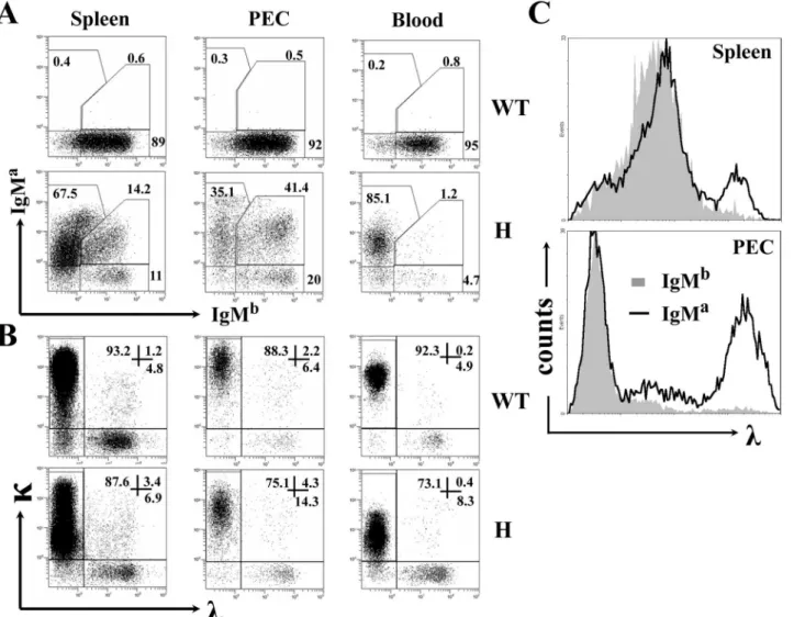
advantage of the allotypic difference between the transgenic IgMaand endogenous IgMbof C57BL/6 mice. As shown inFig 1A(left panels), most splenic B cells from TgVH3B4I mice
ex-pressed IgMaand a fraction (>14%) expressed both IgMaand IgMbsimultaneously, when compared with B cells from C57BL/6 mice. This proportion was more significant in PEC, as more than 40% of the B cells in the PEC of TgVH3B4I mice expressed both IgMaand IgMb,
while B cells expressing IgMbonly increased marginally (Fig 1A, middle panels). Interestingly, we found only a few B cells with H chain allelic inclusion (~1%) in the blood (Fig 1A, right pan-els) and BM (data not shown) of TgVH3B4I mice. These results indicated that peripheral B
cells underwent H chain allelic inclusion in TgVH3B4I mice.
Receptor editing can be elicited by the biased expression of L chains. We found that receptor editing occurred in a significant percentage of each B cell population in TgVH3B4I mice, as
evi-denced by the augmented expression of theλchain (Fig 1B). Notably, mostλ+B cells from PECs of TgVH3B4I mice were IgMa+that constituted the autoreactive BCR (Fig 1C, lower
B cells with H chain allelic inclusion develop preferentially into MZ and
B-1a cells in TgV
H3B4I mice
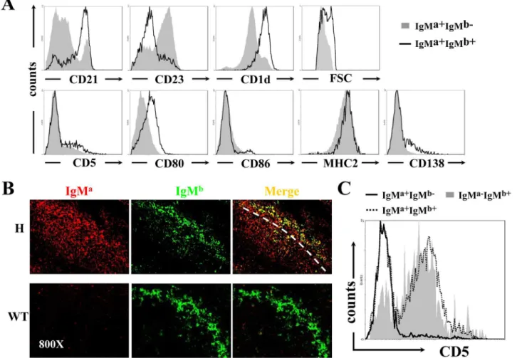
To investigate the effects of H chain allelic inclusion on B cell development in TgVH3B4I mice,
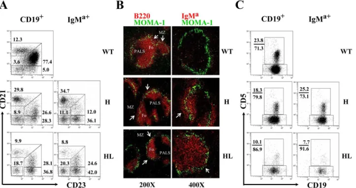
we examined the phenotypes of IgMa+B cells, which could be divided into IgMa+IgMb-(allelic excluded) and IgMa+IgMb+(allelic included) subpopulations in the spleen and PEC. In the spleen, IgMa+IgMb+B cells expressed high level of CD21 and CD1d, but lower level of CD23 (Fig 2A, upper panels), a phenotype identical to that of marginal zone (MZ) B cells. In contrast, most IgMa+IgMb-B cells had a follicular (Fo) B cell-like phenotype of CD21lowCD23high CD1-dlow. Histological analysis using anti-IgMaand anti-IgMbconfirmed that B cells with allelic in-clusion were mainly located at the MZ of the spleen in TgVH3B4I mice when compared with
C57BL/6 mice (Fig 2B). Thus, B cells with H chain allelic inclusion, which were most likely poly-reactive, mainly developed into MZ B cells in the spleen of TgVH3B4I mice. Moreover, Fig 1. H and L chain expression on peripheral B cells from TgVH3B4I.(A) Allelic inclusion of IgH chain on peripheral B cells. Cells from spleen, PEC and
peripheral blood of the indicated mice were labeled with anti-IgMa, anti-IgMb, and anti-CD19, and were analyzed by FACS. CD19+cells were gated on lymphocytes. The numbers indicate the percentage of cells in total CD19+cells within the indicated gates. (B) L chain expression in B cells from TgVH3B4I. Cells were stained with anti-κ, anti-λand anti-CD19 antibodies and were analyzed, as in A. (C)λ-expressing cells are mainly IgMa+in TgV
H3B4I mice. Histograms showλchain expression in IgMb+(solid fill) and IgMa+(no fill, including IgMa+IgMb+and IgMa+IgMb-) cells from splenocytes and PEC of TgVH3B4I mice. Data are representative of more than eight independent experiments with at least three mice in each group.
allelic included B cells expressed slightly higher level of activation markers, such as CD80 and MHC-II (Fig 2A, lower panels), and had larger cellular size than IgMasingle positive B cells (Fig 2A), suggesting BCR engagement or activation [31,32].
In the PEC of TgVH3B4I mice, almost all of the IgMa+IgMb-B cells were CD5-(Fig 2C),
in-dicating that they belonged to B-2 and B-1b subsets. In contrast, the majority of IgMa+IgMb+B cells were CD5+B-1a cells (Fig 2C). These results suggested that B cells with allelic inclusion preferentially developed into B-1a cells in PECs of TgVH3B4I mice.
Analysis of B cell development in the BM of TgV
H3B4I and TgV
H/L3B4
mice
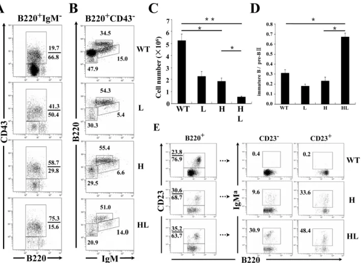
Our data showed that transgenic expression of 3B4 H chain resulted in significant receptor ed-iting and H chain allelic inclusion. Next, we investigated the BM development of original 3B4 B cells. The 3B4κchain transgenic mouse line TgVL3B4 was established and was bred with
TgVH3B4I to generate TgVH/L3B4 mice. In the BM, where clonal deletion and receptor editing
of autoreactive B cells mainly takes place [33,34], both the percentage and absolute number of
Fig 2. Accumulation of B cells with allelic inclusion in the MZ of spleen and their differentiation into B-1a subset in PEC of TgVH3B4I mice.(A)
Phenotypic analysis of IgMa+IgMb-(solid fill) and IgMa+IgMb+(no fill) B cells for the indicated cell surface markers by flow cytometry from splenocytes of TgVH3B4I mice. (B) IgMa+IgMb+B cells localized in the MZ of spleen of TgVH3B4I. Analysis of frozen spleen sections from TgVH3B4I mice, stained with anti-IgMa(red) and anti-IgMb(green), using confocal microscopy. (C) CD5 was expressed predominantly on IgMa-IgMb+and allelic included IgMa+IgMb+B cells. Representative histograms show the surface expression of CD5 on IgMa-IgMb+(solid fill), IgMa+IgMb-(solid line) and IgMa+IgMb+(dotted line) B cells of PEC from TgVH3B4I mice. Data are representative of more than five independent experiments with three mice per group.
total B cells were significantly reduced in different transgenic lines compared to C57BL/6 mice (Fig3A–3C), most likely due to the early expression of the Ig transgenes [35,36]. Indeed, this decrease was mainly attributable to the reduction of early B cells, because IgM-B cell precur-sors decreased most significantly (Fig3A–3Cand data not shown). There was also a severe re-duction of preBII cells (IgM-CD43-) (Fig 3A) in TgVH/L3B4 mice, as evidenced by the highest
ratio of pro/preBI (IgM-CD43+) to preBII cells in this line (Fig 3D). This might explain the ob-served significant reduction in total B cells number in the BM of TgVH/L3B4 mice (Fig 3C),
be-cause the preBII stage is critical to the proliferation of BM B cells.
B220intIgMhighCD43-immature B cells were comparable between TgVH/L3B4 (~14%) and
control littermates (~15%), (Fig 3B). In contrast, the proportion of immature B cells decreased significantly in TgVH3B4I and TgVL3B4 single transgenic mice (Fig 3B), consistent with a
de-velopmental block at the immature stage in B cells expressing single transgenic L or H chains. A higher ratio of immature B to preBII was noticed in TgVH/L3B4 mice, suggesting that more
immature B cells were generated in this line compared with the H or L chain single transgenic
Fig 3. Assessment of B cell development in the BM of transgenic mice.BM cells from the indicated mouse strains were stained with B220, anti-CD43, and anti-IgM, and were analyzed by FACS. (A) The gates in panels represent Hardy fractions A-D (proB to preBII), and can be divided into fraction A-C (proB to preBI, upper rectangle) and fraction D (preBII, lower rectangle) by the expression of CD43. The numbers indicate the percentage of cells within the gates. (B) Hardy fractions D (preBII, lower left panels), F (circulating mature B cells, upper middle panels) and E (immature B cells, lower right panels) of the indicated mice. (C) Decreased absolute numbers of total BM B cells in transgenic mice. Numbers of B220+cells were calculated and shown. (D) Numbers of preBII and immature B cells in B were calculated, and the ratios of immature cells to preBII cells were calculated as shown. Error bars represent means±SD,*P<0.05,**P<0.01, n = 5. (E) FACS analysis of BM B cells from the indicated transgenic mice. CD23-(newly formed) and CD23+(mature) B cells in the B220+gate are shown in the boxes. Data are representative of more than five independent experiments.
mice (Fig 3D). Similarly, in the CD23-immature B cell stage, the percentage of IgMa+cells was much higher in TgVH/L3B4 than in TgVH3B4I (Fig 3E). These results suggested that B cells
ex-pressing the 3B4 natural poly-reactive BCR could be positively selected in the BM.
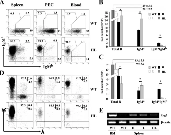
Higher allelic exclusion efficiency in the B cells from TgV
H/L3B4 mice
Over 90% of splenic, PEC and blood B cells in TgVH/L3B4 mice expressed IgMaalone, while
less than 3% expressed IgMb(Fig 4A). Fewer number of B cells expressed both IgMaand IgMb in the spleen and PEC in TgVH/L3B4 mice, in contrast to TgVH3B4I mice in which more B
cells underwent allelic inclusion in the spleen and PEC (Fig4Band4C). This suggested that al-lelic inclusion was suppressed in TgVH/L3B4 mice compared to the H chain transgenic mice.
Consistently, the number of IgD+B cells was more than three folds lower in spleen and PEC of TgVH/L3B4 than that of TgVH3B4I mice (S2 Fig).
Fig 4. High allelic exclusion of H and L chains in peripheral B cells of TgVH/L3B4 mice.(A) Expression of H chains in the peripheral B cells. Cells from
spleen, PEC and peripheral blood of TgVH/L3B4 mice were stained and analyzed as inFig 1A. (B-C) Bar graph shows the absolute numbers of total, IgMa +
IgMb-and IgMa+IgMb+B cells as gated in A in the spleen (B) and PEC (C) of the indicated mice. Error bars show mean±SD,*P<0.05. (D) L chain expression in B cells from TgVH/L3B4 mice. Cells were stained with anti-κ, anti-λand anti-CD19, and were analyzed by FACS. (E) RT-PCR analysis of Rag2 mRNA expression in B cells of spleen and BM from transgenic mice and littermates. Data are representatives of five independent experiments.
Lower percentage of B cells expressedλL chain in the spleen, PEC and blood in TgVH/L3B4
mice compared to controls (Fig 4D). Theλ+B cells were even lower than that in TgVL3B4 mice
(data not shown), suggesting reducedλchain inclusion and receptor editing. Consistently, RAG2 [37] mRNA transcript level increased in the splenic B cells of TgVH3B4I and TgVL3B4
mice, but was comparable to wild type in TgVH/L3B4 mice (Fig 4E). These results indicated
that receptor editing and allelic inclusion were induced in B cells of single H or L chain trans-genic mice, but were suppressed in B cells overexpressing the intact 3B4 BCR.
Evaluation of B cell development in the periphery of TgV
H/L3B4 mice
The percentage of MZ B cells (CD19+CD21highCD23low) increased significantly in TgVH3B4I
mice, but this population decreased in TgVH/L3B4 compared to WT littermates (Fig 5A).
Con-sistently, immunofluorescence analysis of spleen sections showed that compared to TgVH3B4I
mice, with normal or somewhat enlarged MZ B cell area, in TgVH/L3B4 mice the MZ B cell
area was significantly reduced (Fig 5B). However, Fo B cells were significantly reduced in both the mouse lines, likely due to the reduced BM cellular export (S3 Fig). We did not observe a sig-nificant difference in the apoptosis of Fo B cells and MZ B cells of TgVH3B4I and TgVH/L3B4
mice (S4 Fig), suggesting that the differential development of splenic B cells in TgVH3B4 and
TgVH/L3B4 mice was most likely due to altered positive selection. Moreover, a majority of
splenic B cells from TgVH3B4I and TgVH/L3B4 mice developed into a particular subset with a
phenotype of CD21-CD23highCD24low, distinct from the typical T2 phenotype of CD21 int-high-CD23highCD24high(Fig 5Aand data not shown). Most of these cells termed as T2’, were IgMa+
Fig 5. Analysis of B cells development in spleen and PEC of transgenic mice.(A) The phenotype of B cells in the spleen of TgVH3B4I and TgVH/L3B4 mice. CD19+or IgMa+splenic B cells from indicated mice were analyzed for the expression of CD21 and CD23 by FACS. Numbers next to each gate indicate the percentage of cells in that gate in total CD19+or IgMa+cells. At least 7 mice from each genotype were analyzed. (B) Spleen sections from the indicated mice were stained with anti-MOMA1-FITC and anti-B220-Biotin (left panels) or anti-IgMa-biotin (right panel) followed by streptavidin-Cy3, and acquired using fluorescence microscopy. Follicular (Fo) areas around PALS are shown and MZ is indicated with arrow. The original magnitude was ×200 or ×400 as indicated. (C) CD19+or IgMa+B cells in PEC of indicated mice were evaluated for the expression of CD5 by FACS. At least 5 mice from each genotype were analyzed. Numbers next to each gate indicate the percentage of cells in that gate in total CD19+or IgMa+cells.
in both TgVH3B4I and TgVH/L3B4 (Fig5Aand5B, right panels). Similarly, B cells in the blood
of TgVH3B4I and TgVH/L3B4 were also mainly composed of T2’cells, which were distinct
from circulating B cells of the controls (data not shown). These results indicated that 3B4 poly-reactive B cells developed into special B cell subsets.
In the PEC of TgVH3B4I mice, the percentage of B-1 cells (CD19+Mac1+) increased but
CD5+B-1a cells decreased (data not shown). However, within the IgMa+population of TgVH3B4I mice, the percentage of CD5+B-1a cells increased notably (Fig 5C, middle panels).
As discussed previously, majority of these CD5+IgMa+cells were B cells with H chain allelic in-clusion (Fig 2C). In TgVH/L3B4 mice, while B-2 cells (CD19+Mac1-) were comparable to the
lit-termate controls, more than 80% of B-1 cells were B-1b cells (CD19+Mac1+CD5-) (data not shown). IgMa+CD5+cells decreased more significantly (to about 10% of B cells) in TgVH/L3B4
mice when compared to TgVH3B4I mice (Fig 5C). The apparent reduction of B cell subsets in
the PEC of TgVH3B4I or TgVH/L3B4 mice could be the result of lower B cell production in the
BM or due to their negative selection in the PEC. These data were further supported by the lower number of B-1a and B-1b cells in PEC of TgVH/L3B4 compared with TgVH3B4I mice
(S3B Fig), in which B cells possibly escaped negative selection through allelic inclusion. Consis-tent with this opinion, the ratio of apoptotic CD5+and CD5-B cells was significantly higher in PEC of TgVH/L3B4 mice (21%/35%) compared with the littermate controls (5%/16%).
Howev-er, this ratio remained low in TgVH3B4I mice (8%/25%) (S4B Fig).
B cells can secrete poly-reactive autoantibodies independent of H chain
allelic inclusion
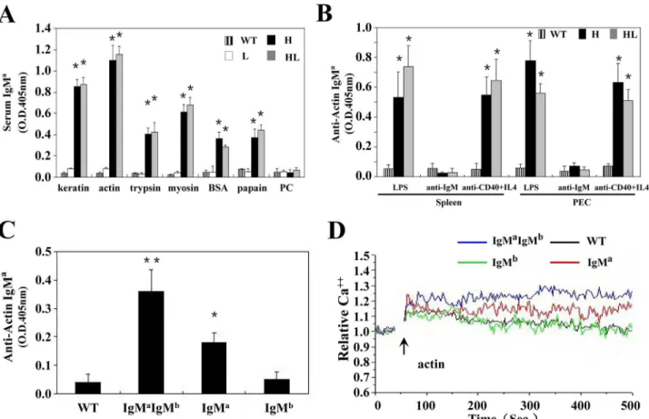
We next characterized the poly-reactivity of antibodies produced by cultured B cells from mice with allelic inclusion (TgVH3B4I) or not (TgVH/L3B4) by ELISA. B cells in both of TgVH3B4I
and TgVH/L3B4 mice produced high titer of IgMain the serum, with antigenic reactivity similar
to that of 3B4 hybridoma (Fig 6A), suggesting that the 3B4 H chain determined the poly-reac-tivity of these B cells in conjunction with 3B4 or allelic included L chains. Moreover, B cells from both spleen and PEC of TgVH3B4I and TgVH/L3B4 mice produced high concentration of
autoantibodies upon in-vitro stimulation with LPS or anti-CD40 (Fig 6B).
IgMa+b-, IgMa+b+, and IgMa-b+B cells, which represented H chain allelic excluded, included, and wild type B cells, respectively, were sorted from splenocytes of TgVH3B4I mice, and tested
for autoantibody production. IgMa+IgMb+cells secreted anactin autoantibodies at higher ti-ters than IgMa+b-cells upon LPS stimulation (Fig 6C), suggesting that autoantibody production was significantly contributed by the B cells that underwent allelic inclusion in the spleen of TgVH3B4I mice. Consistently, IgMa+IgMb+cells showed a relatively sustained and elevated
cal-cium flux than IgMa+b-B cells when stimulated with actin (Fig 6D). These effects were not ob-served in the IgMa-b+cells or wild type control. These data suggested that B cells in TgVH3B4I
mice with H chain allelic inclusion had a considerable ability to produce autoantibodies.
Discussion
In the present study, we compared B cells development in mice bearing H chain, L chain or H +L chains from 3B4 hybridoma, which produces poly-reactive antibodies recognizing autoanti-gens as well. Our results suggest that in TgVH3B4I mice, B cell development is accompanied by
allelic inclusion, which results from receptor editing, leading to the maturation of B cells secret-ing poly-reactive antibodies. B cells in TgVH3B4I might undergo negative selection at the first
tolerance checkpoint (the immature B cells stage), as we observed a remarkable decrease in im-mature B cells in TgVH3B4I mice. However, poly-reactive B cells entering the second tolerance
results in allelic inclusion. Allelic inclusion might rescue poly-reactive B cells from negative se-lection by creating dual receptor expression on their surface. Moreover, H chain allelic inclu-sion may serve as a special mechanism of positive selection to permit the survival of
autoreactive B cells in the periphery [38]. We also found that receptor editing occurred signifi-cantly in B cells of TgVH3B4I mice, as evidenced by the augmented expression of theλchain
(Fig 1B). Sirac has reported that light chain exclusion prevalent in normal B cells is neither tightly ensured by a stringent cell selection process nor absolutely required for normal B cell function [39]. Similarly, B cells of TgVH3B4I mice can further differentiate into mature B cell
compartments and produce poly-reactive antibodies. These findings in TgVH3B4I mice are
consistent with previous reports [23] showing that autoreactive antibodies in the serum of nor-mal mice and humans may be the“fellow travelers”in B cells with allelic inclusion.
When the original 3B4 L chain was introduced into TgVH3B4I mice, receptor editing was
severely suppressed. Few, if any,λ+B cells were found in BM or the periphery in TgVH/L3B4
mice. However, TgVH/L3B4 mice still produced high titers of poly-reactive antibodies in the
serum, indicating that the 3B4 natural poly-reactive B cells could develop into mature B cells without receptor editing. The low avidity of the 3B4 natural autoantibody could potentially help these B cells to escape negative selection mechanisms in both the BM and periphery. Meanwhile, 3B4 poly-reactive B cells were also positively selected through specific
Fig 6. Antibody production by allelic included B cells in TgVH3B4I.(A) ELISA. Sera from at least 5 mice from each genotype were collected, and
analyzed with ELISA using standard protocols. (B) In vitro secretion of autoantibodies by B cells. Purified B cells from spleen and PEC of indicated mice were treated with LPS, anti-IgM and anti-CD40+IL-4 for 3 days. Supernatants were collected and the concentration of secreted antibodies was assessed by ELISA. (C) B cells were sorted from spleens of TgVH3B4I mice and were treated with LPS, and antibody production was tested as in B. Error bars show means±SD.*P<0.05,**P<0.01, n = 5. (D) Calcium mobilization analysis. Sorted splenic B cells from TgVH3B4 mice and littermate controls were stained with Fluo3. Cells were stimulation with actin (20μg/ml) and analyzed by FACS.
recognition of certain autoantigens [40]. Our data suggest that in TgVH3B4I mice, the
incor-poration of secondary non-original L chains at early stages of B cell development would change the original avidity and/or specificity of the BCR, and subsequently trigger receptor editing in the BM and periphery. Alternately, in TgVH/L3B4 mice, B cells with intact 3B4
BCR would directly differentiate into mature B cell subsets without further receptor editing, and secrete poly-reactive autoantibodies. Therefore, receptor editing plays a minor role in the development of 3B4 poly-reactive B cells.
We observed that a majority of 3B4 B cells developed into a special B cells subset that we re-ferred to as T2’cells in the spleen of TgVH/L3B4 mice. These B cells were sensitive to the BCR
crosslink-induced apoptosis [41] (Fig 6Band data not shown), suggesting that they underwent negative selection before maturing into poly-reactive antibody secreting B cells. Although 3B4 hybridoma was originally developed from splenocytes [30], we could not define the phenotype of original 3B4 B cells. However, the T2’phenotype could result from the overexpression of 3B4 BCR on the B cell surface and consequently increased tonic signaling. In the PEC, the 3B4 B cells exhibited a phenotype reminiscent of CD5-“B-1b”cells, a B cells subset providing pro-tection from pathogens [42]. This could be a result of positive selection, depending on BCR sig-nal strength [43], as reported previously [44–46]. A similar situation might also exist in TgVH3B4I mice, in which the frequency of B cells with H chain allelic inclusion is high in the
MZ and B-1a cell compartments.
In conclusion, our findings suggest that receptor editing plays a minor role in the positive selection of B cells expressing natural poly-reactive BCRs, and these B cells can be positively se-lected through heavy chain allelic inclusion to retain their poly-reactivity in the periphery.
Supporting Information
S1 Fig. Generation of VH/L3B4 transgenic mouse.(A) The structure of the 3B4VLtransgene
with exons and introns represented by boxes and lines respectively. (B) PCR analysis of the ge-nome DNA from tails of the offspring of TgVH3B4I and TgVL3B4 mice. Each genotype
is indicated. (TIF)
S2 Fig. IgM and IgD expression in transgenic mice.Cells from spleen, peritoneal cavity and
blood of indicated mice were labeled with a combination of anti-IgM, anti-IgD, and anti-CD19 mAbs and analyzed by flow cytometry with the same strategy asFig 1A. Values indicate the percentage of events in each quadrant relative to the total number of CD19+cells. Data are rep-resentative of more than eight independent experiments.
(TIF)
S3 Fig. Absolute B cells number of different B cell compartments.(A) Comparison between
splenic B cell subsets from indicated mice. The bar chart summarize the absolute numbers cal-culated from dot plot data represented mainly inFig 5A. (B) Comparison between B cell sub-sets of peritoneal cavity from the indicated mice. The bar charts summarize the absolute cell numbers calculated from dot plot data represented inFig 5Cand data not shown.
(TIF)
S4 Fig. Apoptosis analysis of peripheral B cells subsets in transgenic mice.(A) CD21
apoptosis as described above. Data represented three independent experiments with at least three mice in each genotype.
(TIF)
Acknowledgments
We are grateful to Dr. G. Williams and Dr. R. R. Hardy for the plasmids.
Author Contributions
Conceived and designed the experiments: WL YX HH. Performed the experiments: YX QJ YL MF JG PZ XH LF. Analyzed the data: YX HH YL. Contributed reagents/materials/analysis tools: YFL HH WL. Wrote the paper: YX HH WL.
References
1. Tonegawa S (1983) Somatic generation of antibody diversity. Nature 302: 575–581. PMID:6300689 2. Wardemann H, Yurasov S, Schaefer A, Young J W, Meffre E, Nussenzweig MC (2003) Predominant
autoantibody production by early human B cell precursors. Science 301: 1374–1377. PMID:12920303 3. Tiegs SL, Russell DM, Nemazee D (1993) Receptor editing in self-reactive bone marrow B cells. J Exp
Med 177: 1009–1020. PMID:8459201
4. Gay D, Saunders T, Camper S, Weigert M (1993) Receptor editing: an approach by autoreactive B cells to escape tolerance. J Exp Med 177: 999–1008. PMID:8459227
5. Okamoto M, Murakami M, Shimizu A, Ozaki S, Tsubata T, Kumagai S, et al. (1992) A transgenic model of autoimmune hemolytic anemia. J Exp Med 175: 71–79. PMID:1730928
6. Nemazee D A, Burki K (1989) Clonal deletion of B lymphocytes in a transgenic mouse bearing anti-MHC class I antibody genes. Nature 337: 562–566. PMID:2783762
7. Goodnow CC, Crosbie J, Adelstein S, Lavoie TB, Smith-Gill SJ, Brink RA, et al. (1988) Altered immuno-globulin expression and functional silencing of self-reactive B lymphocytes in transgenic mice. Nature 334: 676–682. PMID:3261841
8. Nemazee D (2006) Receptor editing in lymphocyte development and central tolerance. Nat Rev Immu-nol 6: 728–740. PMID:16998507
9. Edry E, Melamed D (2004) Receptor editing in positive and negative selection of B lymphopoiesis. J Immunol 173: 4265–4271. PMID:15383554
10. Pelanda R, Torres R M (2006) Receptor editing for better or for worse. Curr Opin Immunol 18: 184–
190. PMID:16460922
11. Verkoczy LK, Martensson AS, Nemazee D (2004) The scope of receptor editing and its association with autoimmunity. Curr Opin Immunol 16: 808–814. PMID:15511677
12. Lamoureux JL, Watson LC, Cherrier M, Skog P, Nemazee D, Feeney AJ. (2007) Reduced receptor ed-iting in lupus-prone MRL/lpr mice. J Exp Med 204: 2853–2864. PMID:17967905
13. Yurasov S, Tiller T, Tsuiji M, Velinzon K, Pascual V, Wardemann H, et al. (2006) Persistent expression of autoantibodies in SLE patients in remission. J Exp Med 203: 2255–2261. PMID:16966430 14. Yurasov S, Wardemann H, Hammersen J, Tsuiji M, Meffre E, Pascual V, et al. (2005) Defective B cell
tolerance checkpoints in systemic lupus erythematosus. J Exp Med 201: 703–711. PMID:15738055 15. Yachimovich-Cohen N, Fischel R, Bachar N, Yarkoni Y, Eilat D (2003) Autoimmune NZB/NZW F1 mice
utilize B cell receptor editing for generating high-affinity anti-dsDNA autoantibodies from low-affinity precursors. Eur J Immunol 33: 2469–2478. PMID:12938223
16. Sekiguchi DR, Eisenberg RA, Weigert M (2003) Secondary heavy chain rearrangement: a mechanism for generating anti-double-stranded DNA B cells. J Exp Med 197: 27–39. PMID:12515811
17. Li Y, Li H, Ni D, Weigert M (2002) Anti-DNA B cells in MRL/lpr mice show altered differentiation and edit-ing pattern. J Exp Med 196: 1543–1552. PMID:12486097
19. Imai H, Suzuki S, Uchida K, Kikuchi K, Sugiyama H, Kohno H, et al. (1994) Natural autoantibody against apolipoprotein A-I. Detection and characterization of the monoclonal antibody established from normal unimmunized BALB/c mice. J Immunol 153: 2290–2301. PMID:8051425
20. Li H, Jiang Y, Prak EL, Radic M, Weigert M (2001) Editors and editing of anti-DNA receptors. Immunity 15: 947–957. PMID:11754816
21. Liu S, Velez MG, Humann J, Rowland S, Conrad FJ, Halverson R, et al. (2005) Receptor editing can lead to allelic inclusion and development of B cells that retain antibodies reacting with high avidity auto-antigens. J Immunol 175: 5067–5076. PMID:16210610
22. Li Y, Li H, Weigert M (2002) Autoreactive B cells in the marginal zone that express dual receptors. J Exp Med 195: 181–188. PMID:11805145
23. Gerdes T, Wabl M (2004) Autoreactivity and allelic inclusion in a B cell nuclear transfer mouse. Nat Immunol 5: 1282–1287. PMID:15516926
24. Heltemes-Harris L, Liu X, Manser T (2005) An antibody VH gene that promotes marginal zone B cell de-velopment and heavy chain allelic inclusion. Int Immunol 17: 1447–1461. PMID:16204304
25. Sonoda E, Pewzner-Jung Y, Schwers S, Taki S, Jung S, Eilat D, et al. (1997) B cell development under the condition of allelic inclusion. Immunity 6: 225–233. PMID:9075923
26. Li W, Fu M, An JG, Xing Y, Zhang P, Zhang X, et al. (2007) Host defence against C. albicans infections in IgH transgenic mice with V(H) derived from a natural anti-keratin antibody. Cell Microbiol 9: 306–
315. PMID:16925788
27. Meyer KB, Sharpe MJ, Surani MA, Neuberger MS (1990) The importance of the 3'-enhancer region in immunoglobulin kappa gene expression. Nucleic Acids Res 18: 5609–5615. PMID:2120679 28. Li YS, Hayakawa K, Hardy RR (1993) The regulated expression of B lineage associated genes during
B cell differentiation in bone marrow and fetal liver. J Exp Med 178: 951–960. PMID:8350062 29. Yu W, Nagaoka H, Jankovic M, Misulovin Z, Suh H, Rolink A, et al. (1999) Continued RAG expression
in late stages of B cell development and no apparent re-induction after immunization. Nature 400: 682–
687. PMID:10458165
30. Fu M, Fan PS, Li W, Li CX, Xing Y, An JG, et al. (2007) Identification of poly-reactive natural IgM anti-body that recognizes late apoptotic cells and promotes phagocytosis of the cells. Apoptosis 12: 355–
362. PMID:17191117
31. Wortis HH, Teutsch M, Higer M, Zheng J, Parker DC (1995) B-cell activation by crosslinking of surface IgM or ligation of CD40 involves alternative signal pathways and results in different B-cell phenotypes. Proc Natl Acad Sci USA 92: 3348–3352. PMID:7536930
32. Roehm NW, Leibsonb HJ, Zlotnik A, Kappler J, Marrack P, Cambier JC. (1984) Interleukinduced in-crease in Ia expression by normal mouse B cells. J Exp Med 160: 679–694. PMID:6432933 33. Chen C, Nagy Z, Radic MZ, Hardy RR, Huszar D, Camper SA, et al. (1995) The site and stage of
anti-DNA B-cell deletion. Nature 373: 252–255. PMID:7816141
34. Radic MZ, Erikson J, Litwin S, Weigert M (1993) B lymphocytes may escape tolerance by revising their antigen receptors. J Exp Med 177: 1165–1173. PMID:8459210
35. Era T, Ogawa M, Nishikawa S, Okamoto M, Honjo T, Akagi K, et al. (1991) Differentiation of growth sig-nal requirement of B lymphocyte precursor is directed by expression of immunoglobulin. EMBO J 10: 337–342. PMID:1899373
36. Arakawa H, Takeda S (1996) Early expression of Ig mu chain from a transgene significantly reduces the duration of the pro-B stage but does not affect the small pre-B stage. Int Immunol 8: 1319–1328. PMID:8918701
37. Jankovic M, Casellas R, Yannoutsos N, Wardemann H, Nussenzweig MC (2004) RAGs and regulation of autoantibodies. Annu Rev Immunol 22: 485–501. PMID:15032586
38. Chu YP, Spatz L, Diamond B (2004) A second heavy chain permits survival of high affinity autoreactive B cells. Autoimmunity 37: 27–32. PMID:15115308
39. Sirac C, Carrion C, Duchez S, Comte I, Cogné M. (2006) Light chain inclusion permits terminal B cell differentiation and does not necessarily result in autoreactivity. Proc Natl Acad Sci U S A. 103:7747–
7752. PMID:16682638
40. Levine MH, Haberman AM, Sant'Angelo DB, Hannum LG, Cancro MP, Janeway CA Jr, et al. (2000) A B-cell receptor-specific selection step governs immature to mature B cell differentiation. Proc Natl Acad Sci USA 97: 2743–2748. PMID:10688906
41. Carsetti R, Kohler G, Lamers MC (1995) Transitional B cells are the target of negative selection in the B cell compartment. J Exp Med 181: 2129–2140. PMID:7760002
43. Pillai S, Cariappa A, Moran ST (2004) Positive selection and lineage commitment during peripheral B-lymphocyte development. Immunol Rev 197: 206–218. PMID:14962197
44. Lam KP, Rajewsky K (1999) B cell antigen receptor specificity and surface density together determine B-1 versus B-2 cell development. J Exp Med 190: 471–477. PMID:10449518
45. Watanabe N, Nisitani S, Ikuta K, Suzuki M, Chiba T, Honjo T. (1999) Expression levels of B cell surface immunoglobulin regulate efficiency of allelic exclusion and size of autoreactive B-1 cell compartment. J Exp Med 190: 461–469. PMID:10449517