Comparative Genomics of Emerging
Human Ehrlichiosis Agents
Julie C. Dunning Hotopp1*, Mingqun Lin2, Ramana Madupu1, Jonathan Crabtree1, Samuel V. Angiuoli1, Jonathan Eisen1, Rekha Seshadri1, Qinghu Ren1, Martin Wu1, Teresa R. Utterback3, Shannon Smith3, Matthew Lewis3, Hoda Khouri1, Chunbin Zhang2, Hua Niu2, Quan Lin2,¤a, Norio Ohashi2,¤b, Ning Zhi2,¤c, William Nelson1, Lauren M. Brinkac1, Robert J. Dodson1, M. J. Rosovitz1, Jaideep Sundaram1, Sean C. Daugherty1, Tanja Davidsen1, Anthony S. Durkin1, Michelle Gwinn1, Daniel H. Haft1, Jeremy D. Selengut1, Steven A. Sullivan1, Nikhat Zafar1, Liwei Zhou1,
Faiza Benahmed1, Heather Forberger1, Rebecca Halpin1, Stephanie Mulligan1,¤d, Jeffrey Robinson1, Owen White1, Yasuko Rikihisa2, Herve´ Tettelin1
1The Institute for Genomic Research, Rockville, Maryland, United States of America,2Department of Veterinary Biosciences, College of Veterinary Medicine, The Ohio State University, Columbus, Ohio, United States of America,3J. Craig Venter Joint Technology Center, Rockville, Maryland, United States of America
Anaplasma(formerly Ehrlichia) phagocytophilum, Ehrlichia chaffeensis,andNeorickettsia(formerlyEhrlichia) sennetsu
are intracellular vector-borne pathogens that cause human ehrlichiosis, an emerging infectious disease. We present the complete genome sequences of these organisms along with comparisons to other organisms in the Rickettsiales order.
Ehrlichiaspp. andAnaplasmaspp. display a unique large expansion of immunodominant outer membrane proteins facilitating antigenic variation. All Rickettsiales have a diminished ability to synthesize amino acids compared to their closest free-living relatives. Unlike members of the Rickettsiaceae family, these pathogenic Anaplasmataceae are capable of making all major vitamins, cofactors, and nucleotides, which could confer a beneficial role in the invertebrate vector or the vertebrate host. Further analysis identified proteins potentially involved in vacuole confinement of the Anaplasmataceae, a life cycle involving a hematophagous vector, vertebrate pathogenesis, human pathogenesis, and lack of transovarial transmission. These discoveries provide significant insights into the biology of these obligate intracellular pathogens.
Citation: Dunning Hotopp JC, Lin M, Madupu R, Crabtree J, Angiuoli SV, et al. (2006) Comparative genomics of emerging human ehrlichiosis agents. PLoS Genet 2(2): e21.
Introduction
Anaplasma phagocytophilum, Ehrlichia chaffeensis, and Neorick-ettsia sennetsuare small (approximately 0.4–1.5lm),
pleomor-phica-Proteobacteria. These bacteria are human pathogens
that replicate in membrane-bound compartments inside host
granulocytes (A. phagocytophilum) or monocytes/macrophages
(E. chaffeensisand N. sennetsu)[1–3]. They are obligate intra-cellular pathogens with a life cycle that involves both
vertebrate and invertebrate hosts. A. phagocytophilum and E.
chaffeensisdepend on hematophagous ticks as vectors and wild mammals as reservoir hosts (Table 1) [2,4]. Unknown trematodes are suspected to be the vector and reservoir of N. sennetsu [1]. No vaccine exists for any of these human pathogens.
A. phagocytophilum is the causative agent of human
granulocytic anaplasmosis (HGA), formerly recognized as human granulocytic ehrlichiosis (HGE) [5,6]. Infection with A. phagocytophilumcauses fever, headache, myalgia, anorexia, and chills [7]. Prior to 1994, only ruminant and equine
ehrlichiosis were known to be caused by this organism [1].A.
phagocytophilum is transmitted by Ixodes spp. Cases of HGA
correspond to the distribution ofIxodesspp. being identified
in New England, the mid-Atlantic region, the upper Midwest, and northern California in the United States, as
well as in parts of Europe.A. phagocytophilum is one of the
leading causes of ehrlichiosis in the world. Recent serolog-ical data suggest that as much as 15%–36% of the population in endemic areas has been infected [8]. Far
fewer individuals are diagnosed with a symptomatic infec-tion that varies in severity from fever to death [8]. Half of all symptomatic patients require hospitalization, and 5%–7% require intensive care [8].
Human monocytic ehrlichiosis (HME), caused by E.
chaffeensis,was discovered in 1986 [9–11]. HME is a systemic
disease indistinguishable from HGA [12]. E. chaffeensis has
Editor:Paul M. Richardson, The US DoE Joint Genome Institute, United States of America
ReceivedOctober 20, 2005;AcceptedJanuary 9, 2006;PublishedFebruary 17, 2006
DOI:10.1371/journal.pgen.0020021
Copyright:Ó2006 Dunning Hotopp. This is an open-access article distributed under the terms of the Creative Commons Attribution License, which permits unrestricted use, distribution, and reproduction in any medium, provided the original author and source are credited.
Abbreviations:HGA, human granulocytic anaplasmosis; HGE, human granulocytic ehrlichiosis; HME, human monocytic ehrlichiosis; NER, nucleotide excision repair; OMP, outer membrane protein; ORF, open reading frame
* To whom correspondence should be addressed. E-mail: [email protected]
¤a Current address: Developmental and Regenerative Neurobiology Program, Department of Neurology, Institute of Molecular Medicine and Genetics, Medical College of Georgia, Augusta, Georgia, United States of America
¤b Current address: Laboratory of Environmental Microbiology, Institute for Environmental Sciences, Suruga, Shizuoka, Japan
¤c Current address: National Heart, Lung, and Blood Institute, National Institutes of Health, Bethesda, Maryland, United States of America
been most commonly identified in the Lone Star tick (Amblyomma americanum), with white-tailed deer considered to be the major reservoir. Over 500 cases of HME were diagnosed from 1986 to 1997, predominantly in the south-central and southeastern United States [12]. The recogni-tion and increased prevalence of the disease has been proposed to be related to changes in the host-vector ecology [12]. As with all emerging diseases, it is likely outbreaks occurred in the preceding decades. Notably, 1,000 troops training in Texas contracted an unexplained disease with similar symptoms after exposure to the vector from 1942 to 1943 [12].
N. sennetsuis a monocytotropic species that causes sennetsu ehrlichiosis, an infectious mononucleosis-like disease with fever, fatigue, general malaise, and lymphadenopathy [1,13].
Less is known about the distribution of N. sennetsu when
compared toAnaplasmaandEhrlichia. However, sequencing of
its genome allows for interesting comparisons, since tissue tropism and clinical symptoms are similar but the vector (unknown trematodes) is different. Additionally, in the United States and Canada, domestic animals infected with
the closely relatedN. risticiidevelop Potomac horse fever, an
acute febrile disease accompanied by diarrhea with high
morbidity and mortality [14,15]. The related N. helminthoeca
causes acute and highly fatal salmon-poisoning disease of domestic and wild canines [14,16].
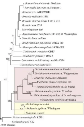
Along with Wolbachia, these bacteria are members of the
Anaplasmataceae family (Figure 1) [3]. Wolbachia infect
arthropods and filarial nematodes, but have not been shown to infect vertebrates directly.
Together with the Rickettsiaceae, the Anaplasmataceae are members of the order Rickettsiales (Figure 1) [3]. The
Rickettsiaceae include the obligate intracellular Rickettsia
spp. Like the Anaplasmataceae, the Rickettsiaceae are obligate intracellular pathogens with a life cycle that involves both vertebrate and invertebrate hosts, but they replicate directly in the cytosol of endothelial cells. All organisms in the order Rickettsiales have relatively small genomes (0.8–1.5 Mb) that have arisen through reductive evolution as they developed dependence on the host cell for necessary
functions [17]. The Rickettsiales and othera-Proteobacteria
also have an unresolved evolutionary relationship with the progenitor of the mitochondria [18,19].
Three Rickettsiaceae genomes have been published:
Rick-ettsia prowazekii, R. conorii, and R. typhi [18,20,21]. Four Anaplasmataceae genomes have been published: the insect
parasiteW. pipientis wMel, the filarial nematode endosymbiont
Wolbachiasp.wBm, the bovine pathogenAnaplasma marginale,
and the bovine pathogenEhrlichia ruminantium[19,22–24].
We present here a comparison of the previously completed Rickettsiales genomes to the first complete genomes of three
representative Anaplasmataceae human pathogens: A.
phag-ocytophilum, E. chaffeensis, and N. sennetsu. The complete genome sequence of these human pathogens will enhance the opportunities for investigation of virulence factors, pathogenesis, immune modulation, and novel targets for antimicrobial therapy and vaccines.
Results/Discussion
Genome AnatomyA. phagocytophilum, E. chaffeensis, andN. sennetsueach have a single circular chromosome (Figure S1). Most genomic
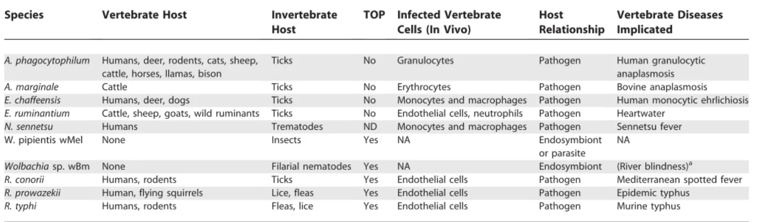
Table 1.Biological Characteristics of the Rickettsiales
Species Vertebrate Host Invertebrate
Host
TOP Infected Vertebrate Cells (In Vivo)
Host Relationship
Vertebrate Diseases Implicated
A. phagocytophilum Humans, deer, rodents, cats, sheep, cattle, horses, llamas, bison
Ticks No Granulocytes Pathogen Human granulocytic
anaplasmosis
A. marginale Cattle Ticks No Erythrocytes Pathogen Bovine anaplasmosis
E. chaffeensis Humans, deer, dogs Ticks No Monocytes and macrophages Pathogen Human monocytic ehrlichiosis E. ruminantium Cattle, sheep, goats, wild ruminants Ticks No Endothelial cells, neutrophils Pathogen Heartwater
N. sennetsu Humans Trematodes ND Monocytes and macrophages Pathogen Sennetsu fever
W. pipientis wMel None Insects Yes NA Endosymbiont
or parasite NA
Wolbachiasp. wBm None Filarial nematodes Yes NA Endosymbiont (River blindness)a R. conorii Humans, rodents Ticks Yes Endothelial cells Pathogen Mediterranean spotted fever R. prowazekii Human, flying squirrels Lice, fleas Yes Endothelial cells Pathogen Epidemic typhus
R. typhi Humans, rodents Fleas, lice Yes Endothelial cells Pathogen Murine typhus
aWolbachiasp. endosymbionts of filarial nematodes cannot directly infect eukaryotes, but theWolbachiaendosymbiont plays a role in immunological response.
TOP, transovarial passage; ND, not determined; NA, not applicable. DOI: 10.1371/journal.pgen.0020021.t001
Synopsis
features are typical of the sequenced Rickettsiales (Table 2). W. pipientis wMel,Ehrlichiaspp., andAnaplasmaspp., which are most closely related, all have numerous repeats in their
genomes. In contrast,N. sennetsuandR. prowazekii have only
six repeats in their respective genomes (Table 2). The
repetitive nature of the Ehrlichia and Anaplasma genomes is
exemplified by the expansion of outer membrane proteins of the OMP-1/P44/Msp2 family (discussed below). In addition numerous other functionally important genes are duplicated including those involved in type IV secretion and vitamin/ cofactor biosynthesis.
The origin of replication was not experimentally deter-mined in any of the genomes. As with other Rickettsiales [18],
genes typically clustered near the origin (dnaA, gyrA, gyrB,
rpmH, dnaN, parA,and parB) were dispersed throughout the
genomes. ForE. chaffeensisandN. sennetsu,a clear shift in
GC-skew occurs near parA and parB (Figure 2). Therefore,
basepair 1 was set in the intergenic region between the two
genes. InA. phagocytophilum,none of these genes were found
near the GC-skew transition. Therefore, basepair 1 was set in
the intergenic region near polA. For E. chaffeensis and A.
phagocytophilum, these predictions coincide with the
predic-tions forE. ruminantium[24] andA. marginale[23].
Only three islands of synteny over 10 kb in length are conserved among all the sequenced Anaplasmataceae, and these islands are shared among all the Rickettsiales (Figure 2). They include two operons of ribosomal proteins and one operon of proteins encoding portions of the type IV secretion system. Similar to the other Rickettsiales sequenced, all three genomes have the equivalent of a single rRNA operon with the 16S rRNA separated from the 23S-5S gene pair, as previously described for this order of bacteria [18].
Of genes typically clustered near the origin,parAandparB
were not identified inA. phagocytophilum. Likewise, parAand
parB are truncated in the Wolbachia sp. wBm. In various
mutational studies in free-living prokaryotes, the effects of
Table 2.Genome Properties
Feature/Property Organisms
APH AMA ECH ERU WOL NSE RPR
ORFs 1,369 965 1,115 920 1,271 935 834
tRNA 37 37 37 36 34 33 33
rRNA 3 3 3 3 3 3 3
sRNA 2 2 2 2 2 2 3
Size 1,471,282 1,197,687 1,176,248 1,516,355 1,267,782 859,006 1,111,523
GC (%) 41.6 49.8 30.1 27.5 35.2 41.1 29.1
Average gene length 775 1,077 840 1,032 855 804 1,005
Coding (%)a 72.2 86.0 79.7 62.0 85.7 87.5 75.4
Prophage 0 0 0 0 3 0 0
Assigned function 747 567 604 758 719 532 523
Conserved hypothetical 82 233 111 50 123 51 NR
Hypothetical proteins 458 151 314 80 337 288 208
Degenerate genes 55 22 13 32 70 3 NR
Repeat families 83 26 61 112 10 6 5
ORFs found in repeatsb 295 128 89 69 218 8 7
Genome in repeats (%) 12.7 5.6 3.8 5.1 10.1 0.4 0.3
a
Percent coding including truncated and frameshifted genes and the P44 silent fragments inA. phagocytophilum.
bAn ORF was determined to be found within a repeat if the repeat had more than 95% nucleotide identity over 10% of the length of either the repeat or the ORF, whichever was shorter.
APH,Anaplasma phagocytophilum; AMA,Anaplasma marginale[23]; ECH,Ehrlichia chaffeensis; ERU,Ehrlichia ruminantium[24]; WOL,Wolbachia pipientis wMel [19]; NSE,Neorickettsia sennetsu; RPR,Rickettsia prowazekii[18]; NR, not reported.
DOI: 10.1371/journal.pgen.0020021.t002
Figure 1.Phylogenetic Tree of thea-Proteobacteria
The protein sequences of select conserved genes were concatenated and aligned, and a phylogenetic tree was inferred of all sequenceda -Proteobacteria (see Materials and Methods). The Anaplasmataceae (purple) and the Rickettsiaceae (yellow) are highlighted.
inactivation of parA or parB range from lethality to production of anucleated cells at low copy number [25,26].
WithoutparA andparB, A. phagocytophilum and theWolbachia
sp. wBm may have random chromosome partitioning, may
require an alternate partitioning factor, or may have inefficient chromosome partitioning.
Of all the sequenced Anaplasmataceae, only theAnaplasma
spp. andEhrlichiaspp. share conserved gene order (synteny)
across their chromosome (Figure 2). E. ruminantium and E.
chaffeensis have a single symmetrical inversion near two duplicate Rho termination factors (Figure 3). Symmetrical inversions around the origin are the most common large-scale rearrangements in microbial genomes [27]. Genomic rearrangements between these Rho termination factors are
also apparent inA. marginale. The presence of the same break
in both theAnaplasmaandEhrlichialineages suggests that the
duplicate Rho termination factors allow for repeated inversions across this region of the genome.
In addition to the synteny breaks near the Rho termination
factors,A. marginalehas rearrangements located near themsp2
and msp3 expression loci and their corresponding
pseudo-genes (Figure 3). Likewise, numerous boundaries of genome
rearrangements are located near the homologous p44
expression locus (p44ES/APH_1221) and silent genes. In
bothAnaplasmaspp., the silentp44andmsp2genes stored in reserve in the genome can recombine into the corresponding expression locus to generate antigenic variation in the immunodominant surface protein (discussed in detail below). These exact, repeated sequences throughout the genome facilitate recombination for antigenic variation and may also provide sites where chromosomal inversions occur.
Genome Comparisons
In order to compare the genomic content of the Rickettsiales to that of other intracellular bacteria, ortholog clusters were delineated for 19 representatives of obligate and facultative intracellular pathogens and endosymbionts (see Materials and Methods). Such comparisons show con-servation of 176 ortholog clusters across these intracellular bacteria (Table S1), most of which correspond to house-keeping functions.
Eleven ortholog clusters present in all the Rickettsiales distinguish the Rickettsiales from other intracellular bacteria examined (Table S2). These include a type I secretion system ATPase, a pyridine nucleotide-disulfide oxidoreductase fam-ily protein, a putative transporter, and type IV secretion system proteins VirB9 and VirB8. Thirteen ortholog clusters composed of 12 conserved hypothetical proteins and a GNAT family acetyltransferase distinguish all the Anaplasmataceae from the Rickettsiales (Table S3).
Five genera in the Rickettsiales order have at least one representative sequenced. In order to compare these five
genera, the following genomes were compared:R. prowazekii,
N. sennetsu, W. pipientis, A. phagocytophilum, and E. chaffeensis. This comparison shows conservation of 423 ortholog clusters (Table S4) generally associated with housekeeping functions. Most genes in the five compared genomes are either conserved among all genomes or unique to a given genome. Indeed, 60% of the two-, three-, and four-way comparisons shared fewer than ten ortholog clusters (Figure 4). In the
three-way comparisons, the BDE (A. phagocytophilum, E.
chaffeensis, and N. sennetsu) and CDE (A. phagocytophilum, E.
chaffeensis,andW. pipientis)intersections harbor more than 20 ortholog clusters (Figure 4). The BDE intersection includes the organisms sequenced here and represents the human pathogens with very similar disease outcomes. Ortholog clusters conserved between these organisms include those for vitamin and cofactor biosynthesis enzymes, a monovalent cation/proton antiporter, a dicarboxylate transporter, and a DNA-binding protein (Table S5). Vitamin and cofactor biosynthesis is specific to the human ehrlichiosis agents, suggesting a niche adaptation or pathogenic trait. The CDE intersection is composed of the most closely related organisms. These ortholog clusters include genes for amino acid, fatty acid and nucleotide biosynthesis, an M48 family peptidase, a cytochrome c-type biogenesis protein, and the type IV secretion system protein VirB4 (Table S6).
In two-way comparisons, the AC (R. prowazekii and W.
pipientis) and DE (A. phagocytophilum and E. chaffeensis) intersections contain more than twenty ortholog clusters.
Genes shared only by R. prowazekii and W. pipientis include
those for cell wall biosynthesis, subunits of cytochrome D ubiquinol oxidase, a biotin transporter, a dinucleoside polyphosphate hydrolase, and an amino acid permease (Table
S7). The presence of genes for cell wall biosynthesis in onlyR.
prowazekiiandW. pipientislikely reflects differences in the cell surface;A. phagocytophilum, E. chaffeensis,andN. sennetsudo not synthesize peptidoglycan [28]. The peptidoglycan biosynthesis
genes are also found in A. marginale, which suggests that if
these genes are expressed,A. marginalemay have a
peptido-glycan layer [23]. Since the peptidopeptido-glycan genes are present in A. marginaleandW. pipientisbut not in the other Anaplasma-taceae, these genes have either been horizontally acquired in these organisms or have been lost numerous times in the Anaplasmataceae. Peptidoglycan binding to the Toll-like
receptor 2 activates leukocytes. Neither A. marginale norW.
pipientis infects the immune cells of a vertebrate host. The peptidoglycan layer may have been lost to allow the organism to successfully infect vertebrate immune cells.
Genes shared only by A. phagocytophilum and E. chaffeensis
include those encoding thiamine biosynthetic proteins, a potassium transporter, a peptide deformylase, and an ankyrin repeat protein (Table S8). Thiamine biosynthesis is distinctly
absent from N. sennetsu, suggesting a possible trematode
niche-specific adaptation.
A. phagocytophilum, E. chaffeensis, and N. sennetsu have 462, 312, and 303 open reading frames (ORFs) or paralog clusters that are unique with respect to the five-organism ortholog cluster analysis, respectively. The vast majority of these unique genes encode hypothetical, conserved hypothetical, and conserved domain proteins, as well as uncharacterized
membrane proteins and lipoproteins. Other A.
phagocytophi-lum-specific genes include those encoding the P44 outer
membrane proteins and the HGE-14 and HGE-2 antigenic
proteins (Table S9).E. chaffeensis-specific genes include those
for the OMP-1 family of proteins, arginine biosynthesis, a major facilitator family transporter, and a variable-length
PCR target protein (Table S10). N. sennetsu-specific genes
include those for an F-type ATPase beta subunit, a cyclo-philin-type peptidyl-prolyl cis-trans isomerase, a branched-chain amino acid transporter, a sensor histidine kinase, a strain-specific surface antigen, thioredoxin, and the type IV secretion system proteins VirB2 and VirB4 (Table S11).
Figure 2.Synteny of the Rickettsiales
comparison, over half were hypothetical proteins, many of which formed genomic islands of hypothetical proteins (Figure 2). The majority of the genes identified as unique were not just unique to the genus, but to the species. Of the 462A. phagocytophilum-unique genes in this comparison, 448
are also unique when compared with A. marginale. The 21
ortholog clusters shared only betweenAnaplasmaspp. include
conserved hypothetical proteins, OMP-1 proteins, membrane
proteins, and HGE-2 (Table S12). Likewise, of the 312 E.
chaffeensis-unique ORFs or paralog clusters in the five-way
comparison, 267 are unique upon comparison with eitherE.
ruminantium strain. The 52 ortholog clusters shared only
between theEhrlichiaspp. include OMP-1 proteins, arginine
biosynthetic proteins, a pyrroline-5-carboxylate reductase, a major facilitator protein, conserved hypothetical proteins, membrane proteins, and lipoproteins (Table S13).
Only one ortholog cluster containing conserved
hypo-thetical proteins is shared between the animal pathogensE.
ruminantium(Erum1840, ERGA_CDS_01780) andA.
margin-ale (AM279) and are absent from the human pathogens E.
chaffeensis, A. phagocytophilum, and N. sennetsu. In addition, a
homolog of these proteins is present in theEhrlichia canisJake
publicly available shotgun sequence. SinceA. phagocytophilum
and E. chaffeensis are maintained in animal reservoirs, presence of this gene is not associated with animal infection. Instead, loss of this protein could be required to establish infection in humans. These conserved hypothetical proteins have some homology to the eukaryotic patatin family of phospholipases. Patatin has been characterized to have phospholipase A-like activity [29].
Except for N. sennetsu, all of the sequenced pathogenic
Anaplasmataceae require an arthropod-vector that feeds on blood (Table 1). Three ortholog clusters, including one for bacterioferritin and two for conserved hypothetical proteins, are absent in all of the tick-, flea-, and louse-borne
Rickettsiales, but are present inWolbachiaspp. andN. sennetsu
(Table S14). The proteins in these ortholog clusters may be correlated to the lack of a blood-sucking arthropod in the life cycles of these organisms.
The tick-borne Anaplasmataceae (Ehrlichia spp. and
Ana-plasmaspp.) are the only Rickettsiales that are not transmitted transovarially in the invertebrate host. One ortholog cluster containing a class II aldolase/adducing domain protein (NSE_0849, RC0678, RP493, RT0479, WD0208) is absent
only from Ehrlichia spp. and Anaplasma spp. Lack of this
aldolase/adducing domain protein may prevent transovarial transmission in the arthropod vector.
Four ortholog clusters of conserved hypothetical proteins are present in all the pathogenic Rickettsiales but none of the endosymbionts. These proteins, which remain to be
charac-Figure 4.Comparison of the Rickettsiales Gene Sets
The composition of ortholog clusters (see Materials and Methods) of representative Rickettsiales (A),Ehrlichiaspp. (B), andAnaplasmaspp. (C) were compared. Numbers within the intersections of different ovals indicate ortholog clusters shared by 2, 3, 4, or 5 organisms. Species compared are indicated in diagram intersections as follows. A, R. prowazekii;B,N. sennetsu;C, W. pipientis;D,A. phagocytophilum;E,E. chaffeensis; F, A. marginale; G, E. ruminantium Gardel; and H, E. ruminantiumWelgevonden.
DOI: 10.1371/journal.pgen.0020021.g004 Figure 3.Synteny betweenAnaplasmaspp. andEhrlichiaspp.
Anaplasmaspp. andEhrlichiaspp. share conserved gene order (synteny) across their chromosomes.E. ruminantium and E. chaffeensis have a single symmetrical inversion near two duplicate Rho termination factors (approximate positions shown in pink). Genomic rearrangements between these Rho termination factors are also apparent inA. marginale (pink). In addition to the synteny breaks near the Rho termination factors, A. marginalehas rearrangements located near the msp2- and msp3-expression locus and pseudogenes (approximate positions shown in light blue). Likewise, in A. phagocytophilum, numerous changes in genome arrangement are located near the homologousp44expression locus and silent genes (approximate positions shown in lavender). DOI: 10.1371/journal.pgen.0020021.g003
strand, and the GC-skew. Representatives of all the Rickettsiales (A) and representativeEhrlichiaspp. andAnaplasmaspp. (B) were compared separately. AMA,A. marginaleSt. Maries; APH,A. phagocytophilumHZ; ECH,E. chaffeensisArkansas; ERU,E. ruminantiumWelgevonden; NES,N. sennetsuMiyayama; RPR,R. prowazekiiMadrid E; WOL,W. pipientis wMel.
terized, may be essential for pathogenesis or survival in the vertebrate host (Table S15).
A. phagocytophilumStrain Comparison
As an initial effort to use these genome sequences to identify the conserved genomic content of unsequenced members of these species, we conducted microarray-based
comparative genome hybridization analyses with two A.
phagocytophilum strains. Except for four p44 hypervariable regions (discussed below), the genomic content across all
three strains is conserved (ratio,3). AlthoughA.
phagocyto-philumand A. marginale have very different complements of
unique genes, the genomic content within the strains of A.
phagocytophilumis highly conserved. Conservation of the gene content of the strains may explain the similarity of clinical signs of HGA from two geographic regions (New York, Minnesota) and equine ehrlichiosis in California [7].
Free-Living and Obligate-Intracellulara-Proteobacteria
In order to understand the differences between these obligate intracellular pathogens and a closely related free-living organism, the number of genes in each role category was compared between representative Anaplasmataceae and Caulobacter crescentus(Table 3).C. crescentusis a closely related
and sequenced free-living a-Proteobacteria to the
Rickett-siales [30]. The scope of this comparison was limited to only
these fivea-Proteobacteria, as only these organisms had role
categories assigned in an identical manner.
All of the Anaplasmataceae examined have significantly higher percentages of their genomes involved in nucleotide biosynthesis, cofactor and vitamin biosynthesis, and protein synthesis. Enzymes in these biosynthetic pathways are likely to play an important role in interactions with their hosts and intracellular survival, as discussed below. The protein syn-thesis category includes many essential genes such as those encoding ribosomal proteins, tRNA synthetases, RNA mod-ification enzymes, and translation factors. These genes are essential and cannot be sacrificed as the genome reduces.
Therefore, as the genome size decreases, the proportion of genes involved in protein synthesis increases.
All of the Anaplasmataceae examined have a significantly lower coding capacity for central intermediary metabolism, transport, and regulatory functions. The decrease in central intermediary metabolism and transport reflects the differ-ences in acquiring nutrients and energy. Since intracellular bacteria are exposed to a relatively restricted complement of nutrients and energy sources, they have evolved to be specialists in acquiring specific compounds from their hosts. Likewise, these intracellular bacteria live in a homeostatic environment and have fewer regulatory genes. ORFs
encod-ingr70andr32were identified (rpoDandrpoH,respectively),
but r24 and r54 were not detected (rpoE and rpoN,
respectively). Several two-component regulatory systems are retained and may be employed as these bacteria transition between their vertebrate and invertebrate hosts. Despite
being identified in Rickettsiaspp. [21], stringent response (a
global regulatory response) may not be expected in the Anaplasmataceae, since neither RelA nor SpoT proteins were identified.
There are several role categories in which only specific organisms have significant differences from, or similarities to, C. crescentus. All the bacteria except E. chaffeensis have a statistically significant decrease in amino acid biosynthesis.
The difference betweenEhrlichiaspp. and the other
Anaplas-mataceae is due to the presence of lysine and arginine
biosynthesis pathways inEhrlichiaspp., as discussed below.A.
phagocytophilumhas a significant increase in the percentage of genes dedicated to the cell envelope due to expansion of the
OMP-1 family inAnaplasmaspp. (discussed below).W. pipientis
has a significantly higher percentage of its genome involved in mobile and extrachromosomal functions due to the unique
presence of phage and transposons in its genome [19]. E.
chaffeensis, A. phagocytophilum,andN. sennetsuhave a significant decrease in mobile elements, as they have no intact prophage, no transposable elements, and only a few phage core
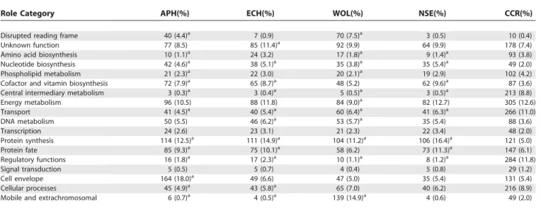
Table 3.Comparison of Role Category Breakdown
Role Category APH(%) ECH(%) WOL(%) NSE(%) CCR(%)
Disrupted reading frame 40 (4.4)a 7 (0.9) 70 (7.5)a 3 (0.5) 10 (0.4)
Unknown function 77 (8.5) 85 (11.4)a 92 (9.9) 64 (9.9) 178 (7.4)
Amino acid biosynthesis 10 (1.1)a 24 (3.2) 17 (1.8)a 9 (1.4)a 93 (3.8)
Nucleotide biosynthesis 42 (4.6)a 38 (5.1)a 35 (3.8)a 35 (5.4)a 49 (2.0)
Phospholipid metabolism 21 (2.3)a 22 (3.0) 20 (2.1)a 19 (2.9) 102 (4.2)
Cofactor and vitamin biosynthesis 72 (7.9)a 65 (8.7)a 48 (5.2) 62 (9.6)a 87 (3.6)
Central intermediary metabolism 3 (0.3)a 3 (0.4)a 5 (0.5)a 3 (0.5)a 213 (8.8)
Energy metabolism 96 (10.5) 88 (11.8) 84 (9.0)a 82 (12.7) 305 (12.6)
Transport 41 (4.5)a 40 (5.4)a 60 (6.4)a 41 (6.3)a 266 (11.0)
DNA metabolism 50 (5.5) 46 (6.2)a 53 (5.7)a 35 (5.4) 88 (3.6)
Transcription 24 (2.6) 23 (3.1) 21 (2.3) 22 (3.4) 48 (2.0)
Protein synthesis 114 (12.5)a 111 (14.9)a 104 (11.2)a 106 (16.4)a 121 (5.0)
Protein fate 85 (9.3)a 75 (10.1)a 58 (6.2) 73 (11.3)a 147 (6.1)
Regulatory functions 16 (1.8)a 17 (2.3)a 10 (1.1)a 8 (1.2)a 284 (11.8)
Signal transduction 5 (0.5) 5 (0.7) 4 (0.4) 5 (0.8) 29 (1.2)
Cell envelope 164 (18.0)a 49 (6.6) 47 (5.0) 35 (5.4) 131 (5.4)
Cellular processes 45 (4.9)a 43 (5.8)a 65 (7.0) 40 (6.2) 216 (8.9)
Mobile and extrachromosomal 6 (0.7)a 4 (0.5)a 139 (14.9)a 4 (0.6) 49 (2.0)
a
Role category composition considered significantly different fromC. crescentus. p-Values less than 0.01 were considered significant. Highly significant or particularly interesting differences are discussed in the text. APH,Anaplasma phagocytophilum;ECH,Ehrlichia chaffeensis;WOL,Wolbachia pipientis wMel [19]; NSE,Neorickettsia sennetsu;CCR,Caulobacter crescentus[30].
components (HK97-like portal, major capsid, and prohead
protease) scattered throughout their genomes. Lastly, A.
phagocytophilum and W. pipientis both have an increased number of disrupted reading frames.
Based on comparisons of the intracellular and free-livinga
-Proteobacteria, the only overall theme that emerges is the conservation of housekeeping genes and the shuffling of the genomes resulting in the loss of many operon structures.
Pathogenesis
Little is known about the genetic determinants required for the Rickettsiales to invade a host and cause disease. Putative pathogenesis genes were identified, including en-zymes to neutralize reactive oxygen species, outer membrane proteins, and protein secretion systems.
Oxidative stress response. Reactive oxygen species have been implicated in both host defense to infection and host
cell injury [31–33]. All of the Rickettsiales containsodB,an
iron superoxide dismutase. This superoxide dismutase may
have an important role in pathogenesis since sodB is
cotranscribed with components of the type IV secretion
system inE. chaffeensisandA. phagocytophilum[34].
Further examination of conserved genes without func-tional annotation (e.g., conserved hypothetical proteins, conserved domain proteins) shows two other ortholog clusters of proteins that may be involved in response to oxidative stress—a putative heme copper oxidase and a putative flavohemoglobin. In both cases, there is no signifi-cant similarity to a protein of known function, but several conserved domains were identified. From a particular combination of domains and conservation of metal/cofactor ligands, a function of response to oxidative stress can be proposed for these proteins [35].
Indeed, ECH_1079, NSE_0121, and APH_1205 each contain the 12 transmembrane segments and six conserved histidine residues consistent with members of the heme-copper oxidase family. Members of this protein family include cytochrome oxidase subunit I, FixN for nitrogen fixation, and NorB for nitric oxide reduction [36]. Each of these organisms is unlikely to be fixing nitrogen and already has a functional subunit I of cytochrome oxidase (ECH_1003, NSE_0622, and APH_1085), so these orthologs may be nitric oxide reduc-tases. Alternatively, there may be another, as yet to be identified, role for this oxidase, which was identified in all
the Rickettsiales genomes except theWolbachiasp.wBm where
it is truncated (an ORF that was not annotated but has genomic coordinates from 536343 to 536534).
APH_0545, NSE_0661, and ECH_0778 encode proteins with three functional motifs similar to flavohemoglobins—a heme binding site, an FAD binding domain, and an NAD
binding domain. The biological function of theEscherichia coli
flavohemoglobin has not been elucidated, but it has been shown to be an efficient alkylhydroperoxide reductase [37] and a nitric oxide reductase [38]. This putative
flavohemo-globin is conserved among the Anaplasmataceae, but
Wolba-chiaspp. are missing the NAD oxidoreductase domain, andR.
prowazekii is missing the heme ligands. Although the spec-ulation of a role for these genes in pathogenicity is intriguing, the precise function of each of these proteins will need to be elucidated experimentally.
The OMP-1/MSP2/P44 protein superfamily. The Anaplas-mataceae all have a diverse complement of outer membrane
proteins. Many of these outer membrane proteins (OMPs) are members of Pfam PF01617 [39] and constitute the OMP-1/
MSP2/P44 family.Anaplasma, Ehrlichia,andWolbachiahave each
undergone variable levels of expansion of their omp-1/msp2
gene families (Figure S2). TheN. sennetsugenome has only one
uncharacterized protein from this family (NSE_0875). W.
pipientis wMel and the Wolbachia sp. wBm have the smallest
expansion with three wsp genes scattered throughout each
genome. The largest expansion of this family is inEhrlichiaspp.
andAnaplasmaspp. These organisms cannot be transovarially inherited in their arthropod hosts. Instead, ticks acquire Ehrlichia or Anaplasma by feeding on an infected vertebrate reservoir animal. The expansion of this family may allow persistence in the vertebrate reservoir by providing antigenic variation, thus allowing for effective tick transmission.
E. chaffeensis, E. canis, and E. ruminantium have 17–22 paralogous tandemly arranged genes from this family that
are flanked by a transcription regulator(tr1)and a preprotein
translocase(secA)[40–42]. These genes all have signal peptides
and are likely to be secreted across the cytoplasmic membrane by SecA [42]. They encode immunodominant major outer membrane proteins that are differentially expressed in ticks and experimentally infected animals [43].
A. marginale St. Maries is reported to have 56 genes that
have been placed into this superfamily, including eightmsp2,
eightmsp3,onemsp4,threeopag,15omp-1,12orfX,sevenorfY,
and two msp3 remnants [23]. These genes are scattered
throughout the genome with a bias in location toward the origin of replication. MSP2 and MSP3 are the
immunodo-minant proteins [44]. The msp2and msp3gene subsets each
include one full-length expression locus and seven reserve/ silent sequences that are thought to recombine into the expression locus to generate antigenic variation [23].
The A. phagocytophilum genome has threeomp-1,one msp2,
twomsp2homologs, onemsp4,and 113p44loci belonging to
the OMP-1/MSP2/P44 superfamily. Although both Anaplasma
spp. msp2 genes are members of PF01617 and the OMP1/
MSP2/P44 superfamily, theA. marginale msp2 gene is distinct
from the A. phagocytophilum msp2 gene. In addition, the
previously identified omp-1N is not a member of this Pfam,
but is homologous to E. chaffeensis omp-1N and the msp2
operon-associated gene 3 ofA. marginale[45].
The largest expansion of this family is that ofp44genes in
A. phagocytophilum. Only 36 copies ofp44are in this Pfam, but many smaller regions were identified, resulting in a total of
113 annotated p44 loci (Table S16). The p44s consist of a
central hypervariable region of approximately 280 bp containing a signature of four conserved amino acid regions (C, C, WP, A) and conserved flanking sequences longer than
50 bp. Diversep44paralogs(p44–1top44–65)are expressed in
mammals and ticks and confer antigenic environmental adaptation, especially during tick transmission [46–49]. The
genomic loci of all 65 previously described p44 genes were
determined in the present study (Figure S3). Twenty-three
novelp44genes(p44-66top44–88)were identified by genome
sequencing, but have not yet been experimentally identified as being expressed.
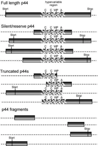
The p44s were annotated as full-length, silent/reserve,
truncated, and fragments (Figure 5). There are 22 full-length
p44s identified that have ORFs longer than 1.0 kb with
conserved start and stop codons. By locating highly conserved
the hypervariable region, 64 shorter p44s were identified. These ORFs lack a translational start codon and likely serve as
reserve/silentp44s that can be expressed after recombining
into the previously described p44-expression locus (p44ES/
APH_1221) [45,50]. The full-length and silent/reserve p44
genes are preferentially located near the replication origin
(Figure S3) and symmetrically located around the p44
expression locus. Localization near the origin, where multiple replication forks coexist, may facilitate recombination
between the expression locus and the reserve/silentp44genes.
In addition to the full-length and silent/reservep44genes,
21 59and 39fragments and six truncations ofp44genes larger
than 60 nucleotides have been identified in the genome. Truncations include portions of a hypervariable region;
fragments did not. The p44s annotated as truncated and
fragments do not contain both conserved regions flanking the
hypervariable region. These p44s are not expected to
recombine through the homologous recombination model
deduced by previous analyses of recombinedp44s [49–52].
Microarray-based comparative genomic hybridization
re-veals that expansion of thep44family is a common feature in
A. phagocytophilum strains. All but four of the p44 unique hypervariable sequences used as targets on the microarray are
present in the human isolate A. phagocytophilumMN and the
horse isolate A. phagocytophilum California MRK (Figure S3;
Table 4). The p44-12 and p44-9 unique regions are either
absent or divergent only in strain MN. Thep44-4andp44-1
unique regions are absent or divergent in strains MN and MRK. This confirms previous results demonstrating that the p44–1unique region is absent/divergent in MN and MRK [52].
Other important outer membrane proteins.N. sennetsuhas a
singlep51gene (NSE_0242) encoding its immunodominant
P51 major outer membrane protein [14]. The p51 gene is
highly conserved among N. risticii, N. sennetsu, and the
Stellantchasmus falcatusfluke agent, but not inN. helminthoeca, the agent causing an acute, highly fatal salmon-poisoning disease of domestic and wild canines [14]. Although a full-length, highly conserved homolog for P51 was not found in the Rickettsiales genome sequences, P51 was placed in an ortholog cluster of genes conserved among all the Rick-ettsiales due to short regions of similarity, particularly in a C-terminal region that may include a secretion peptide motif.
Other outer membrane proteins have been reported inA.
marginale,includingmsp5, msp1a,andmsp1b. Themsp5gene (a SCO1/SenC family protein) is found in all the Rickettsiales,
whereasmsp1aandmsp1bare unique toA. marginale.
OnlyE. chaffeensisand E. canisencode a 120-kDa immuno-dominant surface protein (ECH_0039) [53]. The
variable-length PCR target useful in distinguishing various strains ofE.
chaffeensis [54] is present only in the genome ofE. chaffeensis Arkansas (ECH_0170).
Protein secretion systems.All of the strains sequenced here contain both a Sec-dependent and Sec-independent protein export pathway for secretion of proteins across the inner membrane. The Sec-independent pathway (Tat pathway) has been implicated in the transport of phospholipases in Pseudomonas aeruginosa[55]. All of the strains sequenced here also contain two components of a putative type I secretion system, potentially for transporting toxins or proteases carrying a C-terminal secretion signal.
All of the Rickettsiales have a type IVa secretion system that uses a complex of transmembrane proteins and a pilus to deliver effector macromolecules from prokaryotic to eukary-otic cells. The reference Type IVa secretion system is that of Agrobacterium tumefaciens,which contains 11 genes in thevirB
locus and one gene in thevirD locus. Several components of
Figure 5.Representative Illustrations ofp44Genes
Full-lengthp44genes contain conserved start and stop codons, an ORF longer than 1,000 bp, and a central hypervariable region of approx-imately 280 bp containing a signature of four conserved amino acid regions (C, C, WP, A). These genes can be expressed at their respective current genome location or can recombine into the expression locus (p44ES/APH_1221). A silent/reservep44is less than 1,000 bp. It may have either the conserved or alternative start and/or stop codons. A silent/ reservep44is not likely to be expressed at its current genome location, but can recombine into the expression locus (p44ES/APH_1221). Truncatedp44s carry the complete hypervariable region, or a portion thereof, but only one of the two conserved regions. Fragments ofp44 have only a conserved region and no hypervariable region. Each annotatedp44is longer than 60 bp. It should be noted that smaller fragments can be identified throughout the genome. These, as well as p44truncations and fragments, are likely to be nonfunctional remnants of previous recombination events.
DOI: 10.1371/journal.pgen.0020021.g005
Table 4.SelectedA. phagocytophilumCGH Results
Locus Ratio MN Ratio MRK p44Unique Region
APH_1195 24.066 5.52 p44-1
APH_1154 3.281 4.15 p44-4
APH_1391 9.803 2.83 p44-9
APH_1249 3.503 1.97 p44-12
theA. tumefacienstype IVa secretion system are conserved inA. phagocytophilum, E. chaffeensis,andN. sennetsu. LikeR. prowazekii and W. pipientis, the three organisms sequenced here are lackingvirB1,virB5, andvirB7. All butN. sennetsulackvirB2.
ThevirB3,virB4, andvirB6 homologs are contiguous at one
locus (Figure S4). Neighboring this locus in all of these
organisms are three or fourvirB6 homologs. Contiguous at a
second locus arevirB8,virB9,virB10,virB11, andvirD4. The
type IV secretion system is one of the few sets of genes syntenic between all of the Rickettsiales sequenced, suggest-ing that tight coordination of expression of these genes is critical.
InA. tumefaciens,translocated type IV effector proteins have
the consensus sequence R-X7-R-X-R-X-R-X-Xn, where lysine
can substitute for arginine with no noticeable effect [56]. In addition, effector molecules are often localized to a region of the chromosome near the type IV secretion apparatus. Examination of the regions around the type IV operons in
A. phagocytophilum revealed numerous genes encoding HGE-14, which contain C-terminal sequences similar, but not identical, to this motif (Table S17), suggesting that it may be an excreted effector molecule. Subsequent searches of the Anaplasmataceae genomes with motifs like that found in HGE-14 did not reveal other potential effector molecules.
Metabolism
The metabolic potentials ofA. phagocytophilum, E. chaffeensis,
andN. sennetsuwere compared to that ofR. prowazekiiandW. pipientis [18,19]. Overall, the Anaplasmataceae have very similar metabolic pathways but are quite distinct from those ofR. prowazekii(Figure 6).W. pipientisdiffers from the other Anaplasmataceae in its inability to synthesize some cofactors.
Nucleotide and cofactor biosynthesis. E. chaffeensis, A. phagocytophilum, N. sennetsu, and W. pipientis have the ability
to synthesize all nucleotides. This differs fromR. prowazekii,
which cannot make purines or pyrimidines, and therefore
Figure 6.Comparative Metabolic Potential of Select Rickettsiales
Metabolic pathways ofE. chaffeensis(magenta arrows),A. phagocytophilum(green arrows),N. sennetsu(gold arrows),W. pipientis(lavender arrows), and R. prowazekii(cyan arrows) were reconstructed and compared. The networks of some of the more important pathways are shown with metabolites color coded: red and purple, central and intermediary metabolites; blue, cofactors; green, amino acids; and black, cell structures. Transporters are shown in the membrane and are grouped by predicted substrate specificity: green, inorganic cations; magenta, inorganic anions; red, carbohydrates and carboxylates; blue, amino acids/peptides/amines; yellow, nucleotides/nucleosides; and black, drug/polysaccharide efflux or unknown.
must rely on nucleotide translocases and interconversion of
the bases to obtain the full complement of nucleotides [18].E.
chaffeensis, A. phagocytophilum, and N. sennetsu are able to synthesize most vitamins and cofactors. In contrast to the
other Anaplasmataceae,W. pipientishas lost some of its ability
to synthesize cofactors, and it has completely lost the biosynthetic pathways for biotin, thiamine, and NAD. In addition, it may be in the process of losing the ability to
synthesize folate. R. prowazekii has also lost the ability to
synthesize these cofactors as well as FAD, pantothenate, and pyridoxine-phosphate.
Biotin is one of the essential cofactors only synthesized by the vertebrate-infecting Anaplasmataceae. In most organ-isms, biotin is required for many carboxylation reactions, but is not synthesized by many multicellular eukaryotes. RT-PCR analysis showed that all four genes in the biotin biosynthesis
pathway (BioA/B/D/F) were expressed by E. chaffeensis and A.
phagocytophilum in THP-1 and HL-60 cells, respectively, at both 2 d and 3 d post infection (Figure S5).
The presence of nucleotide, vitamin, and cofactor
bio-synthetic pathway inE. chaffeensis, A. phagocytophilum, and N.
sennetsusuggests that they do not need to compete with the host cell for, and may even supply host cells with, essential vitamins and nucleotides. It has been previously proposed thatWigglesworthia glossinidia supplies its host with vitamins
that are rare in the blood meal of its arthropod host (tsetse
fly) [57]. Interestingly, Ehrlichia spp. andAnaplasmaspp., the
two tick-borne intracellular pathogens sequenced, both have a complement of pathways for cofactor and amino acid
biosynthesis similar toW. glossinidia(Table 5). This raises the
possibility that these pathogens may currently be, or historically have been, able to provide a benefit to their tick hosts by providing necessary cofactors.
Amino acid biosynthesis. The Rickettsiales have a very limited ability to synthesize amino acids and must rely on transporting them from the host (Figure 6). All four of the Anaplasmataceae sequenced have the ability to make glycine,
glutamine, glutamate, and aspartate. Additionally,E.
chaffeen-sisis predicted to be able to synthesize arginine and lysine like
E. ruminantium [24]. One possible role for arginine biosyn-thesis may be to recover an intracellular arginine pool after exposure to inducible host nitric oxide. Nitric oxide is synthesized by nitric oxide synthases that convert arginine to citrulline and nitric oxide [58]. The production of nitric oxide is likely to deplete the intracellular pool of arginine, further hampering intracellular growth. The presence of an arginine biosynthesis pathway and putative nitric oxide
reductase(s) may allowEhrlichiaspp. to recover more rapidly
and subvert the host immune response. This would be similar to the proposed retention of select tryptophan biosynthetic
Table 5.Amino Acid and Cofactor Biosynthesis in Intracellular Bacteria
Class Metabolite Present in:
APH AMA ECH ERU WOL NSE RPR BAP BBP BFL WGL
Amino acids Alanine
Arginine þ þ
Asparagine
Aspartate þ þ þ þ þ þ þ þ þ
Cysteine þ þ
Diaminopimelate þ þ þ þ þ þ þ þ þ
Glycine þ þ þ þ þ þ þ þ þ þ þ
Glutamate þ þ
Glutamine þ þ þ þ þ þ þ þ þ
Histidine þ þ þ
Leucine þ þ þ
Lysine þ þ þ þ þ
Isoleucine þ þ þ
Methionine þ
Phenylalanine þ þ þ
Proline þ þ þ þ þ
Serine
Threonine þ þ þ
Tryptophan þ þ þ
Tyrosine þ
Valine þ þ þ
Cofactors Biotin þ þ þ þ þ þ þ
FAD þ þ þ þ þ þ þ þ þ
Folate þa þ þ þ ? þ þ þ
Lipoate þ þ þ þ þ þ þ þ þ þ
NAD þ þ þ þ þ þ þ
Pantothenate and CoA þ þ þ þ þ þ þ
Protoheme þ þ þ þ þ þ þ þ
Pyridoxine phosphate þ þ þ þ þ þ þ þ
Thiamine þ þ þ þ þ
Ubiquinone þ þ þ þ þ þ þ þ þ
a
The pathway is complete, except that a homolog offolAcould not be identified.
genes in Chlamydia spp. in order to replenish tryptophan pools after host enzymatic degradation of tryptophan in
response to IFN-c[59].
Glycolysis, tricarboxylic acid cycle, pentose phosphate, and respiration.A complete pyruvate dehydrogenase,
tricarbox-ylic acid cycle, F0F1-ATPase, and electron transport chain
were found in all of the organisms. All five organisms are likely to use host-derived carboxylates and amino acids, but none of these organisms can obtain carbon or energy from fatty acids or actively carry out glycolysis. The glycolysis enzymes present are limited to those that produce glycer-aldehyde-3-phosphate and dihydroxyacetone phosphate from phosphoenolpyruvate (Figure 6). The glyceraldehyde-3-phos-phate produced in this manner is used in the nonoxidative pentose phosphate pathway, resulting in the production of pentoses needed for cofactor and nucleotide biosynthesis.
Consistent with this role for the glycolytic enzymes, R.
prowazekiiandR. conoriiretain neither the glycolytic enzymes nor the enzymes needed for the biosynthesis of nucleotides or cofactors from pentose. Similarly, dihydroxyacetone phos-phate from these glycolytic enzymes can be converted to glycerol-3-phosphate for phospholipid biosynthesis in the
Anaplasmataceae. Without the glycolytic enzymes,Rickettsia
spp. must obtain glycerol-3-phosphate from the host via a glycerol-3-phosphate transporter.
Evolution and DNA Repair
A genome-scale phylogenetic analysis using a concatenated alignment of core proteins is consistent with rRNA studies and current taxonomic assignments. This indicates that AnaplasmaandEhrlichiaare sister genera that share a common
ancestor withWolbachia(Figure 1).Neorickettsiais the
deepest-branching lineage in the group.
The branch lengths on the whole genome tree can be used to get an indication of the relative rates of evolution of these organisms. In general, the branch lengths for these intra-cellular organisms are longer than those of their free-living relatives. This may be due to either differences in DNA repair or population genetic and selection-related force. For example, many intracellular organisms go through more stringent population bottlenecks, which in turn increase the amount of genetic drift and possibly the rate of accumulation of deleterious mutations.
Analysis of the genome ofW. pipientis wMel revealed that it
had a longer branch length than the closely relatedRickettsia;
theRickettsiahave higher rates of evolution than free-living organisms [19]. Wu et al. [19] ascribed this increase to features ofWolbachiabiology. However, there appears to be a general increase in the rate for all of the Anaplasmataceae (Figure 1).
Thus, the increase reported forWolbachia[19] is not likely due
to the specific biology of Wolbachia, but instead to some
feature shared by all Anaplasmataceae.
Examination of the putative DNA-repair capabilities of the different species does not reveal any significant differences
between the Anaplasmataceae and theRickettsiaspp. (Table
S18). Interestingly, within the Anaplasmataceae, N. sennetsu
appears to have the longest branch length and the most limited suite of DNA repair genes within the group. For
example, N. sennetsu is missing various glycosylases and
exonucleases that contribute to repair, including uvrABC,
which is involved in nucleotide excision repair. It is possible
that the faster rate of evolution in this organism is related to the absence of some of these repair pathways.
The absence of uvrABC in N. sennetsuand the absence of
uvrBCin the Ehrlichiaspp. suggest that these species do not have nucleotide excision repair (NER). NER is used by other organisms, including bacteria, archaea, and eukaryotes, as a general repair process to remove sections of DNA with gross abnormalities. One important role of NER is in the repair of UV-induced DNA damage, and defects in NER in other species lead to great increases in UV sensitivity. It appears thatNeorickettsiahas compensated for this by acquiring a gene homologous to DNA photolyases, an alternative mechanism
for repairing UV damage. TheNeorickettsia photolyase is not
particularly closely related to known photolyases from a
-Proteobacteria but is instead most closely related to a
photolyase from Coxiella burnetii, a c-Proteobacteria. The
Ehrlichiaspp., however, do not encode a photolyase homolog, and thus these species may be highly UV-sensitive.
Conclusions
The dual existence of members of Anaplasma spp. and
Ehrlichia spp. as invertebrate symbionts or commensals and effective human and animal pathogen requires flexibility, a fact reflected in the genome. Both organisms display an expansive inventory of paralogous genes encoding diverse functions that promote survival and success in different
environments when compared to Neorickettsia spp. and
Wolbachiaspp., which do not require a mammalian host. This capacity is evident from the large repertoire of outer membrane proteins, and partial duplication of some of the virulence determinants (e.g., components of the type IV protein secretion system).
The large number of paralogous genes encoding
immuno-dominant outer membrane proteins in Anaplasma spp. and
Ehrlichia spp. has important implications for the study of pathogenesis and in the development of vaccination strat-egies. Adaptability in the human host may underlie significant disease manifestations. Genomic-level characterization of the full complement of variable antigens will facilitate the future development of more specific and sensitive diagnostic targets. In light of the growing recognition of the increased global burden of ehrlichiosis, development of such diagnostic targets will impact public health.
Between pairwise comparisons of different species within a single genus, there are hundreds of genes that are not shared. Often these gene differences are immunodominant outer membrane proteins, but the vast majority are genes that are not functionally characterized in any organism. Some are likely to be involved in zoonosis or specific disease
character-istics. For instance,A. phagocytophilum is the only sequenced
Rickettsiale that infects neutrophils. Therefore, some of the A. phagocytophilum-unique genes (e.g., genes encoding P44 and HGE-14) may be involved in neutrophil invasion.
Materials and Methods
Intracellular bacteria purification and DNA preparation. Organ-isms (infecting ;1 3109 host cells; 50–100 175-cm2 flasks) were cultured in synchrony in respective host cells (E. chaffeensisin DH82 cells,A. phagocytophilumin HL-60 cells, andN. sennetsuin P388D1cells). Bacterial cells were liberated from the infected host cells using Dounce homogenization, differential centrifugation, and Percoll density gradient centrifugation [60]. Any specimens with host nuclei contamination were excluded. From these isolated bacteria, phenol extraction was used to purify DNA that was minimally fragmented and free of host-cell DNA. Levels of host DNA contamination were verified to be less than 0.001% by PCR using host G3PDH-specific primers. This method was highly successful, with only 14 sequencing reads identified as being of human origin from a total of over 57,000 good sequencing reads.
Sequencing and annotation.The complete genome sequences were determined using the whole-genome shotgun sequencing approach [61], sequences were assembled into contigs using the Celera Assembler [62], and all gaps were closed [63]. ORFs from each genome were predicted and annotated using a suite of automated tools that combine Glimmer gene prediction [64,65], ORF and non-ORF feature identification (e.g., protein motifs), and assignment of database matches and functional role categories to genes [63]. Frameshifts and point mutations were detected and corrected where appropriate; those remaining were annotated as‘‘authentic frame-shift’’or‘‘authentic point mutation.’’Repeats were identified using RepeatFinder [66,67] and were manually curated. The complete genome sequences forA. phagocytophilum HZ,E. chaffeensisArkansas,
andN. sennetsuMiyayama have been deposited in GenBank.
Annotation of the p44 genes. Full-length p44s were defined as having ORFs greater than 1,000 bp with conserved start codon and stop codons. For shorter silent/reservep44s, the ORFs were initially identified by locating highly conserved 59 and 39 sequences and signature sequences within the hypervariable region. Since these silent/reservep44s lack a start and stop codon, the 59and 39ends were annotated on the basis of conserved genome features found in full-lengthp44genes [50,68]. The annotatedp44fragments are at least 60 nucleotides in length, have either 59or 39conserved sequences, and may contain a partial hypervariable region (Figure 5).
Genome comparisons. Ortholog clusters were delineated forR.
prowazekiiMadrid E [18],R. typhiWilmington [21],R. conoriiMalish 7
[20],N. sennetsuMiyayama,Wolbachiasp.wBm [22],W. pipientis wMel [19], E. chaffeensis Arkansas, E. ruminantium Gardel (GenBank CR925677.1),E. ruminantiumWelgevonden [24],A. marginaleSt. Maries [23],A. phagocytophilumHZ,Brucella suis 1330 [69],Bartonella henselae
Houston-1 [70],Coxiella burnetiiRSA 493 [71],Tropheryma whippleiTwist [72], Blochmannia floridanus [73], Buchnera sp. APS [74], Chlamydia
pneumoniaeAR39 [75], andW. glossinidia brevipalpis[76]. ForWolbachia
sp.wBm, the ORFs used in these comparisons were uncurated ORFs predicted and annotated using a suite of automated tools that combine Glimmer gene prediction [64,65], ORF and non-ORF feature identification (e.g., protein motifs), and assignment of database matches and functional role categories to genes [63]. Upon release of the annotated genome [22], these uncurated ORFs were paired with the corresponding curated ones where possible, with exceptions noted in the text.
Paralog clusters within each of the genomes were identified using the Jaccard algorithm with the following parameters: 80% or greater identity and Jaccard coefficient 0.6 or higher [77, Text S1]. Members of paralog clusters were then organized into ortholog clusters by allowing any member of a paralog cluster to contribute to the reciprocal best matches used to construct the ortholog clusters. The conservation of ortholog clusters across the various genomes analyzed was determined using Sybil, a web-based software package for comparative genomics developed at TIGR (http://sybil. sourceforge.net). The database of these clusters and corresponding tools can be accessed through TIGR (http://www.tigr.org/sybil/rcd). Metabolic pathways and transporters were compared across genomes using (1) these calculated ortholog clusters, (2) Genome Properties [78], (3) TransportDB [79], and (4) Biocyc [80].
Significant differences in the role category composition was determined usingv2calculated using the Yates continuity correction. Ap-value less than 0.01 was considered significant.
GC-skew and origin prediction.The GC-skew was calculated as (C G)/(CþG) in windows of 1,000 bp along the chromosome [81]. The origin of replication was not experimentally determined in any of the genomes. ForE. chaffeensisandN. sennetsu,a clear shift in GC-skew occurs near parA and parB. Therefore basepair 1 was set in the intergenic region between the two genes. InA. phagocytophilum,a
GC-skew transition occurs nearpolA.Therefore, basepair 1 was set in the intergenic region nearpolA.
Atypical nucleotide composition. Regions of atypical nucleotide composition were identified by thev2analysis: the distribution of all 64 trinucleotides was computed for the complete genome in all six reading frames, followed by the trinucleotide distribution in 5,000-bp windows overlapping by 500 bp. For each window, thev2statistic was computed based on the difference between the trinucleotide content in that window and that of the whole genome. Peaks indicate regions of atypical nucleotide composition.
Genome tree construction.Protein sequences of 31 housekeeping genes(frr, infC, nusA, pgk, pyrG, rplA, rplB, rplC, rplD, rplE, rplF, rplK, rplL, rplM, rplN, rplP, rplS, rplT, rpmA, rpoB, rpsB, rpsC, rpsE, rpsI, rpsJ, rpsK,
rpsM, rpsS, smpB,andtsf) from completea-Proteobacteria genomes
were aligned to predefined HMM models and ambiguous regions were autotrimmed according to an embedded mask. Concatenated alignments were then used to build a maximum likelihood tree with bootstrapping using PHYML [82]. Thec-ProteobacteriaE. coliand the b-ProteobacteriaNeisseria meningitidiswere used as outgroups to root the tree.
Array construction and hybridizations.Oligonucleotides (70-mer) were designed from the unique ORFs of each of the three genomes. The oligonucleotides (Illumina, San Diego, California, United States) were diluted to 25lM in DMSO and spotted in quadruplicate onto UltraGap slides (Corning, Acton, Massachusetts, United States). Cy3 and Cy5 probes were synthesized from genomic DNA as previously described [83]. In order to obtain enough DNA for microarray analysis, small amounts of DNA were prepared in the manner described above for genome sequencing. This DNA was then quantitatively amplified using GenomiPhi (Amersham, Piscataway, New Jersey, United States).
Appropriately labeled query and reference probes were hybridized overnight, washed, and scanned using an Axon GenePix 4000B scanner (Axon Instruments, Union City, California, United States). The corresponding images were analyzed with TIGR Spotfinder [84]. Log mode centering was used to normalize the data alleviating the bias of expression microarray normalization methods, which expect a normal distribution of data. Briefly, a Perl script was designed to construct the histogram of the log2 of the ratio and adjust the histogram mode to zero. The data presented are the geometric means of the normalized ratios from at least two slides with different reference Cy dyes and with oligonucleotides printed in quadruplicate. Transcript analysis of biotin biosynthetic genes. Total RNA was extracted fromE. chaffeensisorA. phagocytophilum-infected THP-1 or HL-60 cells at 2 d or 3 d postinfection using RNeasy (Qiagen, Valencia, California, United States). RNA was DNase I treated (Invitrogen, Carlsbad, California, United States) in the presence of 40 U of RNaseOUT (Invitrogen) for 15 min at room temperature, followed by inactivation at 658C in the presence of 2.5 mM EDTA for 10 min. For cDNA synthesis, total RNA (0.5 lg) was reverse-transcribed at 428C for 1 h in 50 mM Tris-HCl (pH 8.3), 75 mM KCl, 3 mM MgCl2, 0.5 mM of each dNTP, 1 U of RNase inhibitor (Invitrogen), 1.5 lM random hexamers (Invitrogen), and 10 U of Superscript II reverse transcriptase (Invitrogen). The reaction was terminated by heat inactivation at 708C for 15 min. To ensure the absence of DNA contamination in the RNA preparations, the assay was duplicated without reverse transcriptase. The subsequent amplification was conducted with standard conditions for 25 cycles of 958C for 45 s, 548C for 45 s, and 728C for 1 min and with the PCR primer pair (Table S19).
Supporting Information
Figure S1. Linear Representation of E. chaffeensis Arkansas, A.
phagocytophilumHZ, andN. sennetsuMiyayama Genomes