Vol.53, n. 3: pp.513-518, May-June 2010
ISSN 1516-8913 Printed in Brazil BRAZILIAN ARCHIVES OF
BIOLOGY AND TECHNOLOGY
A N I N T E R N A T I O N A L J O U R N A L
Isolation and Characterization by Asymmetric PCR of the
ENDO1
Gene for Glucan endo-1,3-
ββββ
-D-glucosidase in
Phytophthora
cinnamomi
Associated with the ink Disease of
Castanea sativa
Mill.
Sofia Meirinho
1, Marisa Carvalho
1, Ángel Dominguez
2and Altino Choupina
1*1Departamento de Biologia e Microbiologia; Escola Superior Agrária de Bragança; Centro de Investigação de
Montanha; Apartado 1172; 5301-854;Bragança - Portugal. 2Departamento de Microbiología y Genética; CIETUS, IMB/CSIC, Universidad de Salamanca, Plaza de los Dres. de la Reina, s/n; 37007; Salamanca - Spain
ABSTRACT
Ink disease is one of the most destructive diseases in Castanea sativa. The most common symptoms are root necrosies and a reduction in root growth, which invariably lead to the death of the trees. Phytophthora cinnamomi
is an oomycete associated with this disease whose life cycle develops integrally in the soil. In the present work, was a fragment with 1231bp of the glucan endo-1,3-β-D-glucosidase gene obtained by amplification, using conserved primers and the full-length gene sequence by flanking this known sequence by asymmetric PCR. This fragment was obtained from genomic DNA of Phytophthora cinnamomi isolated in the European Regions ofCastilla-Leon (Spain) and Trás-os-Montes (Portugal) and associated with the ink disease of Castanea sativaMill.
Key words: Phytophthora cinnamomi; glucan endo-1,3-β-D-glucosidase; asymmetric PCR
* Author for correspondence: [email protected]
INTRODUCTION
The genus Phytophthora belongs to the oomycetes in the eukaryotic stramenopile lineage and comprises over 65 species, all of which are plant pathogens for a wide range of dicotyledons. P. cinnamomi is a widespread soil-borne pathogen that infects woody plant hosts. It requires moist soil conditions and warm temperatures to be active, but the damage caused by the disease occurs most often in summer, when plants are drought stressed. P. cinnamomi is one of the most destructive species of Phytophthora, and it has been associated with the decline of several forestry, ornamental, and fruit species.
sporulating diploids. In plants, 1,3-β-glucanases have been characterized for their major role in plant defense, as well as for their involvement in germination, microsporogenesis and embryogenesis. In oomycetes, glucanases have been studied at biochemical level for their possible role in hyphal tip growth and branching, where there is thought to be a delicate balance between the cell wall synthesis and hydrolysies. They have also been studied for their role in Ca2+- induced sporulation and their role in the host pathogen interaction (McLeod, A., 2002).
In the present work, was a fragment with 1231bp of the endo-1,3-β-glucanase gene obtained by standard PCR, using conserved primers, and the whole genomic sequence, with 2586 bp, by amplifiying the previous sequence by asymmetric PCR.
MATERIALS AND METHODS
Oomycete growth conditions
P. cinnamomi mycelia for genomic DNA extraction were obtained by growth in cellophane-PDA media. The C-cellophane-PDA plates were inoculated by placing a plug of mycelium (0.5 cm in diameter) on new plates and incubating in the dark for 4-6 days to AT 22-25ºC.
PCR Amplification
The polymerase chain reaction (PCR) was used to
amplify a 1231bp fragment of the glucan endo-1,3-β-D-glucosidase gene. The conserved primers Gendo1F and Gendo1R were applied (McLeod et al., 2003). PCR was performed with 10X amplification buffer (Promega, Madison, Wis.); 100ng DNA template; 0.2 mM dATP, dCTP, dGTP, and dTTP (each); 0.2µM each primer; 1.5mM MgCl2; and 1U Taq DNA polymerase (Promega), in a final reaction volume of 50 µl. Amplification consisted of: one cycle of 1 min at 94ºC, and 36 cycles of denaturing for 1 min at 94ºC; annealing for 1 min at 60ºC, and extension of 30 s at 72ºC. A final extension step of 5 min at 72ºC was performed for one cycle.
TAIL-PCR Procedure
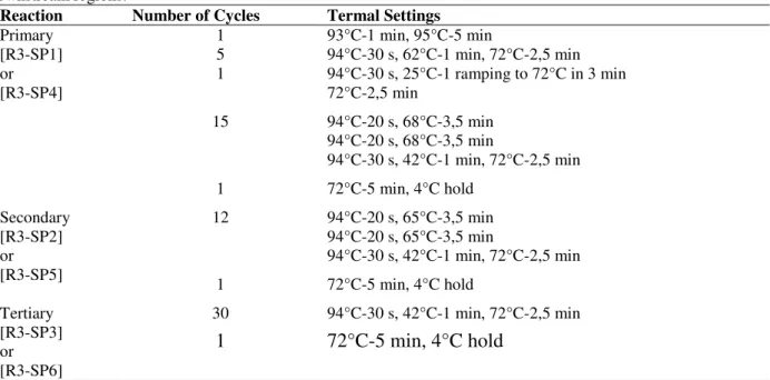
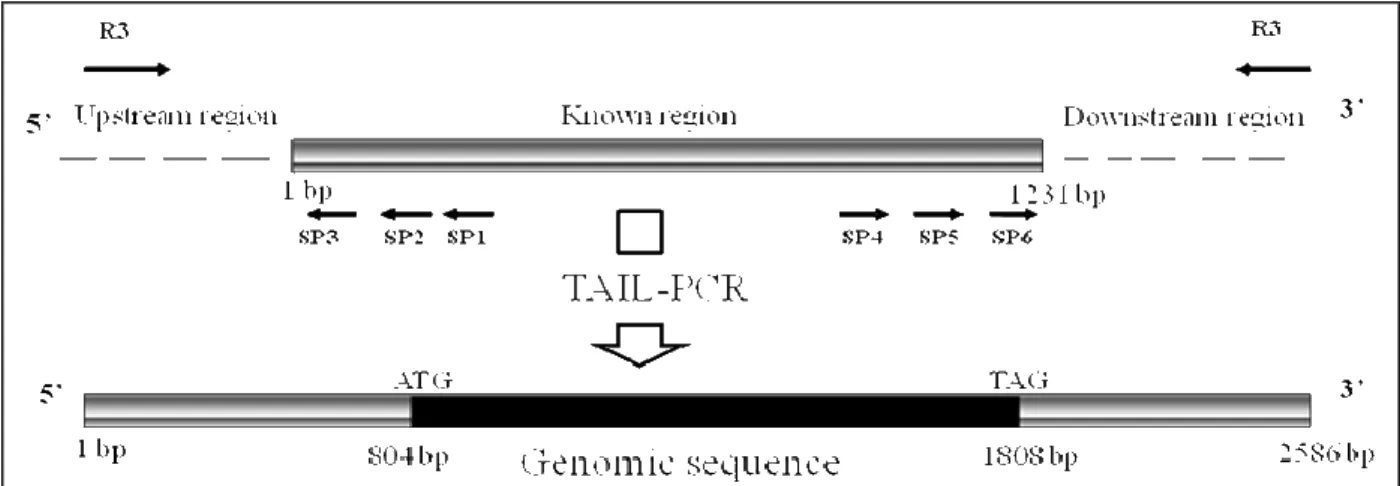
Gene-specific primers SP1 (5’– GGT ACA CGG CCT CGC TGC CAA CG – 3’), SP2 (5’ – ACG AGT CCA CCC ACA TGC CGA GC – 3’), SP3 (5’ – CTC GGT GGC AAT CTG CGA GGC GG – 3’) were used for flanking the 5’ region and SP4 (5’ – ACG GTG GCG TAC TTC GGC CT – 3’), SP5 (5’ – CAC CCA TCA CTG CCG TCG GCG CG – 3’), SP6 (5’ – TCG GCC GAC GCC ACT TCC ACG GA – 3’) were used for flanking the 3’ region. Degenerated primer R3 (5’-WGT GNA GWA NCA NAG A-3’) was applied (Fig. 1). Three rounds of PCR were performed on a MyCyclerTM Thermal Cycler (Bio-Rad) using the product of the previous PCR as the template for the next one. A detailed cycler program is given in Table1.
Table 1 - TAIL-PCR cycle setting for genomic DNA to walk from a short known sequence to isolate upstream or
downstream regions.
Reaction Number of Cycles Termal Settings
1 5 1
93°C-1 min, 95°C-5 min
94°C-30 s, 62°C-1 min, 72°C-2,5 min 94°C-30 s, 25°C-1 ramping to 72°C in 3 min 72°C-2,5 min
15 94°C-20 s, 68°C-3,5 min 94°C-20 s, 68°C-3,5 min
94°C-30 s, 42°C-1 min, 72°C-2,5 min Primary
[R3-SP1] or [R3-SP4]
1 72°C-5 min, 4°C hold
12 94°C-20 s, 65°C-3,5 min 94°C-20 s, 65°C-3,5 min
94°C-30 s, 42°C-1 min, 72°C-2,5 min Secondary
[R3-SP2] or [R3-SP5]
1 72°C-5 min, 4°C hold
30 94°C-30 s, 42°C-1 min, 72°C-2,5 min Tertiary
[R3-SP3] or [R3-SP6]
The primary PCR was performed in 50µl containing 80ng of genomic DNA; 0.2 µM of primer SP1; 2 µM of primer R3; 0.2mM of each dNTP; 1U Taq DNA polymerase (Promega) and 10X amplification buffer supplied with the enzyme. The secondary PCR was performed with primer SP2 (0.2 µM) and the same random primer R3 (2 µM) as used in the primary reaction. One 1 l of 1/50 a dilution of the primary PCR was used as a template. Single-step annealing-extension PCR consisting of a combined annealing and extension step at 65ºC or 68ºC was used in the primary and secondary PCR reactions. The tertiary reaction was carried out with 1 µl of a 1/10 dilution of the secondary reaction, 0.2 µM of primer SP3, 0.2 µM of random primer R3, 0.2 mM of each dNTP, 1U DNA Taq polymerase (Promega) and 10X amplification buffer. In the tertiary reaction was included the control SP3-SP3, SP6-SP6 and R3-R3, to exclude further non-specific amplification.
The "DNA and Gel Band Purification kit" (GE Healthere) protocol was used to purify the fragments interest.
DNA Sequencing
DNA fragments were sequenced in an automatic capillary sequencing ABI Prism 377TM at the University of Salamanca.
Sequences Analysis
The "European Bioinformatics Institute" was accessed to extract the sequences from the databases (EMBL - www.ebi.ac.uk), and from the "National Center of Biotechnology Information" (NCBI - www.ncbi.nlm). These servers allowed the comparison of DNA sequences and of proteins by several programs: Clustal, Fasta, and Blast. The BioEdit, Sequencher and DNASTARTM (SeqMan and EditSeq) programs were used to analyze the sequences.
Figure 1 - Schematic representation of TAIL-PCR amplification.
RESULTS AND DISCUSSION
The polymerase chain reaction (PCR) was used to amplify a 1231 bp sequence of the glucan endo-1,3-D-glucosidase gene. For total amplification of the gene, the Thermal Asymmetric InterLaced (TAIL)-PCR method was used, which allowed the amplification of DNA fragments adjacent to a known sequence. Tail-PCR uses three nested specific primers in successive reactions together with a shorter arbitrary degenerate (R3) primer so that the relative amplification efficiencies of
specific and non-specific products can be thermally controlled.
and sequenced. In the upstream region, a fragment of approximately 800 bp was obtained, while in
the downstream region, one of approximately 600 bp was collected.
800pb
M R3 M
SP3 R3 SP3 SP3 SP3 R3 R3 2 600pb
M R3 M
SP6 R3 SP6 SP6 SP6 R3 R3 1 600pb
M R3 M
SP6 R3 SP6 SP6 SP6 R3 R3 1 600pb
M R3 M
SP6 R3 SP6 SP6 SP6 R3 R3
M R3 M
SP6 R3 SP6 SP6 SP6 R3 R3 2 800pb
M R3 M
SP3 R3 SP3 SP3 SP3 R3 R3 2 800 bp
M R3 M
SP3 R3 SP3 SP3 SP3 R3 R3
M R3 M
SP3 R3 SP3 SP3 SP3 R3 R3 1
I
II
III
IV
V
VI
VII
VIII
600 bp
Figure 2 - Products of the tertiary PCR with controls. [1] Agarose gel analysis of tertiary TAIL-PCR
products using degenerated primer R3 to isolate the upstream region of the endo1 gene of
P. cinnamomi. The gene-specific primer used was SP3. [2] Agarose gel analysis of tertiary TAIL-PCR products using degenerated primer R3 to isolate the downstream region of the
endo1 gene of P. cinnamomi. The gene-specific primer used was SP6. M is a Promega 1kb DNA Ladder. Lanes I and V - reaction control without DNA. Lanes II and VI - amplification product. Lanes III, IV, VII and VIII - tertiary reaction control.
The complete sequence of the glucan endo-1,3-D-glucosidase gene was compared with the sequences deposited in the NCBI and EMBL databases. According to the BLASTX search program, the glucan endo-1,3-D-glucosidase gene of P. cinnamomi had the homology with a putative endo-1,3-beta-glucanase of P. infestans (E-value = 8e-135), an endo-1,3-beta-glucanase of Saprolegnia parasitica (E-value = 7e-56), an endo-beta-1,3-glucanase of Tetrapisispora phaffii (E-value = 8e
-25
) and an endo-beta-1,3-glucanase of Saccharomyces cerevisiae (E-value = 1e-24). This sequence presented the conserved domain Exo-beta-1,3-glucanase an carbohydrate transport and metabolism(COG5309), and homology with a glycoside hidrolase family 17 (InterProSan – EMBL).
Alignment of glucan endo-1,3-D-glucosidase gene with several endo-1,3-β-glucanases from GH family 17 revealed that the conserved domain of this family contained ([LIVMKS] -X- [LIVMFYWA](3)- [STAG] -E- [STACVI] -G- [WY] -P- [STN] -X- [SAGQ]), (Fig. 3). The
Figure 3 - Alignment of the amino acid sequences of glycoside hydrolase family 17 fungal
endo-1,3-β-glucanases and plant endo-1,3-β- and 1,3; 1,4-β-glucanses, as well as an endo-1,3-β -glucanases of P. infestans and P. cinnamomi. (∗) Is an active site residue. (1) Characteristic domain of GH family 17 ([LIVMKS]-X-[LIVMFYWA](3)-[STAG]-E-[STACVI]-G-[WY]-P-[STN]-X-[SAGQ]).
Figure 4 - Phylogram of glycoside hydrolase family 17 fungal endo-1,3-β-glucanase and oomycete
endo-1,3-β-glucanase.
Figure 5 - Alignment of the 5’ upstream region of Phytophthora glucanases genes to a conserved
sequence GCTCATTYYNCAWTT found in a promoter region of oomycete genes. [a]
CONCLUSIONS
The P. cinnamomi glucan 1,3-beta-D-glucosidase gene (PcENDO1) was closely related to glycoside hydrolases of family 17 because it also contained the conserved domain of this family (Fig. 3 and 4). A putative conserved 16 nt core sequence (GCTCATTYYNCAWTT) possibly used as an initiation of transcription point in oomycetes has been also identified in Pcendo1 (Fig. 5).
ACKNOWLEDGEMENTS
The Project COMBATINTA/SP2.P11/02 Interreg IIIA – Cross-Border Cooperation Spain-Portugal, financed by The European Regional Development Fund, supported this work.
RESUMO
Doença da tinta é um das doenças mais destrutivas em Castanea sativa. Os sintomas mais comuns são necroses e uma redução em crescimento da raiz que invariavelmente leva à morte das plantas. Phytophthora cinnamomi é o oomycete associado a esta doença cujo ciclo de vida acontece integralmente no solo.
Foi obtido um fragmento com 1231pb do gene glucan endo-1,3-β-D-glucosidase por amplificação usando oligonucleotidos conservados e a sequência completa do gene foi obtido flanqueando esta sequência conhecida por PCR assimétrico. Este fragmento foi obtido de ADN
genómico de Phytophthora cinnamomi isolado por nós nas Regiões Europeias de Castilla-Léon (Espanha) e Trás-os-Montes (Portugal) e associado à doença da tinta da Castanea sativa Mill.
REFERENCES
Cruz-Ortega, R., Cushman, J.G., Ownby, J.D.,(1995). Nucleotide Sequence of cDNA for a 1,3-Beta-Glucanase Associated with Aluminum Toxicity in Wheat Roots. Plant Physiol,. 109, 722.
McLeod, A., Smart, C.D., Fry, W.E., (2003).
Characterization of 1,3-beta-glucanase and 1,3; 1,4-beta-glucanase genes from Phytophthora infestans. Fungal Genetics and Biology,38, 250-263.
Michiels, A.N., Tucker, M., Ende, W.V.D., Laere, A. V., (2003). Chromosomal walking of flanking regions from short known sequences in GC-Rich plant genomic DNA. Plant Molecular Reporter, 21, 295-302.
Liu, Y.G., Mitsukawa, N., Oosumi, T., Whittier, R.F., (1995). Efficient isolation and mapping of
Arabidopsis thaliana T-DNA insert junction by termal asymmetric interlaced PCR. The Plant Jounal,
8 (3): 457-463.
Kamoun, S., (2003). Molecular Genetics of Pathogenic Oomycetes. Eukaryotic Cell, p. 191-199.
Kamoun, S.; Van West, P.; Vleeshouwers, V.; Groot, K.; Govers, F., (1998), “Resistance of Nicotiana benthamiana to Phytophthora infestans is mediated by the recognition of the elicitor protein INF1” The Plant Cell,10, 1413-1425).
![Figure 2 - Products of the tertiary PCR with controls. [1] Agarose gel analysis of tertiary TAIL-PCR products using degenerated primer R3 to isolate the upstream region of the endo1 gene of P](https://thumb-eu.123doks.com/thumbv2/123dok_br/15960228.683931/4.892.113.802.171.421/products-tertiary-controls-agarose-analysis-tertiary-products-degenerated.webp)

