Study conducted at the Department of Pediatrics, Botucatu Medical School, Universidade Estadual Paulista “Júlio de Mesquita Filho” – UNESP – Botucatu (SP), Brazil.
(1) Department of Pediatrics, Botucatu Medical School, Universidade Estadual Paulista “Júlio de Mesquita Filho” – UNESP – Botucatu (SP), Brazil. (2) Department of Otorhinolaryngology, Botucatu School of Medicine, Universidade Estadual Paulista “Júlio de Mesquita Filho” – UNESP – Botucatu (SP), Brazil. Conflict of interests: None
Authors’ contributions: CFCF and CSM participated in the protocol design, data collection, analysis, interpretation of results and manuscript writing. JCM participated on the assessment and interpretation of hearing tests. All the authors have read and approved the manuscript.
Correspondence address: Claudia Saad Magalhães. Departamento de Pediatria, Faculdade de Medicina de Botucatu, Universidade Estadual Paulista – UNESP, Distrito de Rubião Junior, s/n, Botucatu (SP), Brazil, CEP: 18618-970. E-mail: claudi@fmb.unesp.br
Received: 2/6/2012; Accepted: 2/4/2013
Hearing loss assessment in patients with pediatric
rheumatic disorders
Avaliação audiológica em pacientes com doenças reumáticas
pediátricas
Carolina Ferreira Campos-Flumian1, Jair Cortez Montovani2, Claudia Saad Magalhães1
ABSTRACT
Purpose: To perform audiological assessment in children and ado-lescents followed up at a reference outpatient clinic for autoimmune diseases, regardless of specific diagnoses. Methods: A single-blind case-control study was conducted. Participants were 48 patients with ages from 5 to 19 years and one to 151 months follow-up, categorized into three groups: 15 control individuals with pain in limbs and no au-toimmune disorders, 23 individuals with Juvenile Idiopathic Arthritis, and ten cases diagnosed with other autoimmune disorders. All subjects were submitted to clinical, otological, and audiological assessments (tympanometry, acoustic reflex, audiometry, speech audiometry, oto-acoustic emissions, and auditory brainstem response test with click stimuli). Results: The group with other autoimmune disorders had a greater proportion of patients with symptoms and more altered results in the otoacoustic emission test, when compared with the control group and the group with Juvenile Idiopathic Arthritis. In the group with other autoimmune disorders, 50% of the subjects with no symptoms presented impaired acoustic reflexes, alterations in audiometry and in otoacoustic emissions. In the audiometry, the group with Juvenile Idiopathic Ar-thritis presented more alterations in higher frequencies, and the group with other autoimmune disorders, in lower frequencies. Conclusion: Symptoms related to hearing loss and audiological alterations were more frequent in children and adolescents with Juvenile Idiopathic Arthritis and other autoimmune disorders. The hearing alterations also occurred in patients with no symptoms, indicating the need for systematic hearing assessment for these patients in their clinical routine.
Keywords: Autoimmunity; Audiology; Hearing loss; Rheumatic dise-ases; Pediatrics
RESUMO
Objetivo: Realizar avaliação audiológica em crianças e adolescentes acompanhados em um ambulatório de referência para doenças autoi-munes, independentemente do diagnóstico específico. Métodos: Foi realizado um estudo cego simples do tipo caso-controle. Foram incluídos 48 pacientes com idades entre 5 e 19 anos e tempo de seguimento de um a 151 meses, divididos em três grupos: 15 pacientes controle com diagnóstico de dor em membros e exclusão de doença autoimune, 23 pacientes com Artrite Idiopática Juvenil, e dez pacientes com outras doenças autoimunes. Os voluntários foram submetidos a avaliações clínica, otológica e audiológica (timpanometria, pesquisa dos reflexos acústicos, audiometria, índice de reconhecimento de fala, emissões otoacústicas e potenciais evocados auditivos de tronco encefálico com estímulo click). Resultados: O grupo com outras doenças autoimunes teve maior número de pacientes sintomáticos e maior número de ore-lhas alteradas no teste de emissões otoacústicas em comparação com o grupo controle e com o grupo com Artrite Idiopática Juvenil. Ainda no grupo com outras doenças autoimunes, 50% dos sujeitos assintomáticos apresentaram alterações na pesquisa dos reflexos, na audiometria e nas emissões otoacústicas. Na audiometria, o grupo com Artrite Idiopática Juvenil apresentou mais alterações nas frequências altas, e o grupo com outras doenças autoimunes, nas frequências baixas. Conclusão: Houve maior número de sintomas relacionados à perda auditiva e a alterações audiológicas em crianças e adolescentes com Artrite Idiopática Juvenil e outras doenças autoimunes. As alterações auditivas ocorreram também em pacientes assintomáticos, justificando-se a avaliação audiológica como rotina clínica desses pacientes.
INTRODUCTION
Evidence of hearing loss or poor sound discrimination, tinnitus and vertigo has been verified in adults affected by au-toimmune rheumatic disorders(1-10); however, pediatric studies
are scarce(11-14). Isolated autoimune hearing loss is reported in
both adults and children, with normal otoscopy and clinical hearing assessment, and sudden or progressive asymmetric bila-teral hearing loss. In autoimune sensorial hearing loss the main histological findings are atrophy of the stria vascularis, loss
of spiral ganglion cells, with various degrees of hair cell loss and loss of the membranous labyrinth(15). Early treatment with
prednisone can result in full recovery, and prevent progressive hearing loss for what is considered one of the rare reversible sensorineural hearing loss conditions(2). Autoimmune rheumatic
disorders are usually responsive to prednisone(16).
It is possible that systemic autoimmune diseases may simi-larly affect the inner ear. Autoimmune rheumatic diseases cause structural damage or dysfunction in multiple organs and sys-tems due to inflammation, hence the outcome is variable(1,2,17).
In children, the discrimination of symptoms due to eye or ear involvement is limited. Therefore, monitoring of those subjects with minimal or no symptoms is important.
To our knowledge, there are only a few previous reports of systematic assessment of hearing in pediatric autoimmune diseases. Due to the low prevalence of these diseases and the importance of hearing during development, this study had the aim to systematically assess the hearing function in a series of cases from a reference outpatient clinic for Pediatric Rheumatology.
METHODS
A single-blind case-control study was conducted in a case series of pediatric rheumatic disorders, in which otological and audiological assessments were conducted, according to a standard protocol. The protocol was approved by the Research Ethics Committee of the Botucatu Medical School, Universidade Estadual Paulista “Júlio de Mesquita Filho” (UNESP) (no. 374/2004), signed informed consent by parents
or legal guardians.
Inclusion criteria were: age between 2 and 19 years, diag-nosis confirmed by a specialist, and follow-up of at least two consultations. Individuals with history of congenital hearing loss, infection-related hearing loss, presence of ear discharge or middle ear effusion, familial deafness, genetic or congenital diseases, chronic neurological disorders, tympanic membrane abnormalities, fever, pain or discomfort during the tests were excluded.
No other case selection criteria occurred, apart from the clinical attendance required by the patient and the agreement to participate, confirmed by the consent term. Upon completion of all independent evaluations by one of the authors (CFCF) and
diagnosis disclosure by another author (CSM), the individuals were divided into three groups:
- Control group: healthy children with benign muscu-loskeletal complaints and the exclusion of autoimmune disorders(18);
- Group with Juvenile idiopathic arthritis (JIA): the cases werediagnosed according to the International League of Associations for Rheumatology (ILAR) criteria(19);
- Group with other autoimmune disorders (AID): represen-ted bycases of Juvenile Systemic Lupus Erythematosus, Scleroderma, and Primary Sjögren’s Syndrome. All the cases were diagnosed according to established clinical criteria(17,20-22).
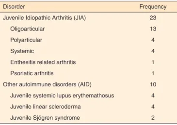
Forty-eight individuals with ages between 5 and 19 years were included. The Control group had 15 subjects, the group with JIA presented 23 subjects, and the AID group, ten subjects. The specific diagnoses are presented in Table 1.
Audiological assessments were performed by the same investigator (CFCF) throughout the study. The otological assessments included a complete investigation of auditory symptoms and clinical assessments by an independent otorhi-nolaryngologist author (JCM).
Audiological tests
Acoustic immittance measurements (tympanometry/stape-dial reflex), standard pure tone audiometry, speech audiometry, transient evoked otoacoustic emission (TEOAE), and auditory brainstem response (ABR) tests were performed in all patients.
The tympanometry was performed with an Amplaid® 775
admittance-meter at a probe tone frequency of 226 Hz. The tympanograms were classified according to Jerger types A, As, Ad, B, and C(23).
The acoustic reflex thresholds were tested for the frequen-cies of 500 Hz, 1, 2 and 4 kHz. Results were considered to be normal when the thresholds were between 70 and 90 dB SL.
An Amplaid® A321 twin-channel audiometer and TDH 49P
Table 1. Frequency of specific diagnoses of rheumatologic disorders
Disorder Frequency
Juvenile Idiopathic Arthritis (JIA) 23
Oligoarticular 13
Polyarticular 4
Systemic 4
Enthesitis related arthritis 1
Psoriatic arthritis 1
Other autoimmune disorders (AID) 10
Juvenile systemic lupus erythemathosus 4
Juvenile linear scleroderma 4
earphones were used for air conduction measurements, and a B-71 vibrator was used for bone conduction measurements in the audiometry. Air conduction thresholds were obtained from 250 Hz to 8 kHz, whereas bone conduction thresholds and air-bone gaps were obtained from 500 Hz to 4 kHz. Hearing loss was defined when pure tone results exceeded 15 dBHL for air conduction threshold and 10 dBHL for bone conduc-tion threshold(24).
The speech audiometry was carried out by a speech rec-ognition index, and the frequency of correct responses was considered within the normal range when equal to or higher than 92%(25).
The TEOAE test was performed with an Otodinamic ILO-92 setting ILO-88 connected to a computer. Click stimuli from 1 to 4 kHz were used at intensities between 75 and 85 dB SPL. A TEOAE response was considered positive when the repro-ducibility rate was greater than 50%, the probe stability was greater than 75%, and the signal-to-noise ratio was higher than 6 dB SPL in at least three bands.
The ABR was obtained using a Neuropack2 Nihon Kohden®, which was properly grounded to minimize
electro-magnetic interference. The parameters used were click stimuli at an intensity of 85 dB HL, rarified polarity, period of analysis of 10 ms, with 1,000 clicks. The contralateral ear to the sound stimulus was masked with -40 dBHL. The duplication of each recording was conducted in order to ensure wave reproduc-ibility and reliability. The ABR was accepted as normal with the presence of three consecutive peaks (waves I, III and V). The absolute latency values were considered normal when: (I) from 1.3 to 1.8 ms; (III) from 3.5 to 4.0 ms; (V) from 5.2 to 5.9 ms; and the interpeak latency values were normal when: (I-III) from 1.8 to 2.3 ms; (III-V) from 1.7 to 2.2 ms (I-V) from 3.7 to 4.5 ms. Interaural latency difference in wave V <0.4 ms was considered normal.
Statistical analysis
Demographic and clinical data are descriptively presen-ted. Statistical comparisons between groups were performed using the Student t test, the variance analysis (ANOVA), or the Kruskal-Wallis followed by multiple comparison tests. Associations between categorical variables were determined by the Chi-square and the Fisher exact tests.
RESULTS
Clinical presentation and hearing assessment
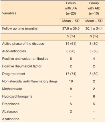
Clinical and immunological features and the treatment administered to patients with JIA and AID are presented in Table 2. A significant difference (p=0.02; x2=5.0516) was
verified between those on nonsteroidal antiinflammatory drug treatment. Observation confirmed that most patients (61% from
the JIA and 80% from the AID group) were taking antirheu-matic medication during the hearing assessment consultation.
Otological and audiological assessments
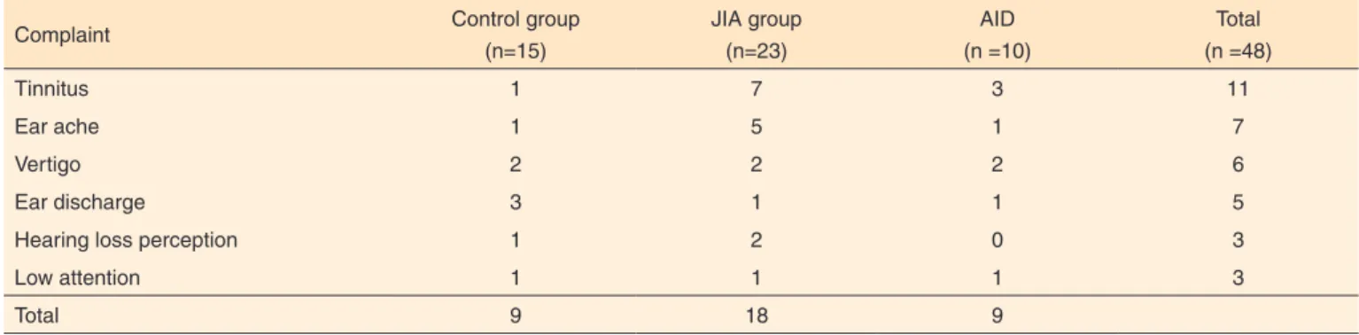
During clinical assessments, 21 out of the 48 subjects eva-luated had at least one of the otologic complaints listed. The frequency of symptoms is presented in Table 3.
The otological assessments indicated middle ear invol-vement in 17% of the patients, who were then referred for treatment. Further assessment was required in 13% of the subjects in the Control group, 17% of the JIA, and 20% of the AID subjects.
Subjects in the JIA and AID groups had the same complaints (tinnitus, ear ache, vertigo, ear discharge, perception of hearing loss, low attention). The symptoms survey showed that 43% of the JIA group and 60% of the AID group presented at least one of the otologic complaints listed. Tinnitus was the most frequent complaint, followed by ear ache and vertigo. There was a higher frequency of patients for overall complaints in the AID group, while a higher frequency of otologic complaints was verified in the JIA group.
Each ear was independently evaluated. The frequency of abnormal audiological findings in the three groups is presented in Table 4. There were differences between groups regarding the abnormal findings in the transient evoked otoacoustic
Table 2. Clinical features and treatment of patients with pediatric rheu-matologic disorders by the time of hearing assessment
Variables
Group with JIA
(n=23)
Group with AID
(n=10)
Mean ± SD Mean ± SD
Follow up time (months) 37.9 ± 39.6 30.1 ± 34.4
n (%) n (%)
Active phase of the disease 14 (61) 8 (80)
Auto-antibodies 6 (26) 5 (50)
Positive antinuclear antibodies 6 4
Positive rheumatoid factor 2 2
Drug treatment 17 (74) 8 (80)
Non-steroidal antiinflammatory drugs 16 2
Methotrexate 8 2
Hydroxychloroquine - 6
Prednisone 5 5
Abatacept 2
-Azatioprine - 1
emissions (TEOAE), with higher frequency of alterations in both the JIA and AID groups, compared to the Control group (Chi-square test: p=0.004).
The abnormal tympanometry findings were classified as “As”, which means stiffness of the middle ear bones. The audiometry revealed a higher number of ears classified with sensorineural hearing loss(26) with thresholds up to 35 dB in both
the JIA and AID groups. In the JIA group, hearing impairments for low (250, 500 Hz and 1 kHz), medium (2 and 3 kHz) and high (4, 6 and 8 kHz) frequencies were verified, while in the AID group, only low and high frequency impairments were identified. Overall, high frequencies were more frequently affected in the JIA group, and low frequencies were more frequently affected in the AID group.
The Auditory Brainstem Response (ABR) test was within normal limits for all groups, and waves I, III and V had normal waveform morphology. The speech audiometry test was also within the normal range for all patients.
DISCUSSION
A comprehensive otological and audiological assessment was conducted, selecting the cases during standard practice. However, due to the low prevalence of pediatric rheumatic diseases, a limited number of cases was included. The results obtained indicated more otological symptoms and hearing
impairment in children and adolescents affected by rheumatic diseases, compared with a control group in which rheumatic diseases were ruled out. A full hearing testing comprising otological symptoms survey combined to otological and au-diological systematic assessments revealed comparable rates of hearing impairment in adults with the same rheumatic disease spectrum(1-10).
Although hearing impairment was more frequent in pa-tients with otological complaints, it was verified that 46% of the patients with no symptoms in the JIA group and 50% of those in the AID group presented some degree of impairment in the hearing tests.
Tinnitus is a common complaint in patients with autoimmu-ne diseases(1,10). Complaints of tinnitus and also vertigo were
recorded in our patients. Tinnitus was present in both the JIA and AID groups, but it was not possible to determine its rela-tionship with adverse events of nonsteroidal antiinflammatory drugs used very often by those patients regardless of their diagnoses, such as nonsteroidal antiinflammatory drugs. The presence of tinnitus could be caused by biochemical changes preceding structural damage.
Tympanometric and stapedial reflex findings in the JIA group might be due to the involvement of the small joints of middle ear bones in the disease process and arthritis, resul-ting in otological complaints and abnormal tympanometric findings(11).
Table 3. Frequency of otological complaints in patients from Control, Juvenile Idiopathic Arthritis and Other autoimmune disorders groups
Complaint Control group
(n=15)
JIA group (n=23)
AID (n =10)
Total (n =48)
Tinnitus 1 7 3 11
Ear ache 1 5 1 7
Vertigo 2 2 2 6
Ear discharge 3 1 1 5
Hearing loss perception 1 2 0 3
Low attention 1 1 1 3
Total 9 18 9
Chi-square test (p>0,05)
Note: JIA = juvenile idiopathic arthritis; AID = other autoimmune disorders
Table 4. Frequency of altered results in each of the ears from patients in the Control, Juvenile Idiopathic Arthritis and Other autoimmune disorders groups
Test Control
(n=30)
JIA group (n=46)
AID group (n=20)
Total (n=96)
Tympanometry 1 2 1 4
Stapedial reflex 0 7 2 9
Audiometry 6 16 7 29
Otoacoustic emission 2 8 9* 19
Chi-square test (p<0.05)
* Control x AID (p=0.004; x2=8.1634) JIA x AID (p=0.04; x2=4.2063)
Tympanometric findings did not affect otoacoustic emis-sions responses. Otoacoustic emisemis-sions were detected in ears that presented type As curves; however, the otoacoustic emissions testing revealed a higher number of impaired results in both the JIA and AID groups, indicating possible cochlear involvement(7,10). The number of ears with altered results in
the otoacoustic emissions was higher than observed in the audiometry, which may be due to its greater sensitivity for detecting minor changes in external hair cells, prior to their detection by audiometry. This was further illustrated by one of the cases with localized scleroderma en coup de sabre affecting the right side of the face, for whom the audiometry results were normal, while the otoacustic emissions were altered on the right ear(16).
In our study, we found sensorineural hearing loss, which has been observed by different studies in both adults(2,3,9,10,16)
and children(12,13). Audiometry also detected alterations in all
frequencies, with thresholds up to 35 dB, however, the JIA group presented mainly high frequency impairments, and the AID group, low frequency impairments. Speech audiometry was within the normal range for all patients. Auditory brain-stem response results in our patients ruled out retrocochlear involvement previously described in patients with rheumato-logic diseases(9).
The results of this study indicated that the hearing impair-ment was of cochlear origin, since the ABR with clicks pre-sented absolute and interpeak latency values of waves I, III and V within the normal range, and the audiometry and/or the OAE presented altered results. Current electrophysiological proce-dures that assess with specificity by frequency, like the ABR use “tone burst” stimuli and Auditory Steady State Response (ASSR) were not used. These tests assist diagnosis when abnormalities in ABR results are detected(27,28), which was not
the case in our study.
It seems likely that cortical auditory evoked potentials could benefit research involving patients with autoimmune diseases, detecting possible alterations in the central auditory nervous system, since the pathophysiology of the disease could cause changes in its functional origin with different manifestations in different brain regions. However, such tests are only performed in children over 12 years of age, and since the age range of our participants was from 2 to 19 years old, these tests were not performed.
All the tests performed should be interpreted together, as they are complementary. Although the otoacoustic emissions test seems to show greater sensitivity for detecting cochlear involvement, it is limited by the frequencies assessed. None of the tests performed independently is sufficient to establish an accurate diagnosis.
There are numerous limitations to this study, including its cross-sectional design, the low number of participants, and a convenience sample of patients recruited during standard prac-tice. The mean duration of the disease in our series of cases was
30 months, a period that may not be sufficient to detect cumu-lative auditory system damage. The possibility remains that auditory system damage due to immune mechanisms is different in pediatric patients, when compared to adults with the same disorders, emphasizing the need for systematic assessment.
Hearing impairment can be related to several factors: the inflammatory process alone affecting small joints in the middle ear bones, adverse events of medication such as an-tiinflammatory and immunossupressive drugs, including non steroidal antiiinflammatories, prednisone, methotrexate, hidro-xicloroquine, azatioprine or biologic agentes. The pathogenic mechanisms of inflammation affecting the auditory system remain unclear, indicating the need for further controlled lon-gitudinal assessments.
CONCLUSION
Symptoms related to hearing loss and audiological alte-rations were more frequent in children and adolescents with Juvenile Idiopathic Arthritis and other autoimmune disorders. The hearing alterations also occurred in patients with no symp-toms, indicating the need for systematic hearing assessment for these patients in their clinical routine.
ACKNOWLEDGEMENTS
This work was developed for a Master’s thesis. Carolina Ferreira Campos-Flumian was granted a Fundação de Amparo à Pesquisa do Estado de São Paulo (FAPESP) scholarship (process number 04/12950-4). The authors acknowledge Ms. Josilene Luciene Duarte, Audiologist, for her advice on inter-pretation of hearing tests.
REFERENCES
1. Stone JH, Francis HW. Immune-mediated inner ear disease. Curr Opin Rheumatol. 2000Jan;12(1):32-40.
2. Ruckenstein MJ. Autoimmune inner ear disease. Curr Opin Otolaryngol Head Neck Surg. 2004Oct;12(5):426-30.
3. Piccirillo JF. Steroids for idiopathic sudden sensorineural hearing loss: some questions answered, others remain. JAMA. 2011May;305(20):2114-5.
4. Kastanioudakis I, Ziavra N, Voulgari PV, Exarcharkos G, Skevas A, Drosos AA. Ear involvement in systemic lupus erythematosus patients: a comparative study. J Laryngol Otol 2002Feb;116(2):103-7.
5. Iskandar SB, Loyd S, Roy TM. Cranial nerve VIII involvement in a patient with progressive systemic sclerosis. Tenn Med. 2004Mar;97(3):117-9.
7. Ziavra N, Politi EN, Kastanioudakis I, Skevas A, Drosos AA. Hearing loss in Sjögren’s syndrome patients. A comparative study. Clin Exp Rheumatol. 2000Nov-Dec;18(6):725-8.
8. Konttinen YT, Ramsay H, Hietanen J, Sorsa T, Nordstrom D. Otitis externa sicca/fibrotising external otitis (FEO) as complication of Sjögren’s syndrome. Clin Exp Rheumatol. 2000Nov-Dec:18(6):746-8.
9. Boki KA, Ioannidis JP, Segas JV, Maragkoudakis PV, Petrou D, Adamopoulos GK, et al. How significant is sensorineural hearing loss in primary Sjögren’s syndrome? An individually matched case-control study. J Rheumatol. 2001Apr;28(4):798-801.
10. Papadimitraki ED, Kyrmizakis DE, Kritikos I, Boumpas DT. Ear-nose-throat manifestations of autoimmune rheumatic diseases. Clin Exp Rheumatol 2004Jul-Aug;22(4):485-94.
11. Giannini P, Marciano E, Saulino C, Strano CG, Alesio M, Marceli V, et al. Middle ear involvement in children with chronic rheumatoid juvenile arthritis. Eur Arch Otorhinolaryngol. 1997;254(Suppl.1):S30-3.
12. Berrettini S, Ravecca F, Sellari-Franceschini S, Matteucci F, Siciliano G, Ursino F. Progressive sensorineural hearing loss in childhood. Pediatr Neurol 1999Feb;20(2):130-6.
13. Tomasi JP, Lona A, Deggouj N, Gersdorff M. Autoimmune sensorineural hearing loss in young patients: an exploratory study. Laryngoscope 2001Nov;111(11Pt1):2050-3.
14. Ikis AO, Unsal E, Kirkim G, Erdag TK, Guneri EA. Hearing loss and middle ear involvement in patients with juvenile idiopathic arthitis. Int J Pediatr Otorhinolaryngol. 2007Jul;71(7):1079-85.
15. Sone M, Schachern PA, Paparella MM, Mirizono, N. Study of systemic lupus erythematosus in temporal bones. Ann Otol Rhinol Laryngol. 1999Apr;108(4):338-44.
16. Rauch SD, Halpin CF, Antonelli PJ, Babu S, Carey JP, Gantz BJ, et al. Oral vs intratympanic corticosteroid therapy for idiopathic sudden sensorineural hearing loss: a randomized trial. JAMA. 2011May;305(20):2071-9.
17. Saad-Magalhães C, Medeiros PBS, Sato JO, Domingues MAC.
Clinical presentation and salivary gland histopathology of paediatric primary Sjögren’s syndrome. Clinical Exp Rheumatol. 2011May;29(3):589-93.
18. Malleson PN, Connell H, Bennett SM, Eccleston C. Chronic musculoskeletal and other idiopathic pain syndromes. Arch Dis Child. 2001Mar;84(3):189-92.
19. Petty RE, Southwood TR, Manners P, Baum J, Glass DN, Goldenberg J, et al. International League of Associations for Rheumatology classification of juvenile idiopathic arthritis: second revision, Edmonton 2001. J Rheumatol. 2004Feb;31(2):390-2. 20. Hochberg MC. Updating the American College of Rheumatology
revised criteria for the classification of systemic lupus erythematosus. Arthritis Rheum 1997Sep;40(9):1725.
21. Athreya BH. Juvenile Scleroderma. Curr Opin Rheumatol. 2002Sep;14(5):553-61.
22. Kassan SS, Moutsopoulos HM. Clinical manifestations and early diagnosis of Sjögren’s syndrome. Arch Intern Med. 2004Jun;164(12):1275-84.
23. Jerger JF. Clinical experience with impedance audiometry. Arch Otolaryngol 1970Oct;92(4):311-24.
24. Roeser RJ. Manual de consulta rápida em audiologia: um guia prático. Rio de Janeiro: Revinter; 2001.
25. Jerger J, Speacks C, Trammell JL. A new approach to speech audiometry. J Speech Hear Disord. 1968Nov;33(4):318-28. 26. Silman S, Silverman CA. Basic audiologic testing. In: Silmann S,
Silverman CA. Auditory diagnosis: principles and applications. San Diego: Singular Publishing Group, 1997; 44-52.
27. Duarte JL. A utilização da resposta auditiva de estado estável para estimar limiares auditivos em indivíduos com perda auditiva neurossensorial [dissertação]. Bauru: Universidade de São Paulo – Faculdade de Odontologia de Bauru; 2007.