Article
Increased Sclerostin Levels after Further Ablation of
Remnant Estrogen by Aromatase Inhibitors
Wonjin Kim1, Yoonjung Chung2, Se Hwa Kim3, Sehee Park1, Jae Hyun Bae1, Gyuri Kim1, Su Jin Lee1, Jo Eun Kim1, Byeong-Woo Park4, Sung-Kil Lim1, Yumie Rhee1
1Department of Internal Medicine, Endocrine Research Institute, Severance Hospital, Yonsei University College of Medicine; 2
The Graduated School Yonsei University, Seoul; 3
Department of Internal Medicine, Kwandong University College of Medicine, Incheon; 4
Department of General Surgery, Severance Hospital, Yonsei University College of Medicine, Seoul, Korea
Background: Sclerostin is a secreted Wnt inhibitor produced almost exclusively by osteocytes, which inhibits bone formation. Aromatase inhibitors (AIs), which reduce the conversion of steroids to estrogen, are used to treat endocrine-responsive breast cancer. As AIs lower estrogen levels, they increase bone turnover and lower bone mass. We analyzed changes in serum sclerostin levels in Korean women with breast cancer who were treated with an AI.
Methods: We included postmenopausal women with endocrine-responsive breast cancer (n=90; mean age, 57.7 years) treated with an AI, and compared them to healthy premenopausal women (n=36; mean age, 28.0 years). The subjects were randomly as-signed to take either 5 mg alendronate with 0.5 µg calcitriol (n=46), or placebo (n=44) for 6 months.
Results: Postmenopausal women with breast cancer had significantly higher sclerostin levels compared to those in premenopaus-al women (27.8±13.6 pmol/L vs. 23.1±4.8 pmol/L, P<0.05). Baseline sclerostin levels positively correlated with either lumbar spine or total hip bone mineral density only in postmenopausal women (r=0.218 and r=0.233; P<0.05, respectively). Serum sclerostin levels increased by 39.9%±10.2% 6 months after AI use in postmenopausal women; however, no difference was ob-served between the alendronate and placebo groups (39.9%±10.2% vs. 55.9%±9.13%, P>0.05).
Conclusion: Serum sclerostin levels increased with absolute deficiency of residual estrogens in postmenopausal women with en-docrine-responsive breast cancer who underwent AI therapy with concurrent bone loss.
Keywords: Sclerostin; Breast neoplasms; Aromatase inhibitors; Osteoporosis
INTRODUCTION
Bone remodeling is a tightly regulated process leading to bal-anced resorption and formation of skeletal tissue with the coor-dinated action of osteocytes [1,2]. Sclerostin is a glycoprotein secreted by osteocytes and a potent Wnt inhibitor. Sclerostin travels through osteocyte canaliculi to the bone surface where
it binds to the coreceptors lipoprotein receptor-related protein (LRP) 5 and LRP 6; thus, it prevents colocalization with Friz-zled and Wnt proteins, and reduces osteoblastogenesis and bone formation [3]. The clinical relevance of sclerostin in bone metabolism was initially recognized in patients with scleroste-osis or van Buchem’s disease with sclerostin mutations, result-ing in excessive bone formation leadresult-ing to osteosclerosis [4].
Received: 1 May 2014, Revised: 5 June 2014,
Accepted: 18 June 2014
Corresponding author: Yumie Rhee
Department of Internal Medicine, Severance Hospital, Yonsei University College of Medicine, 50-1 Yonsei-ro, Seodaemun-gu, Seoul 120-752, Korea
Tel: +82-2-2228-1973, Fax: +82-2-393-6884, E-mail: YUMIE@yuhs.ac
Copyright © 2015 Korean Endocrine Society
Overexpression of normal human SOST alleles (the scleros-tin gene) in mice results in osteopenia [5]. Mirza et al. [6] compared 20 postmenopausal and 20 premenopausal women and found that the former had significantly higher serum sclerostin levels. Estrogen replacement and intermittent para-thyroid hormone (PTH) therapy in postmenopausal women lead to a significant reduction in sclerostin levels [7,8]. Sever-al studies have reveSever-aled the molecular mechanism of de-creased sclerostin production by estrogens. Estrogens bind to estrogen receptors and interact with the Wnt/β-catenin signal -ing pathway, which downregulates osteocyte production of sclerostin via estrogen receptor-induced prostaglandin E2 [9]. Aromatase inhibitors (AIs) are used to treat endocrine-re-sponsive breast cancer by lowering remnant 17β-estradiol (E2) or estrione levels to prevent cancer recurrence [10]. This is the preferred adjuvant therapy for postmenopausal women with estrogen receptor/progesterone receptor-positive breast cancer, because it reduces breast cancer recurrence up to 50% com-pared to tamoxifen [11-13]. AIs affect bone metabolism by in-creasing bone turnover rates and accelerating bone loss, lead-ing to increased fracture risk [2,14-16]. Bisphosphonates are the preferred treatment for AI-induced bone loss [2], and cal-citriol, an active metabolite of vitamin D, in combination with an AI is effective for inhibiting tumors [10]. In a previous study [2], we reported that the combination of alendronate (5 mg) and calcitriol (0.5 µg) (Maxmarvil, Yuyu Co., Seoul, Ko-rea) was quite beneficial for preventing the bone loss that oc-curs with AI administration in patients with endocrine-respon-sive breast cancer.
Here, we analyzed serum sclerostin levels in patients with endocrine-responsive breast cancer treated with an AI to as-sess the changes in serum sclerostin levels following treatment with alendronate or placebo.
METHODS
Study design and subjects
This study was a randomized, placebo-controlled, double-blind study performed at a single center. A cross-sectional comparison of sclerostin was performed between premenopausal and post-menopausal women. Changes in sclerostin levels were analyzed in 90 endocrine-responsive breast cancer patients during new AI administration over 24 weeks. Forty-four patients received 5 mg alendronate with 0.5 µg calcitriol, calcium, and vitamin D, and the remaining 46 patients received only calcium and vi-tamin D [2].
The diagnosis of breast cancer and indication for adjuvant chemotherapy were made according to the guidelines of the National Comprehensive Cancer Network [10]. Patients who met the inclusion criteria were reviewed from March 2010 to March 2011. Postmenopausal women who were taking an AI after breast cancer surgery were included, and the definitions of menopause in these patients were as follows: (1) prior bilat-eral oophorectomy; (2) amenorrhea for ≥12 months in the ab-sence of tamoxifen, toremifene, or ovarian suppression; and (3) follicle-stimulating hormone and E2 levels in the post-menopausal range.
Thirty-six healthy premenopausal women were recruited by advertisement and served as controls. All subjects were in-formed of the nature of the study, and consent was obtained from each participant. This protocol was approved by the In-stitutional Review Board of Severance Hospital.
Biochemical measurements
Blood samples were collected in the morning after an overnight fast. Routine serum chemistry determinations including calci-um and phosphate were performed by standard automated techniques. Bone turnover markers were measured using the following methods: osteocalcin (OCN; by enzyme-linked im-munosorbent assay [ELISA], CIS Bio International, Gif-sur-Yvette, France; intra-assay coefficient of variation [CV], <2.0%; interassay CV, <5.0%), C-telopeptide of type I collagen (CTx; Osteomark, Ostex International, Seattle, WA, USA; intra-assay CV, <5.8%; interassay CV, <5.9%), intact PTH (by immuno-radiometric assay, Biosource, Nivelles Belgium; intra-assay CV, 2.7%; interassay CV, <3.5%), and 25-hydroxyvitamin D (25[OH]D by D3-radioimmunoassay-coated tube, Biosource; intra-assay CV, 11.0%; interassay CV, 12.5%).
Serum sclerostin concentrations were measured using a hu-man sclerostin ELISA kit (Biomedica Co., Wien, Austria) ac-cording to the manufacturer’s instructions. Intraassay and inter-assay CVs were 4% to 6% and 5% to 7%, respectively. Scleros-tin measurements are reported in pmol/L, and the lower limit of detection was <8.9 pmol/L. Measurements were taken twice at intervals of 24 weeks in postmenopausal women with endo-crine-responsive breast cancer. Sclerostin was measured only at baseline in the premenopausal control group.
Assessment of bone mineral density
12.6, Hologic, Waltham, MA, USA). The measurements were performed at baseline and after 24 weeks of treatment.
Statistical analysis
Differences in continuous variables between the premenopaus-al and postmenopauspremenopaus-al endocrine-responsive breast cancer groups were determined using Student t test. The correlation between continuous variables was analyzed by Pearson corre-lation analysis. Student t test and the paired t test were used to compare sclerostin levels and bone turnover markers in the postmenopausal breast cancer group. A P<0.05 was considered statistically significant. All analyses were performed using IBM SPSS version 18.0 (IBM Co., Armonk, NY, USA).
RESULTS
Baseline clinical and biochemical characteristics
This study included 90 postmenopausal women who were di-agnosed with endocrine-responsive breast cancer and 36 healthy premenopausal control women. The postmenopausal women had a mean age of 57.7 years, and the premenopausal women had a mean age of 28.0 years (Table 1). Among the 90 postmenopausal women, nine (10%) were diagnosed with os-teoporosis.
Lumbar spine and total hip BMD were lower in postmeno-pausal women than in premenopostmeno-pausal women (0.92±0.14 g/ cm2
vs. 0.99±0.01 g/cm2
; 0.82±0.11 g/cm2
vs. 0.86±0.10 g/ cm2
; postmenopausal vs. premenopausal women; P<0.05,
re-spectively) (Table 1). OCN levels were 74.6% higher and CTx was 52.0% higher in postmenopausal women than in premeno-pausal women (P<0.001, respectively) (Table 1). Baseline se-rum sclerostin levels were 20.3% higher in the postmenopausal group compared to those in the premenopausal group (P<0.05) (Table 1).
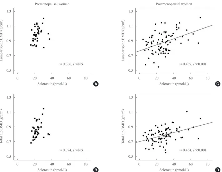
Correlation between sclerostin and bone metabolism Table 2, Fig. 1 show the relationships between sclerostin and various parameters according to Pearson correlation analysis.
Table 1. Baseline Clinical Characteristics of Premenopausal and Postmenopausal Women
Characteristic Premenopause (n=36) Postmenopause (n=90) P value
Age, yr 28.0±6.3 57.7±7.0 <0.001 BMI, kg/m2 21.2±3.1 24.3±3.1 <0.001
BMD L1–L4, g/cm2
0.985±0.1 0.918±0.1 0.002 BMD total hip, g/cm2 0.859±0.1 0.823±0.1 NS
Osteocalcin, ng/mL 12.6±3.2 22.0±9.0 <0.001 C-telopeptide, ng/mL 0.25±0.1 0.38±0.2 0.001 Serum calcium, mg/dL 9.4±0.4 9.2±0.4 0.015 Serum phosphorus, mg/dL 4.0±0.4 3.9±0.6 NS Serum PTH, pg/mL 35.7±11.6 27.0±12.1 <0.001 25(OH)D, ng/mL 25.6±12.0 15.5±7.0 <0.001 Serum sclerostin, pmol/L 23.1±4.8 27.8±13.6 0.029
Values are expressed as mean±SD. P values determined by Student t test.
BMI, body mass index; BMD, bone mineral density; NS, not significant; PTH, parathyroid hormone; 25(OH)D, 25-hydroxyvitamin D.
Table 2. Partial Correlations between Sclerostin and Various Parameters in Premenopausal and Postmenopausal Women Adjusted for Body Mass Index
Parameter Premenopause Postmenopause
ra P value ra P value
Weight 0.028 NS 0.220 0.047 Osteocalcin −0.218 NS 0.131 NS C-telopeptide −0.031 NS −0.093 NS Serum calcium −0.073 NS −0.084 NS Serum phosphorus 0.028 NS 0.028 NS Serum PTH −0.010 NS −0.088 NS 25(OH)D −0.062 NS 0.042 NS
P values calculated by Pearson correlation analysis.
NS, not significant; PTH, parathyroid hormone; 25(OH)D, 25-hy-droxyvitamin D.
Serum baseline sclerostin level positively correlated with lum-bar spine and total hip BMD (r=0.218 and r=0.233; P<0.05, respectively) (Fig. 1), but only in the postmenopausal group. No significant correlations were observed between bone turn-over markers such as CTx and OCN and serum sclerostin (Ta-ble 2). A negative correlation was observed between PTH and sclerostin in postmenopausal women (r=–0.074), but was not significant. Serum calcium, phosphorus, and 25(OH)D had no relevant relationships with sclerostin levels.
Biochemical changes after AI treatment with or without alendronate
We further divided the postmenopausal patients into two sub-groups based on medications: 5 mg alendronate and calcitriol
in one group, and calcium and vitamin D only in the other group. Baseline characteristics were presented in a previous re-port [2]. Baseline features, such as the period after menopause, biochemical parameters, and baseline BMD at all sites were similar between the two groups. Serum OCN and CTx levels increased significantly in both groups after 24 weeks of AI ad-ministration, but increased lesser in the alendronate-treated group by 29.0% and 72.4%, respectively [2].
We also analyzed percent changes in sclerostin levels from baseline to 24 weeks after AI use and found a significant in-crease (48.1%±6.9%, P<0.05) in all postmenopausal women. Sclerostin levels in the placebo group increased significantly after 24 weeks of AI administration (55.9%±9.13%, P< 0.001). Less of an increase was observed in patients treated
1.3
1.1
0.9
0.7
0.5
1.3
1.1
0.9
0.7
0.5
1.3
1.1
0.9
0.7
0.5
1.3
1.1
0.9
0.7
0.5
Lumbar
spine BMD (g/cm
2)
T
otal
hip BMD (g/cm
2)
Lumbar
spine BMD (g/cm
2)
T
otal
hip BMD (g/cm
2)
0 20 40 60 80
0 20 40 60 80
0 20 40 60 80
0 20 40 60 80 Sclerostin (pmol/L)
Sclerostin (pmol/L) Premenopausal women
Sclerostin (pmol/L)
Sclerostin (pmol/L) Postmenopausal women
r=0.066, P=NS
r=0.094, P=NS
r=0.439, P<0.001
r=0.454, P<0.001
A
B
C
D
with alendronate (39.9%±10.2%, P<0.001) (Fig. 2), but this was not significantly different from the increase in the placebo group.
DISCUSSION
We showed higher serum sclerostin levels in postmenopausal women compared to those in premenopausal women. Scleros-tin was positively correlated with BMD in postmenopausal women at all sites. In addition, sclerostin levels increased fur-ther after use of an AI which is a treatment for breast cancer that ablates the remnant estrogen, but were not affected by the use of alendronate.
Sclerostin, encoded by the SOST gene, is a major Wnt antag-onist and a potent inhibitor of bone formation [4]. Previous ani-mal studies have shown that overexpression of the SOST gene causes osteopenia [5]. Moreover, production of sclerostin by osteocytes is dramatically reduced by mechanical loading in rodents [17-19]. Thus, sclerostin may be a potential specific marker for bone metabolism, but the clinical implications are not completely understood. Modder et al. [20] showed that sclerostin levels increase with age and are higher in men com-pared to women. Polyzos et al. [21] reported that serum sclerostin is positively correlated with lumbar spine BMD and
T-score. Arasu et al. [22] also found a positive correlation be-tween sclerostin and BMD. A positive correlation was observed between serum sclerostin and spine and total hip BMD by Gar-nero et al. [23], which was in accordance with our data. They also found negative correlations between sclerostin and CTx, serum intact N-terminal propeptide of type I collagen, and in-tact PTH. However, the risk of hip fractures is higher in pa-tients with a higher serum sclerostin level [22]. A strong asso-ciation was observed between increased sclerostin levels and osteoporosis-related fracture risk in postmenopausal women [24]. The paradoxical positive correlation between sclerostin and BMD suggests that serum sclerostin may reflect the num-ber of osteocytes rather than individual cell activity or individ-ual bone remodeling units [25].
The possible regulators of circulating sclerostin levels are known as such; increasing the level by mechanical unloading, immobilization, male sex, aging, and decreasing by mechani-cal loading, exercise and PTH [17-24,26]. Among them, PTH is probably one of the most important regulators of sclerostin secretion in postmenopausal women [23]. Sclerostin is also af-fected by estrogens. It is well established that estrogen defi-ciency increases serum sclerostin levels and is associated with bone loss [27-29]. Modder et al. [7] reported that 4 weeks of estrogen treatment in postmenopausal women decreased se-rum sclerostin levels. Endogenous estrogen and testosterone production were ablated in 59 elderly men by administering gonadotropin-releasing hormone, after which these patients were given physiological levels of testosterone and estrogen. As a result, estrogen, but not testosterone, prevented an increase in sclerostin following induction of sex steroid deficiency [7]. Consistent with previous studies, our study also revealed a negative relationship between estrogen and sclerostin. The possible mechanism of how estrogen suppresses sclerostin production is that estrogen interacts with the Wnt/β-catenin signaling pathway, a main pathway of sclerostin action, by binding to the estrogen receptor via several factors such as prostaglandin E2 [9,25].
In this study, subjects with endocrine-responsive breast can-cer who were treated with an AI showed reduced remnant es-trogens and bone mass [14-16], and showed elevated scleros-tin levels. As mentioned above, estrogens and sclerosscleros-tin have an inverse correlation [27-29]. In addition to this concept, our results revealed that further suppression of remnant estrogens sensitively affected serum sclerostin levels. Thus, we suggest that sclerostin may be a surrogate marker for quantitative and sensitive changes in estrogens and bone remodeling.
Fig. 2. Comparison of changes in sclerostin. Sclerostin was mea-sured at baseline and after 24 weeks of aromatase inhibitor admin-istration. We compared changes in sclerostin in the placebo and alendronate with calcitriol groups, and showed a significant in-crease in sclerostin levels in both groups (placebo group, P<0.05;
alendronate with calcitriol group, P<0.05, by paired t test).
How-ever, the changes in sclerostin levels in the groups were not sig-nificantly different (P=not significant).
100
80
60
40
20
0
Sclerostin
(pmol/L)
Placebo
Baseline After 24 weeks Baseline After 24 weeks
Alendronate+calcitriol
P<0.05
Polyzos et al. [21] reported that serum sclerostin increases significantly after 6 months of risedronate use. However, Chung et al. [30] found that circulating sclerostin levels were suppressed by raloxifene, but not by bisphosphonates. They suggested that sclerostin may mediate the action of estrogens on bone metabolism independently of the anti-resorptive ef-fects. Our results revealed a significant increase in sclerostin levels after 24 weeks of AI administration, with a slightly blunted increase during alendronate treatment, but without statistical significance.
This study had some limitations. We demonstrated differ-ences in bone markers, BMD, and serum sclerostin levels be-tween premenopausal and postmenopausal women. However, the results are difficult to generalize because of the small num-ber of subjects.
In conclusion, our data show that serum sclerostin levels positively correlated with BMD at all sites, and that serum sclerostin increased according to the level of estrogen defi-ciency in postmenopausal women, and was further increased by AI treatment. Additional experimental and clinical studies in a larger population and with other drugs that correlate with sclerostin are needed.
CONFLICTS OF INTEREST
No potential conflict of interest relevant to this article was re-ported.
REFERENCES
1. Parfitt AM. The bone remodeling compartment: a
circula-tory function for bone lining cells. J Bone Miner Res 2001; 16:1583-5.
2. Rhee Y, Song K, Park S, Park HS, Lim SK, Park BW.
Effi-cacy of a combined alendronate and calcitriol agent (Max-marvil®) in Korean postmenopausal women with early breast cancer receiving aromatase inhibitor: a double-blind, randomized, placebo-controlled study. Endocr J 2013;60: 167-72.
3. Kneissel M. The promise of sclerostin inhibition for the
treatment of osteoporosis. IBMS Bonekey 2009;6:259-64. 4. Moester MJ, Papapoulos SE, Lowik CW, van Bezooijen
RL. Sclerostin: current knowledge and future perspectives. Calcif Tissue Int 2010;87:99-107.
5. Winkler DG, Sutherland MK, Geoghegan JC, Yu C, Hayes
T, Skonier JE, Shpektor D, Jonas M, Kovacevich BR,
Staehling-Hampton K, Appleby M, Brunkow ME, Latham JA. Osteocyte control of bone formation via sclerostin, a novel BMP antagonist. EMBO J 2003;22:6267-76. 6. Mirza FS, Padhi ID, Raisz LG, Lorenzo JA. Serum
scleros-tin levels negatively correlate with parathyroid hormone levels and free estrogen index in postmenopausal women. J Clin Endocrinol Metab 2010;95:1991-7.
7. Modder UI, Clowes JA, Hoey K, Peterson JM, McCready
L, Oursler MJ, Riggs BL, Khosla S. Regulation of circulat-ing sclerostin levels by sex steroids in women and in men. J Bone Miner Res 2011;26:27-34.
8. Drake MT, Srinivasan B, Modder UI, Peterson JM,
Mc-Cready LK, Riggs BL, Dwyer D, Stolina M, Kostenuik P, Khosla S. Effects of parathyroid hormone treatment on cir-culating sclerostin levels in postmenopausal women. J Clin Endocrinol Metab 2010;95:5056-62.
9. Galea GL, Sunters A, Meakin LB, Zaman G, Sugiyama T,
Lanyon LE, Price JS. Sost down-regulation by mechanical strain in human osteoblastic cells involves PGE2 signaling via EP4. FEBS Lett 2011;585:2450-4.
10. Carlson RW, Allred DC, Anderson BO, Burstein HJ, Carter
WB, Edge SB, Erban JK, Farrar WB, Goldstein LJ, Gradis-har WJ, Hayes DF, Hudis CA, Jahanzeb M, Kiel K, Ljung BM, Marcom PK, Mayer IA, McCormick B, Nabell LM, Pierce LJ, Reed EC, Smith ML, Somlo G, Theriault RL, Topham NS, Ward JH, Winer EP, Wolff AC; NCCN Breast Cancer Clinical Practice Guidelines Panel. Breast cancer. Clinical practice guidelines in oncology. J Natl Compr Canc Netw 2009;7:122-92.
11. Howell A, Cuzick J, Baum M, Buzdar A, Dowsett M,
Forbes JF, Hoctin-Boes G, Houghton J, Locker GY, Tobias JS; ATAC Trialists’ Group. Results of the ATAC (Arimi-dex, Tamoxifen, Alone or in Combination) trial after com-pletion of 5 years’ adjuvant treatment for breast cancer. Lancet 2005;365:60-2.
12. Goss PE, Ingle JN, Martino S, Robert NJ, Muss HB,
Pic-cart MJ, Castiglione M, Tu D, Shepherd LE, Pritchard KI, Livingston RB, Davidson NE, Norton L, Perez EA, Abrams JS, Cameron DA, Palmer MJ, Pater JL. Randomized trial of letrozole following tamoxifen as extended adjuvant therapy in receptor-positive breast cancer: updated find-ings from NCIC CTG MA.17. J Natl Cancer Inst 2005;97: 1262-71.
13. Coombes RC, Hall E, Gibson LJ, Paridaens R, Jassem J,
Fallow-field LJ, Mickiewicz E, Andersen J, Lønning PE, Cocconi G, Stewart A, Stuart N, Snowdon CF, Carpentieri M, Mas-simini G, Bliss JM, van de Velde C; Intergroup Exemestane Study. A randomized trial of exemestane after two to three years of tamoxifen therapy in postmenopausal women with primary breast cancer. N Engl J Med 2004;350:1081-92. 14. Eastell R, Adams JE, Coleman RE, Howell A, Hannon RA,
Cuzick J, Mackey JR, Beckmann MW, Clack G. Effect of anastrozole on bone mineral density: 5-year results from the anastrozole, tamoxifen, alone or in combination trial 18233230. J Clin Oncol 2008;26:1051-7.
15. Brufsky A, Bundred N, Coleman R, Lambert-Falls R, Mena
R, Hadji P, Jin L, Schenk N, Ericson S, Perez EA; Z-FAST and ZO-FAST Study Groups. Integrated analysis of zole-dronic acid for prevention of aromatase inhibitor-associated bone loss in postmenopausal women with early breast can-cer receiving adjuvant letrozole. Oncologist 2008;13:503-14.
16. Brufsky A, Harker WG, Beck JT, Carroll R, Tan-Chiu E,
Seidler C, Hohneker J, Lacerna L, Petrone S, Perez EA. Zoledronic acid inhibits adjuvant letrozole-induced bone loss in postmenopausal women with early breast cancer. J Clin Oncol 2007;25:829-36.
17. Lin C, Jiang X, Dai Z, Guo X, Weng T, Wang J, Li Y, Feng
G, Gao X, He L. Sclerostin mediates bone response to me-chanical unloading through antagonizing Wnt/beta-catenin signaling. J Bone Miner Res 2009;24:1651-61.
18. Robling AG, Niziolek PJ, Baldridge LA, Condon KW,
Al-len MR, Alam I, Mantila SM, Gluhak-Heinrich J, Bellido TM, Harris SE, Turner CH. Mechanical stimulation of bone in vivo reduces osteocyte expression of Sost/sclerostin. J Biol Chem 2008;283:5866-75.
19. Robling AG, Bellido T, Turner CH. Mechanical
stimula-tion in vivo reduces osteocyte expression of sclerostin. J Musculoskelet Neuronal Interact 2006;6:354.
20. Modder UI, Hoey KA, Amin S, McCready LK, Achenbach
SJ, Riggs BL, Melton LJ 3rd, Khosla S. Relation of age, gender, and bone mass to circulating sclerostin levels in women and men. J Bone Miner Res 2011;26:373-9. 21. Polyzos SA, Anastasilakis AD, Bratengeier C, Woloszczuk
W, Papatheodorou A, Terpos E. Serum sclerostin levels positively correlate with lumbar spinal bone mineral densi-ty in postmenopausal women: the six-month effect of rise-dronate and teriparatide. Osteoporos Int 2012;23:1171-6. 22. Arasu A, Cawthon PM, Lui LY, Do TP, Arora PS, Cauley
JA, Ensrud KE, Cummings SR; Study of Osteoporotic
Frac-tures Research Group. Serum sclerostin and risk of hip frac-ture in older Caucasian women. J Clin Endocrinol Metab 2012;97:2027-32.
23. Garnero P, Sornay-Rendu E, Munoz F, Borel O, Chapurlat
RD. Association of serum sclerostin with bone mineral den-sity, bone turnover, steroid and parathyroid hormones, and fracture risk in postmenopausal women: the OFELY study. Osteoporos Int 2013;24:489-94.
24. Ardawi MS, Rouzi AA, Al-Sibiani SA, Al-Senani NS, Qari
MH, Mousa SA. High serum sclerostin predicts the occur-rence of osteoporotic fractures in postmenopausal women: the Center of Excellence for Osteoporosis Research Study. J Bone Miner Res 2012;27:2592-602.
25. Sapir-Koren R, Livshits G. Is interaction between
age-de-pendent decline in mechanical stimulation and osteocyte-estrogen receptor levels the culprit for postmenopausal-im-paired bone formation? Osteoporos Int 2013;24:1771-89. 26. Rossini M, Gatti D, Adami S. Involvement of WNT/β-catenin
signaling in the treatment of osteoporosis. Calcif Tissue Int 2013;93:121-32.
27. Li X, Ominsky MS, Warmington KS, Morony S, Gong J,
Cao J, Gao Y, Shalhoub V, Tipton B, Haldankar R, Chen Q, Winters A, Boone T, Geng Z, Niu QT, Ke HZ, Kostenuik PJ, Simonet WS, Lacey DL, Paszty C. Sclerostin antibody treatment increases bone formation, bone mass, and bone strength in a rat model of postmenopausal osteoporosis. J Bone Miner Res 2009;24:578-88.
28. Li X, Warmington KS, Niu QT, Asuncion FJ, Barrero M,
Grisanti M, Dwyer D, Stouch B, Thway TM, Stolina M, Ominsky MS, Kostenuik PJ, Simonet WS, Paszty C, Ke HZ. Inhibition of sclerostin by monoclonal antibody in-creases bone formation, bone mass, and bone strength in aged male rats. J Bone Miner Res 2010;25:2647-56. 29. Ominsky MS, Vlasseros F, Jolette J, Smith SY, Stouch B,
Doellgast G, Gong J, Gao Y, Cao J, Graham K, Tipton B, Cai J, Deshpande R, Zhou L, Hale MD, Lightwood DJ, Henry AJ, Popplewell AG, Moore AR, Robinson MK, Lacey DL, Simonet WS, Paszty C. Two doses of sclerostin antibody in cynomolgus monkeys increases bone formation, bone min-eral density, and bone strength. J Bone Miner Res 2010;25: 948-59.
30. Chung YE, Lee SH, Lee SY, Kim SY, Kim HH, Mirza FS,