Vanessa Acúrcio da Silva Gonçalves
Survival of
biofilm-released cells in human blood
and plasma
Universidade do Minho
Escola de Engenharia
Outubro de 2016
V
anessa Acúr
cio da Silva Gonçalv
es
Minho | 20 1 6 UStaphylococcus epidermidis
Sur vival of biofilm-released cells in human blood a
nd plasma
St
aph
Vanessa Acúrcio da Silva Gonçalves
Survival of Staphylococcus epidermidis
biofilm-released cells in human blood and plasma
Master Thesis
Master’s Degree in Bioengineering
Work carried out under the supervision of:
Doctor Nuno M iguel Dias Cerca
co-supervision of:
Doctor Ângela M aria Oliveira de Sousa França
ii
DECLARAÇÃO
Nome: Vanessa Acúrcio da Silva Gonçalves
Endereço eletrónico: v.acurciogoncalves@gmail.com
Cartão do Cidadão: 13434848
Título da dissertação: Survival of Staphylococcus epidermidis biofilm-released cells in human blood and plasma
Orientador:
Doutor Nuno Miguel Dias Cerca Co-orientadora:
Doutora Ângela Maria Oliveira de Sousa França
Ano de conclusão: 2016 Mestrado em Bioengenharia
É AUTORIZADA A REPRODUÇÃO INTEGRAL DESTA DISSERTAÇÃO APENAS PARA EFEITOS DE INVESTIGAÇÃO, MEDIANTE DECLARAÇÃO ESCRITA DO INTERESSADO, QUE A TAL SE COMPROMETE.
Universidade do Minho, 10/ 2016
iii “ To myself I am only a child playing on the
beach, while vast oceans of truth lie undiscovered before me.”
v
A
CKNOWLEDGM ENTS
I would first like to express my sincere gratitude to my supervisor Doctor Nuno Miguel Dias Cerca and co-supervisor Doctor Ângela Maria Oliveira de Sousa França for the immense knowledge, patience, motivation and engagement through the learning process of this master thesis. Without their guidance and persistence help this dissertation would not have been possible.
My sincere thanks also goes to the participants in my study, who have willingly donated their precious blood for my experiments. Without their availability and will this dissertation could not have been successfully conducted.
I would also like to thank to all my colleagues of the team “ Cerca et al” , in particular to my fellow lab mate, Fernando Oliveira, for the amazing work environment, stimulating discussions, friendship and for all the fun we have had in the last year.
Besides the academic support, I have many friends to thank for listening to and, at times, having to tolerate me over these years. Also, I have no words to express my gratitude and appreciation to Diana Faria for her true friendship, personal support and memorable evenings out and in.
Last but not the least, I must express my very profound gratitude to my parents and brother for providing me with unfailing support and continuous encouragement throughout my years of study and through the process of researching and writing this thesis. This accomplishment would not have been possible without them.
vi
This thesis was supported by the Portuguese Foundation for Science and Technology (FCT) under the scope of the strategic funding of UID/ BIO/ 04469/ 2013 unit and COMPETE 2020 (POCI-01-0145-FEDER-006684)
vii
R
ESUM O
Staphylococcus epidermidis, uma bactéria comensal que coloniza a pele e mucosa humanas, tem vindo a manifestar-se como uma das principais causas de infecções nosocomiais, nomeadamente infecções relacionadas com dispositivos médicos como catéteres intravasculares. Estima-se que 22 % das infecções detectadas nas unidades de cuidados intensivos dos EUA, relacionadas com a corrente sanguínea, são causadas por S. epidermdis. O seu potencial patogénico deve-se sobretudo à sua grande capacidade de aderência em superfícies de dispositivos médicos e à formação de biofilmes, na superfície dos mesmos. As células que se libertam destes biofilmes têm sido associadas a infecções graves tais como sepsis e endocardite. No entanto, não obstante a sua importância clínica, muito pouco se sabe acerca da interacção das células libertadas dos biofilmes de S. epidermidis quando expostas ao sistema imune do hospedeiro. Por isso, de forma a compreender melhor a interacção destas células com o sistema imunitário humano, foi avaliada a sua sobrevivência após contacto com sangue e plasma de dadores saudáveis. Foram, também, analizados factores de virulência e a respectiva quantificação por PCR em tempo real. Os resultados confirmaram que as células libertadas dos biofilmes de S. epidermidis são potencialmene virulentas. Curiosamente, foram observadas diferentes respostas entre as estirpes em estudo, e estas variaram consoante o diferente sangue dos dadores. As diferenças de transcriptoma em resposta às células que fazem parte do sistema imunitário entre as estirpes estudadas, realçam as características particulares inerentes a cada estirpe, e, como consequência, comportamentos distintos desenvolvidos após interacção com o sistema imunitário do hospedeiro.
Assim, avaliando as características das celulas libertadas dos biofilmes, este estudo é determinante para conhecer a interação destas células com os componentes do sangue e visa o desenvolvimento de novas estratégias, preventivas e/ ou terapêuticas, contra estas infecções altamente prevalentes na sociedade.
Palavras-chave
: Staphylococcus epidermidis; células libertadas do biofilme; sangue; plasma; expressão genética.ix
A
BSTRACT
Staphylococcus epidermidis colonizes healthy human skin and mucosa as a commensal microbe. It is an opportunistic pathogen, since it requires a major breach in the host innate defence. Also, this bacterial species has become one of the leading nosocomial pathogens, in particular medical devices-related infections such as intravascular catheters. Accordingly, S. epidermidis causes at least 22 % of bloodstream infections, detected in intensive care in the United Sates. The main factor that often sustains the commensal lifestyle of this bacteria is its remarkable capability to adhere to the surfaces of indwelling medical devices and subsequently form biofilms. Once a biofilm is completely developed, cells start to detach from the biofilm. The release of cells from biofilms plays a crucial role in spread of the infection, as they have been associated with acute infections such sepsis and devastating embolic events of endocarditis. However, despite its pathogenicity, the research regarding to the interaction between S.
epidermidis biofilm-released cells (Brc) and human blood lies in an embryonic state. For a better
understanding of the interaction between these cells and host immune system, Brc were characterized upon contact to human blood and plasma. Furthermore, virulence determinants were analyzed and its quantification performed by quantitative PCR. Our results revealed that S. epidermidis Brc display virulence potential. Interestingly, S. epidermidis Brc survival showed different responses between strains as well as donors. The transcriptome differences in response to immune cells between the strains studied enhance the particular characteristics inherent to each strain and, as a consequence, a particular behaviour developed when exposed to the host immune system. Thus, targeting the particular characteristics of Brc is important to prevent the severe acute infections associated with the release of cells from biofilms. In conclusion, the workflow described throughout this thesis provide an important contribution to the knowledge of the Brc, of this important nosocomial pathogen, associated with these serious and prevalent infections.
xi
P
AGE
I
NDEX
ACKNOWLEDGM ENTS v
RESUM O vii
ABSTRACT ix
INDEX OF FIGURES xiii
INDEX OF TABLES xv
ABBREVIATION LIST xvii
INTRODUCTION 1
1 • Staphylococcus epidermidis ... 1
2 • Staphylococcus epidermidis infections ... 4
3 • Biofilms ... 5
3 .1 • Attachment to a surface 6 3 .2 • M aturation 7 3 .3 • Detachment and return to the planktonic growth mode 11 4 • The quorum sensing system ... 13
5 • Survival of Staphylococcus epidermidis in human blood ... 16
6 • Aims and Objectives ... 18
MATERIAL AND METHODS 19 A • Bacterial strains and growth conditions ... 19
B • Blood and plasma collection ... 19
C • Bacterial challenge with human blood and plasma ... 20
D • Biofilm-released cells culturability and viability ... 20
E • Gene expression assays ... 21
F • Statistical analysis ... 24
RESULTS AND DISCUSSION 25 A • The ex vivo model of Staphylococcus epidermidis biofilm-released cells and its virulence determinants ... 25
B • Optimizing the volume of blood for gene expression analysis ... 33
CONCLUSIONS AND FUTURE WORK 35
xiii
I
NDEX OF
F
IGURES
Figure 1 | Staphylococcus epidermidis. ... 1
Figure 2 | Scheme of the molecular architecture of S. epidermidis cell surface. ... 2
Figure 3 | The extracellular substances matrix at different dimensions. ... 5
Figure 4 | Phases of biofilm development. ... 6
Figure 5 | The exopolysaccharide PNAG... 8
Figure 6 | Regulation of PSM s. ... 11
Figure 7 | PSM s function in biofilm detachment. ... 12
Figure 8 | The staphylococcal agr system. ... 13
Figure 9 | Chemical structure of the agr pheromone of the S. epidermidis. ... 14
Figure 1 0 | PSM s activities... 17
Figure 11 | Percentage of survival of S. epidermidis 9 1 42 Brc after incubation with whole human blood and plasma (comparing to T0 h with a concentration of 1 × 10⁹ CFU/ mL). ... 26
Figure 12 | Percentage of survival of S. epidermidis IE1 86 Brc after incubation with whole human blood and plasma (comparing to T0 h with a concentration of 1 × 10⁹ CFU/ mL). ... 27
Figure 13 | Percentage of survival of S. epidermidis PT12 00 3 Brc after incubation with whole human blood and plasma (comparing to T0 h with a concentration of 1 × 10⁹ CFU/ mL). ... 27
Figure 1 4 | Image composition of S. epidermidis 9 14 2 Brc exposed to blood, acquired with a CCD color camera DP7 1 (Olympus). ... 28
Figure 1 5 | Viable brc after incubation with whole human blood or plasma. ... 29
Figure 16 | Relative transcription levels in three strains S. epidermidis of the selected virulence genes in whole human blood and plasma. ... 30
Figure 17 | Variation in relative transcription levels in the strain S. epidermidis IE1 86 of the selected virulence genes in whole human blood using different volumes of blood. ... 33
xv
I
NDEX OF
T
ABLES
xvii
A
BBREVIATION
L
IST
Aap
Accumulation-associated proteinAgr
Acessory gene regulatorAIP
Auto inducing peptideAM P
Antimicrobial peptideANOVA
Analysis of varianceAtlE
Autolysin EBrc
Biofilm-released cellsBp
Base paircDNA
Complementary DNACFU
Colony forming unitsCoNS
Coagulase-negative staphylococciDC
Dendritic cellDEPC
DiethylpyrocarbonateDNA
Deoxyribonucleic acideDNA
Extracellular DNAEDTA
Ethylenediaminetetraacetic acidFW
ForwardGlcNAc
N-acetylglucosamineHssRS
Hemo-sensor systemHrtAB
Heme-regulated transporterIca
Intercellular cluster adhesionLTA
Lipoteichoic acidxviii
M SCRAM M s
Microbial surface components recognizing adhesive matrix moleculesM T
Melting temperatureNAG
N-acetylglucosamineNAM
N-acetylmuramicNHS
Normal human serumNRT
No reverse transcriptase controlNTC
No template controlOD
Optical densityPCD
Programmed cell deathPCR
Polymerase Chain ReactionPGA
Poly-ƴ
-DL-glutamic acidPI
Propidium iodidePIA
Polysaccharide intercellular adhesionPNAG
Poly-N-acetylglucosaminePSM
Phenol soluble modulinsqPCR
Quantitative PCRQS
Quorum sensing systemRNA
Ribonucleic acidRpm
Rotations per minuteRT
Room temperatureRV
ReverseSCCmec
Staphylococcal cassette chromosome mecTSA
Tryptic soy agarTSB
Tryptic soy brothxix
1
I
NTRODUCTION
1 • Staphylococcus epidermidis
Staphylococci are Gram-positive spherical microorganisms, with size between 0.5 µm and 1.5 µm in diameter that form irregular grape-like structures. They are facultative anaerobes, which mean that they can grow in the presence or absence of oxygen and produce catalase and acids due to the degradation of glucose, whether in aerobiose or anaerobiose (Murray et al. 2015). Also, they can growth in the
presence of high salt concentrations and in temperatures ranging from 18
⁰
C to 40⁰
C (Murray et al.2015).
Among the enzymes produced by staphylococci, the enzyme coagulase has particular importance, because it is capable of causing fibrinogen coagulation in plasma (Garrity et al. 2010). Among the human-associated pathogenic species, only Staphylococcus aureus produces the enzyme coagulase. The other staphylococcal species, which do not produce the enzyme coagulase, are designated as coagulase-negative staphylococci (CoNS), from which Staphylococcus epidermidis (Figure 1) is the best studied species (Otto 2009).
The cell wall (Figure 2), the most relevant structural component, surrounds the cytoplasmic membrane and it is formed by a thick layer of peptidoglycan and teichoic acids, which is linked to the cell membrane
Figure 1 | Staphylococcus epidermidis.
2
by diacylglycerol (Shockman & Barrett 1983). The peptidoglycan provides rigidity to the cell wall, being responsible for maintaining the osmotic homeostasis. Its basic structure contains alternating units of N-acetylglucosamine (NAG) and N-acetylmuramic acid (NAM). Teichoic acids are glycopolymers anchored whether to the bacterial membrane or peptidoglycan (Namvar et al. 2014). Their composition is specific for different species, and is made up of polyols (glycerol or ribitol), sugars and/ or N-acetylamino-sugars.
S. aureus contains ribitol teichoic acids whereas S. epidermidis contains glycerol teichoic acids
(Sadovskaya et al. 2004).
These microorganisms live in an intimate relationship of commensalism or mutualism with their hosts (Otto 2009) and are highly abundant on healthy human skin, present mainly in epithelial surfaces such as axillae, head and nares (Kloos & Musselwhite 1975). Among CoNS, S. epidermidis is responsible for
Figure 2 | Scheme of the molecular architecture of S. epidermidis cell surface
.The proteins SdrG and Aap bind to the cell surface through sortase-catalysed covalent interactions and contain a LPXTG motif linked to the peptidoglycan. The SdrG belongs to the Sdr protein family of microbial surface components recognizing adhesive matrix molecules (MSCRAMMs) and has serine/ aspartate (SD) and A regions that reaches the peptidoglycan and binds fibrinogen, respectively. The B repeat places a
Ca2+ connecting EF-hand domain. The accumulation-associated protein (Aap) gathers G5 domains over
Zn2+. G5 domains bind N-acetylglucosamine and may interact with poly-N-acetylglucosamine (PNAG). For
its function, the A repeats and the globular / domain are proteolytically removed. Autolysin AtlE attach non-covalently to the cell surface due to interactions with teichoic acids. They are also bifunctional adhesins and contribute for biofilm formation due to its surface hydrophobicity. Furthermore, lipoteichoic
acids (LTAs), wall teichoic acids (WTAs) and poly-
ƴ
-glutamic acid (PGA), negatively charged polymers,might interact with cationic PNAG. Negative and positive charges are represented by green and blue shading, respectively. (Adapted from Otto, 2009)
3
the greater number of hospital-acquired infections, most frequently among immunocompromised or immunosuppressed individuals as well as among patients with implanted medical devices (A report from the NNIS System 2004). Despite its low pathogenic potential, a few decades ago S. epidermidis was recognized as a major opportunistic pathogen (Goldmann & Pier 1993; von Eiff et al. 2002). This microorganism is able to reach the bloodstream trough the breach of the human skin tissue, in particular during device insertion and it can cause chronic and acute infections (A report from the NNIS System 2004; O’Grady et al. 2002).
4
2 • Staphylococcus epidermidis infections
Due to the significant advantages that biofilm phenotype provide for bacterial subsistence in the host, many clinically relevant infections involve biofilms (Costerton et al. 1999). The increasing use of medical devices in modern medicine has resulted in an upsurge in the number of medical-devices associated infections, being S. epidermidis one of the most important causative agents (Otto 2009). These infections are related to its remarkable capability of adherence and, consequently, to form biofilms on artificial surfaces. A biofilm is defined as a complex, organized and dynamic community of microorganisms attached to a surface as well as to each other (Costerton et al. 1995), being frequently found on intravascular catheters (Donlan; Kong et al. 2006). These infections often start with the introduction of bacteria from skin of the patient or health care personnel during device insertion. At least 22 % of the bloodstream infections detected in the intensive care units in the United States are caused by S.
epidermidis (A report from the NNIS System 2004). Furthermore, S. epidermidis can also be associated
with other foreign medical devices, such as cardiovascular devices, orthopaedic implants, prosthetic joint, vascular graft, surgical site as well as central nervous system shunt (Rogers et al. 2009). For example,
S. epidermidis biofilms are responsible for approximately 30-40 % of intravascular catheter-bloodstream
associated infections (Rupp 2014), 15-40 % of endocarditis and cardiac devices- and vascular grafts-associated infections (Lalani et al. 2006; Lee et al.), 30-43 % of orthopaedic prosthetic device infections (Teterycz et al. 2010), 20-40 % of peritoneal dialysis catheter-associated infections (Vas & Oreopoulos 2001) and 35-60 % of the infections associated with genitourinary prostheses (Carson 2003).
Biofilm formation is indeed recognized as the most important virulent factor involved in the pathogenesis of S. epidermidis (Otto 2008). More virulence factors include, among others, poly-N-acetylglucosamine (PNAG), the main component of the extracellular matrix of biofilm, surface adhesion proteins and phenol-soluble modulins (Namvar et al. 2014).
5
3 • Biofilms
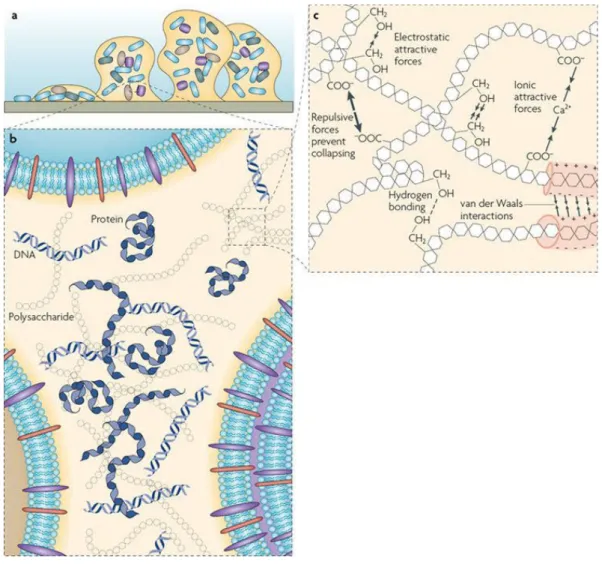
Bacteria within biofilms are surrounded by a self-produced extracellular polymeric matrix (Boles & Horswill 2011; Costerton et al. 1995) (Figure 3). The composition of the matrix depends on a variety of factors, such as the environment where biofilm is formed (Wimpenny 2000), the species involved and nutrients availability (Wimpenny 2000; Flemming & Wingender 2010) as well as the shear forces experienced (Flemming & Wingender 2010). The biofilm matrix is composed, among others, by proteins, polysaccharides and nucleic acids and in which biofilm cells are embedded (Flemming & Wingender 2010).
Figure 3 | The extracellular substances matrix at different dimensions.
a |
An example of a bacterial biofilm attached to a solid surface.b |
Distribution around the cells of themain matrix components, such as polysaccharides, proteins and DNA.
c |
Physicochemical interactionsand entanglement of biopolymers responsible for the stability of the matrix. (Adapted from Flemming et
6
The biofilm matrix plays a crucial role on the subsistence of the bacteria inside the biofilm, thereby acting as a physical protection against external stimuli such as high osmotic pressure (Flemming et al. 2007), it protects bacteria from the action of antibiotics (Høiby et al. 2010) as well helping evading the host immune system defences (Otto 2006). It is also responsible for their adhesion to surfaces as well as for the cohesion in the biofilm (Flemming & Wingender 2010).
A biofilm formation is divided in three stages, starting with the attachment to a surface, followed by maturation and finally biofilm detachment (O’Toole et al. 2000) (Figure 4).
Figure 4 | Phases of biofilm development.
The biofilm development comprises attachment, maturation and detachment. The first phase, attachment, occurs weather in biotic or abiotic surfaces, such as “ conditioning biofilm” built by host matrix proteins or polymeric surface of an indwelling medical device, respectively. The interactions involved are specific, protein-protein interaction, for biotic surfaces whereas for abiotic surfaces are non-specific. Then, biofilm grows and matures due to agglomeration of cells. The predominant molecules or adhesive factors that stick cells together are the polysaccharide intercellular adhesin (PIA), also called poly-N- acetylglucosamine (PNAG), teichoic acids and some proteins such as accumulation-associated protein (Aap). Regarding disruptive factors, they are important in producing channels. Finally, cell clusters detach. This process depends on the expression of phenol-soluble modulins (PSMs). (Adapted from Otto, 2012)
3 .1 • Attachment to a surface
This initial step of colonization is a phenomenon that occurs naturally and depends on the superficial characteristics of the abiotic or biotic surfaces, such as a polymeric surface of an indwelling medical device or the human tissue, respectively (Otto 2009), and the microorganisms involved in the attachment such as superficial charge (Otto 2012b), hydrophobicity (Otto 2012b; Dunne 2002) and superficial
7
tension (Dunne 2002). The attachment of bacteria to an abiotic surface is mediated mostly by non-specific and hydrophobic interactions (Vacheethasanee et al. 1998). However, other molecules such as teichoic acid are involved in this process (Heilmann et al. 1997; Gross et al. 2001).
In addition to the abiotic surfaces, it is known that microorganisms may adhere to biotic surfaces. In this case, the interactions between the surface and bacteria are very specific (Otto 2012b; Patti et al. 1994). Staphylococci express surface-anchored proteins called microbial surface components recognizing adhesive matrix molecules (MSCRAMMs) with a capacity to bind to host matrix proteins such as fibrinogen or fibronectin (Patti et al. 1994) and, as a consequence, are critical to establish infection (Otto 2012a). MSCRAMMs have a domain that is responsible for the covalent and non-covalent attachment to the bacterial surface.
After insertion, the devices are covered by host matrix proteins, thereby the specific interactions between MSCRAMMs and these proteins leads to the colonization of the device (Otto 2008). As the process of adhesion proceeds, bacterial cells change their phenotype in result to the proximity to the surface (Fletcher 1991; Costerton et al. 1995).
3 .2 • M aturation
The maturation process includes the proliferation or agglomeration, in which bacteria attached to a surface begins to growth, and formation of multicellular structures around a surface by adhesive and disruptive forces (O’Toole et al. 2000; Costerton et al. 1995).
3.2.1 • Adhesive forces
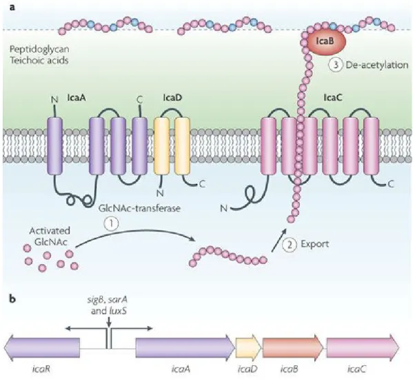
Adhesive forces are determinant for the adherence of bacteria to each other (Otto 2009). It mainly occurs due to the presence of an exopolysaccharide known as polysaccharide intercellular adhesion (PIA) (Mack
et al. 1996). This molecule together with other polymers such as teichoic acids and proteins form a substance often called “ slime” that increases the adherence of bacteria to inert surfaces, developing biofilms and providing protection from whether the immune system or antibiotic tolerance (Costerton et
al. 1999; Costerton et al. 1995). The PNAG molecule sticks the cells together by electrostatic interactions
and is produced by the products of the ica operon, which include icaA, icaD, icaB and icaC genes (Gerke
et al. 1998; Heilmann et al. 1996; Vadyvaloo & Otto 2005). Its biosynthesis is regulated by environmental factors and regulatory proteins (Otto 2008). The N-acetylglucosamine transferases IcaA and IcaD are responsible for the production of the chain of N-acetylglucosamine (GlcNAc) residues. The elongation and
8
the exportation of these residues is carried out by IcaC protein (Gerke et al. 1998). The surface-located PNAG deacetylase IcaB causes partial de-acetylation of the GlcNAc monomers (Vuong, Kocianova, et al. 2004) (Figure 5).
Figure 5 | The exopolysaccharide PNAG.
a |
The PNAG is a partially de-acetylated 1-6-linked N-acetylglucosamine (GlcNAc) homopolymeressential for biofilm formation and immune evasion as well. Its synthesis is accomplished by the products of the ica operon, icaA, icaD, icaB and IcaC. The accessory IcaD activates the membrane-located GlcNAc transferase IcaA (step 1). The expansion of PNAG chain is exported by the IcaC membrane protein (step 2). The cell surface-located enzyme IcaB, after export, removes some of the N-acetyl monomers
introducing positive charges into the polymer, which are crucial for surface attachment (step 3).
b |
TheicaADBC operon and the icaR gene, which encodes a regulatory protein, form the ica gene locus which
encodes the Ica proteins. The IcaA promoter or the production of IcaR, both regulated by global regulatory proteins (SigB, SarA and LuxS), control the expression of the icaADBC operon. Negative and positive charges are represented by green and blue shading, respectively. Abbreviations: C, carboxyl; N, amino. (Adapted from Otto, 2009)
9
Partial de-acetylation introduces positive charges into the otherwise neutral polymer by liberating free amino groups which become charged at neutral or acid environments such as the human skin (Vuong, Kocianova, et al. 2004). Thus, de-acetylation is of major importance for the binding of PNAG to the cell surface as well as for biofilm formation and immune evasion (Vuong, Kocianova, et al. 2004), and has been recognized as key virulent factor in S. epidermidis as well as the production of PNAG (Rupp, Ulphani, Fey & Mack 1999; Rupp, Ulphani, Fey, Bartscht, et al. 1999; Otto 2012b). Although, there are some strains that do not produce PNAG due to the lack of ica operon (Arciola et al. 2006), biofilm formation can still occur due to the presence of adhesive proteins such as accumulation-associated protein (Aap) (Hussain et al. 1997).
In addition to PNAG, teichoic acids has also been shown to contribute for staphylococcal biofilm formation (Gross et al. 2001; Sadovskaya et al. 2005). These molecules contribute to the complex network that forms the cell surface of Gram-positive bacteria (Otto 2012b). Furthermore, teichoic acids occur in two forms, lipoteichoic acids (LTA) and wall teichoic acids (WTA) when anchored to the cell membrane by a membrane-spanning lipid and when anchored to the peptidoglycan, respectively (Glaser 1973). In S.
epidermidis, they are composed of poly(glycerol phosphate) (Sadovskaya et al. 2004), as mentioned in
section 1. In staphylococci, teichoic acids are responsible for maintaining the cells physiology and are involved in pathogenesis of the bacterium since they mediates adhesion to abiotic surfaces and enhances the adhesion to fibronectin covered surfaces (Hussain et al. 2001). In addition they induces inflammation, mediating interactions with the host receptors (Weidenmaier & Peschel 2008).
Similar to teichoic acids, extracellular DNA (eDNA) is part of the complex network of the matrix (Whitchurch et al. 2002) and it is only a minor component of the S. epidermidis biofilms (Izano et al. 2008). The eDNA is released through the lysis of the bacteria that is controlled by the gene cidA and operon IrgAB (Mann et al. 2009; Rice et al. 2007). Together with teichoic acids, DNA play an important role in interacting with other surfaces polymers due to their negative charges (Otto 2008; Otto 2012b).
3.2.2 • Disruptive forces
Besides the importance of adhesive forces in biofilm maturation, disruptive forces are equally crucial for biofilm structuring (Otto 2008; Otto 2009). Biofilm channels are essential for the diffusion of nutrients in to the deeper layers of the biofilm (Costerton et al. 1995; Otto 2008). In addition, these channels are important for the disposal of waste products that are generated as part of the natural activities by the cells (Sutherland 2001).
10
The production of enzymes that degrade specific biofilm matrix components could be one of the mechanisms responsible for biofilm maturation and structuring (Otto 2012b). However, such enzymes appear to contribute for biofilm maturation and structuring in PNAG-independent biofilm formation (Rohde
et al. 2007). Proteases are one of these enzymes acting as accessory facilitators of biofilm formation in
species that form PNAG-dependent biofilms. Nucleases, enzymes that degrade extracellular DNA have also influence in the process of biofilm maturation (Otto 2012b). As an example, DNase I of human serum causes degradation of DNA molecules involved in intercellular adhesion and, subsequently, decreases the number of cells within the biofilm (Kaplan et al. 2012).
Since 2000, the Agr (accessory gene regulator) quorum sensing system has been identified as a primary regulator of biofilm maturation and detachment (Vuong et al. 2000). The Agr is expressed on the surface and also in the deeper layers of the biofilm for the efficient formation of channels (Periasamy et al. 2012). Though, it is not well understood which signals or mechanisms are involved in the expression of agr required for biofilms to reach their typical structure (Otto 2012b).
Additionally to the production of proteases and nucleases, the presence of surfactant molecules may contribute to biofilm formation and structuring (Otto 2012b; Otto 2008). The phenol-soluble modulins (PSMs) are amphipathic -helical peptides with surfactant properties, produced by staphylococci (Mehlin
et al. 1999; Wang et al. 2007) and under control by the Agr quorum sensing system (Otto 2008; Vuong,
Dürr, et al. 2004) (Figure 6). This peptide has proinflammatory ability in the way that promotes neutrophil chemotaxis and cytokine release (Wang et al. 2007; Kretschmer et al. 2010). PSM efficiently lyse important human cells such as neutrophils, monocytes, and erythrocytes (Wang et al. 2007; Cheung et
al. 2010). In addition, some PSM peptides have antibacterial capabilities, having a potential role in
bacterial interference (Cogen et al. 2010; Joo et al. 2011).
S. epidermidis produces the PSM peptides, mainly in a biofilm mode of growth(Yao et al. 2005; Wang et al. 2011). However, interestingly, an isogenic mutant of the PSM operon forms a more compact and
extended biofilm compared to the wild-type strain (Wang et al. 2011; Otto 2008). In contrast to proteases, their contribution for biofilm formation do not depend on the type of staphylococcal biofilm (PNAG-dependent or –in(PNAG-dependent) (Otto 2012b).
11
3 .3 • Detachment and return to the planktonic growth mode
The last stage on the biofilm life-cycle, also called dispersal, is the result of the detachment of single cells or group of cells of the biofilm colony that contributes to the spread of bacteria into the involving environment, allowing the colonization of different sites (Kaplan 2010; Otto 2012b). The detachment may be a result of several factors: i) the flow that increases the superficial tension (Otto 2008), ii) the production of extracellular enzymes that hydrolyse proteins and subsequently destroy the matrix and, iii) the cessation of the production of PSMs (Otto 2009; Otto 2008). When produced excessively, these factors contribute for the detachment of the biofilm surface area (Otto 2008). Furthermore, environmental conditions such as glucose depletion, changes in pH and temperature, among others promote biofilm detachment (Boles & Horswill 2008). In addition, under favorable conditions, biofilms undergo detachment process, being more pronounced after longstanding periods of growth (Yarwood et al. 2004a).
S. epidermidis produces exoproteases with low substrate specificity that are responsible for the degradation of surface proteins, causing detachment of biofilm cells (Teufel & Götz 1993; Ohara-Nemoto
Figure 6 | Regulation of PSM s.
PSMs are regulated by the agr system. The psm and psm genes are directly regulated by AgrA, indicating an early evolutionary link between quorum sensing and PSM production, and suggesting that RNAIII-dependent gene regulation was added later by formation of the RNAIII encoding genetic information around the hld. The psm-mec gene in the staphylococcal cassette chromosome mec (SCCmec) element is also under Agr control. (Adapted from Peschel et al, 2013)
12
et al. 2002; Dubin et al. 2001). In addition, detergent-like molecules may disrupt electrostatic and
hydrophobic interactions between the anionic surface polymer and the cationic PNAG molecule or even between hydrophobic regions on the bacterial surface (Otto 2009). Biofilm detachment determines the biofilm volume, thickness and expansion, related with the absence of -toxin and PSMs (Otto 2009). PSMs have a key role in biofilm detachment leading to the formation of “ holes” in biofilm (Otto 2008) through the disruption of non-covalent interactions (Otto 2012b) (Figure 7). The detachment process plays a crucial role during biofilm-associated infection due to the dissemination of cells from the surface of an indwelling medical device to other sites by the bloodstream and lymph system. Thereby, after bacteria detachment they might form biofilms in other regions of the body (Otto 2012b).
Figure 7 | PSM s function in biofilm detachment.
Cells which express PSMs bind to a surface (step 1). Subsequently, some cell clusters stop producing PSMs, probably owing limited oxygen concentration or lack of responsible proteins for its synthesis (step 2). The other cell clusters, which have the expression of PSMs active, detach, leaving openings in the biofilm structure, characterized by fluid-filled channels and cell towers (step 3). (Adapted from Otto, 2008)
13
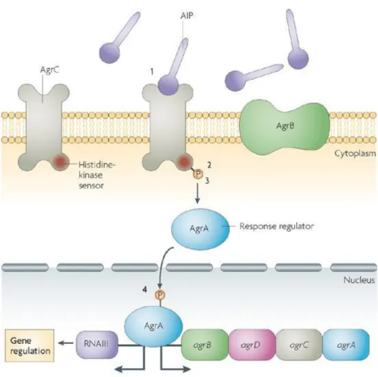
4 • The quorum sensing system
Bacteria are increasingly known as highly interactive microorganisms with complex social lives (Velicer 2003; Foster et al. 2007). In biofilms, bacteria interact and respond to local cell density through a regulatory mechanism known as quorum sensing (QS) (Bassler & Losick 2006; Hall-Stoodley et al. 2004; Nadell et al. 2008) (Figure 8).
Figure 8 | The staphylococcal agr system.
QS in staphylococci is used by the agr locus, comprising the agrA, agrC, agrD and agrB genes and he intracellular effector of the system, RNAIII. The synthesis of an autoinducing peptide (AIP) as well as the expression of RNAIII and the regulation of the QS genes are involved in the regulation of the system. The AIP is accumulated extracellularly and turn on the two-component response system (TCRS), which involves signal recognition through a histidine kinase, AgrC (step 1), before histidine phosphorylation (step 2) and phosphotransfer to a response regulator, AgrA (step 3). Then, the latter binds to the RNAIII transcript which encodes a small RNA to modulate gene expression (step 4). (Adapted from Cegelski et
14
The quorum sensing, or cell-cell communication, controls gene expression in response to increasing cell density, allowing the bacteria to adapt to different environmental conditions, such as low availability of nutrients (Otto 2004a), oxygen levels and the transition from planktonic to biofilm growth (Otto 2004b). It plays a crucial role in synchronising gene expression and functional co-ordination in bacterial communities (Dong & Zhang 2005) as well as to build a well-ordered surface community (McCann et al. 2008).
The staphylococcal agr system consists of two transcription units, RNAII and RNAIII. RNAII encodes the
agr protein components (agrA, agrB, agrC and agrD) and its biosynthesis is regulated by the P2 promoter
(Mack et al. 2007; Otto 2004a) while P3 promoter regulates the transcription of RNAIII, the effector molecule of the agr system, whose synthesis depends on the agr activation (Novick et al. 1995; Yarwood et al. 2004b). In addition, RNAIII regulates the transcription of other genes (McCann et al. 2008; Otto 2004a).
The agr system upregulates the expression of degradative exoenzymes and toxins, and downregulates the expression of surface adhesion proteins (Otto 2012b). It contains a two-component signal transduction system (agrA and agrC), a prepheromone (AgrD), and AgrB, that seems to be responsible for maturation of the post-translationally modified prepheromone peptide (Novick 2003; Otto 2004a). The modified pheromone peptide is the auto inductive signalling molecule of the agr system and contains a thiolactone linkage between the C-terminal carboxyl group and a central conversed cysteine residue (Otto 2004a; McCann et al. 2008) (Figure 9). The thiolactone ring structure is essential for its biological activity (Mayville et al. 1999; Otto et al. 1998).
Figure 9 | Chemical structure of the agr pheromone of the S. epidermidis.
In red is represented the ring size, central cysteine and thiolactone structure, which are conserved regions, while the amino acid sequence and length of the N-terminal peptidyl extension to the ring are variable regions. (Adapted from Otto, 2004)
15
The peptide pheromone or auto inducing peptide (AIP), binds to a membrane-located histidine kinase, AgrC, when a threshold concentration is reached at a certain cell density. AgrC in turn activates the response regulator protein, AgrA that binds to the two promoters, inducing the transcription of the agr operon itself (Ji et al. 1995; Otto 2012b). In addition, AgrA can directly activate expression of specific operons such as the one encoding PSMs (Otto 2012b).
Although this mechanism regulates some of the molecules that are essential for biofilm formation, it does not control the expression of the major and most important component of the biofilm extracellular matrix, the PNAG (Otto 2009; Vuong et al. 2000).
16
5 • Survival of Staphylococcus epidermidis in human blood
Human blood is a complex mixture of soluble factors and immune circulating cells, which are often very active at eradicating foreign organisms. After entering in the human body, the microorganism are confronted with the advanced innate defence mechanisms of the human host (Rooijakkers et al. 2005). However, due to the coevolution with the host, several pathogens developed mechanisms to avoid the high microbicidal properties of human blood. S. epidermidis is capable to overcome the bacterial activity of human blood (França et al. 2014). Bacteria are protected from the host immune system due to the particular physiological changes in the biofilm which reduce the sensitivity to cytokines among other prejudicial molecules, and also by acquiring a non-aggressive state which reduces chemotaxis and inflammation of immune cells (Yao et al. 2005).
PNAG and poly-
ƴ
-glutamic acid (PGA) are known for their role in S. epidermidis evasion to neutrophilskilling through the inhibition of phagocytosis (Kocianova et al. 2005; Vuong, Voyich, et al. 2004). PGA is an extracellular anionic molecule which provides protection against altered environmental conditions, such as high salt concentrations, and mediates resistance to phagocytosis and antimicrobial peptides (Kocianova et al. 2005). Also, its expression seems to be an advantage for those microorganism living in high salt environments which is the case of the human skin (Fey & Olson 2010).
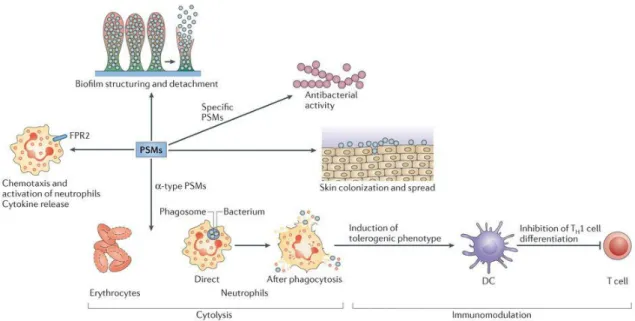
The PSMs are cytolytic toxins with capacity to attract, stimulate and lyse neutrophils (Wang et al. 2007) (Figure 10). However, in S. epidermidis they do not have a significant contribution in neutrophil lysis (Cheung et al. 2010).
One of the aspects that is important for survival in human blood and one of the first lines of its defence is the iron acquisition (Cheung et al. 2010). In the human blood, the concentration of free iron available
for bacteria is about 1011 times lower than the required for bacteria subsistence (Malachowa & DeLeo).
Iron is an important cofactor in metabolic pathways which is essential to microorganisms (Jordan & Reichard 1998; Jakubovics & Jenkinson 2001). Iron acquisition is facilitated by the interaction of catecholamine inotropes, often used in patients in intensive care units, with transferrin and lactoferrin (Lyte et al. 2003) and, as a consequence, leading to an increased number of catheter-associated infections (Neal et al. 2001). While in S. epidermidis little is known regarding the molecular mechanisms of iron metabolism, in S. aureus, the production of siderophores help in the capture iron (Torres et al. 2007), in which there is a two-component Hemo-sensor system (HssRS). The Hemo-sensor system responds to heme exposure, thereby activating the production of the heme-regulated transporter (HrtAB),
17
an efflux pump which controls intracellular level of hemin in order to prevent toxicity and have an essential role in heme homeostasis (Friedman et al. 2006).
Neutrophils cells kill bacteria with non-oxygen-dependent antimicrobial processes and reactive oxygen species, after phagocytosis (Faurschou & Borregaard 2003). Among the non-oxygen-dependent processes, cationic antimicrobial peptides (AMPs) present in all living species, such as defensins and cathelicidins play a crucial role in innate host defences (Hancock & Diamond 2000). In S. epidermidis, the protease SepA has a strong capacity to eliminate human AMPs through proteolysis. Its production is regulated by Agr system (Lai, Amer E. Villaruz, et al. 2007). In addition, S. epidermidis uses a passive defence strategy to evade elimination by innate host defence, preventing S. epidermidis from being seen and remaining in the host. (Cheung et al. 2010).
Figure 1 0 | PSM s activities.
PSMs are able to spread on surfaces and cytolysis, leading to biofilm structuring and detachment, respectively. Some of them have antibacterial activity. The cytolysis is exclusively of the -tipe PSMs and many cells such as erythrocytes as well as neutrophils are destroyed by them due to the receptor-independent manner of the cytolysis. The lysis of neutrophils may occur after phagocytosis, which means PSMs can be an excellent obstacle against innate host defence. PSMs also have an impact on the adaptive immune system due to the induction of a tolerogenic phenotype in dendritic cells (DCs) and restraining T helper 1 (TH1) cell differentiation. The chemotaxis and activation of neutrophils as well as cytokine release occurs after N-formyl-peptide receptor 2 (FPR2) activation by all the PSMs. (Adapted from Peschel et al, 2013)
18
6 • Aims and Objectives
Due to the impact on morbidity, mortality and economic costs of S. epidermidis infections, prevention and clinical management of such infections is of major importance. Since the release of cells from S.
epidermidis biofilms is a critical aspect of infection, assessment of the transcriptional changes that occur in these cells upon contact with host immune system is an essential step to understand the mechanisms of evasion.
Therefore, the purpose of this study was to characterize the survival of biofilm-released cells upon contact with human blood and plasma, over time. Furthermore, the analysis of key genes of interest involved in immune evasion was addressed and its quantification of their expression was performed by quantitative PCR (qPCR).
19
M
ATERIAL AND
M
ETHODS
A • Bacterial strains and growth conditions
For this study, three S. epidermidis biofilm-forming strains were used: 9142 (Mack et al. 1992), IE186 (Cerca et al. 2006) and PT12003 (unpublished clinical isolated from a patient after a stomach surgery). Brc were obtained as described before (França, Carvalhais, et al. 2016). In short, a single colony of each
S. epidermidis strain grown in Tryptic Soy Agar plates (TSB, VWR, Radnor, Pennsylvania, USA, plus 1.5 % Agar, Liofilchem, Roseto degli Abruzzi, Italy) was inoculated into a 10 mL Erlenmeyer flask with 3 mL of Tryptic Soy Broth (TSB, Liofilchem) and incubated at 37 ° C with shaking at 120 rpm overnight in an orbital shaker-incubator ES-20 (10 mm orbit). The starter culture was diluted in TSB (Liofilchem) to obtain a bacterial suspension with an optical density, at a wavelength of 640 nm (OD640), between 0.250 ± 0.05,
corresponding to 1 x 10⁸ colony forming unit (CFU)/ mL, measured in an UV-3100PC Spectrophotometer
(VWR). Hereafter, 10
μ
L of this suspension were inoculated into 1 mL TSB (Liofilchem) supplementedwith 0.4 % (v/ v) of glucose (TSBG) to induce biofilm formation in 24-well plates (Thermo Fisher Scientific, Waltham, Massachusetts, USA). The plates were then incubated at 37 ° C with shaking at 120 rpm. After 24 ± 2 hours, spent medium was carefully removed and biofilms were washed twice with 1 mL of 0.9 % NaCl to remove planktonic or loosely adherent cells. Then, 1 mL of TSBG was carefully added and biofilms allowed to growth for additional 24 ± 2 hours under the same temperature and shaking conditions. Before any of the analyses described below, Brc were gently collected by aspirating the biofilm bulk fluid of 2 wells for each 2.0 mL tube. Hereafter, Brc were harvested by centrifugation at 16 000 g for 10 minutes at 4 ° C. The bacterial pellet was then suspended in 1 mL of 0.9 % of NaCl and sonicated three times for 10 seconds at 35 % of amplitude, in an Ultrasonic Processor (Cole Parmer, Chicago, USA), in order to eliminate bacterial aggregates and to homogenize the suspension (Freitas et al. 2014). The OD640 of the
suspension was measured and then it was prepared a suspension with a concentration of 1 × 10⁹
CFU/ mL.
B • Blood and plasma collection
Whole human blood was collected from healthy female volunteers between 20 years and 30 years of age, under a protocol approved by the Institutional Review Board of the University of Minho (SECVS 002/ 2014)
20
in accordance with the Declaration of Helsinki and Oviedo convention. All donors gave informed written consent prior to blood donation. Blood was collected in to BD Vacutainer® tubes spread-coated with lithium heparin (Becton Dickinson, East Rutherford, New Jersey, USA). Nine hundred µL of whole human blood were transferred into a 2.0 mL tube and the rest was centrifuged in a CL31R Multispeed Centrifuge (Thermo Fisher Scientific) for 20 minutes at 1500 g at room temperature in order to obtain human plasma (NHS). Then, 900 µL of plasma was transferred into a 2.0 mL tube.
C • Bacterial challenge with human blood and plasma
100 µL of the adjusted bacterial suspension at 1 × 10⁹ were added to each tube containing the fluids.
The tubes were incubated for 2 hours ± 15 minutes in a warm room at 37 ° C with agitation in a PSU-10i Orbital Shaker (Biosan, Riga, Latvia) (80 rpm in a 10 mm orbit). Samples were then sonicated once, for 10 seconds at 35 % and centrifuged at 16 000 g for 7 minutes at 4 ° C. This procedure was also performed, in a 96-well cell culture plate, flat bottom (Orange Scientific, Braine-l'Alleud, Belgium) testing different volumes of blood: 100 µL and 180 µL, in which were added, respectively, 10 µL and 20 µL of the adjusted bacterial suspension. Each condition was carried out in triplicates.
D • Biofilm-released cells culturability and viability
After mixing very well, 50 µL of bacterial suspensions of each of the conditions under test were serially diluted in 450 µL of 0.9 % NaCl and plated on TSA for quantification of the number of culturable bacteria. This experiment was performed in triplicates.
In order to count the total viable and dead cells, Brc incubated with whole human blood or plasma were
suspended in 1 mL of 0.9 % NaCl and then stained with LIVE/ DEAD® BacLightTM Bacterial Viability Kit
(L7012) (Thermo Fisher Scientific) for microscopy, following the manufacturer’s instructions. Both positive (cells suspensions without treatment or live cells) and negative controls were prepared in which the latter the cells were incubated, for 15 minutes, at 100 ° C. Cells were observed, trapping 10 µL between a slide and a coverslip of 20 mm, in a OLYMPUS BX51 Epifluorescent Microscope equipped with a DP71 digital color camera (OLYMPUS, Shinjuku, Tokyo, Japan), using a magnification of 60 ×. At least 10 JPEG images for each condition were acquired. Each condition was performed in triplicates (Freitas et al. 2014).
21
E • Gene expression assays
This experimental setup involved several steps, including RNA extraction, DNase treatment, RNA quantification, complementary (c) DNA synthesis, and quantitative PCR (qPCR) run.
RNA extraction
RNA extraction was performed as described before (França et al. 2012) using the E.Z.N.A.® Total RNA Kit I (Omega Bio-tek, Norcross, USA). Brc were suspended in 500 µL of TRK lysis buffer (supplemented with -mercaptoethanol) and 500 µL phenol solution. Suspension were transferred into a 2 mL safe lock tube containing 0.5 g of acid-washed 150 mm – 212 mm silica beads and placed the tubes into the
FastPrep®-24 cell disruptor (MP Biomedicals, Santa Ana, California, USA) with setting 6.5 m/ s for 35
seconds. This cycle was repeated 3 times with intervals of 5 minutes on ice. Afterwards, samples were centrifuged at 12000 g for 2 minutes and the supernatants transferred in to a new tube and mixed with equal volume of 70 % ethanol. Samples were transferred (including any remaining precipitate) to the silica columns and centrifuged at 12000 g for 1 minute. The flow-through was discarded and each column reinserted into a new collection tube. To wash the columns, 500 µL of Wash buffer I were added to each column and centrifuged at 12000 g for 1 minute. Again, the flow-through was discarded and the column inserted into the same collection tube. After that, 500 µL of Wash buffer II were added to each column and centrifuged at 12000 g for 1 minute. Once more, the flow-through was discarded and the column inserted into the same collection tube and then 500 µL of Wash buffer II added to each column and centrifuged at 12000 g for 1 minute. To remove any trace of Wash Buffer II, the flow-through was discarded and the columns were transferred into a new collection tube for a new centrifugation at 12000
g for 2 minutes. The collection tube was discarded and each column inserted into a 1.5 mL
DNase/ RNase-free tube. Finally, to elute total RNA, 40 µL of DEPC-treated water were added to the membrane, incubated for 1 minute and then centrifuged for 2 minutes at 12000 g. RNA solution were immediately placed on ice or stored at – 20 ° C until further use. All steps were performed at room temperature.
DNase treatment
DNase treatment was performed to degrade genomic DNA. Briefly, 2 µL of DNase I (Fermentas, Thermo Fisher Scientific) and 4 µL of DNase I buffer (10 ×) (Fermentas, Thermo Fisher Scientific) were added to a volume of 40 µL, containing total RNA, and incubated at 37 ° C for 30 minutes. Then, DNase I was
22
inactivated by adding 5 µL of 50 mM EDTA (Thermo Fisher Scientific) and by incubation at 65 ° C for 10 minutes (França et al. 2012).
RNA quantification
After genomic DNA degradation, the concentration of total RNA was determined using a NanoDrop 1000TM
Spectrophotometer (Thermo Fisher Scientific) in which nucleic acids, including DNA and RNA, absorb at 260 nm. Before measuring RNA concentration, the nanodrop was switched on to allow the light source warm up and stabilize. The absorbance ratios A260/ A280 and A260/ A230 were used as indicators, respectively, of protein contamination and chemical and polysaccharides contamination. In short, the blank was done and then 2.0 µL of the sample was applied to the pedestal. At the end the blank was confirmed once more, the pedestal washed and the software closed.
Complementary (c) DNA synthesis
The synthesis of cDNA was performed as described before (França et al. 2012). After normalization of RNA concentration, in which total RNA isolated in each independent experiment, using different blood donors, for each strain, were pooled together, 188.4 ng, 92.1 ng and 187.0 ng of total RNA from the strains S epidermidis 9142, IE186 and PT12003, respectively, were converted into cDNA in the presence of the enzyme RevertAid Premium Reverse Transcriptase (Thermo Fisher Scientific). As a priming strategy were used Exo-Resistant Random Hexamer primers (Bioron, Ludwigshafen, Germany). Afterwards,
samples were placed into a C1000TM Thermal Cycler (Bio-Rad, Hercules, California, USA) with the
following cycling parameters: 5 minutes at 25 ° C followed by 60 minutes at 42 ° C, 10 minutes at 70 ° C, and finally 4 ° C until storage. To determine genomic DNA carry-over and contamination of the reagents, it was prepared control reactions lacking reverse transcriptase (no reverse transcriptase control, NRT) and the template (no template control, NTC).
Quantitative PCR
Quantitative PCR was performed as described before (França et al. 2012). For the strains S. epidermidis
9142 and PT12003, the experiment was performed in a CFX96TM (Bio-Rad) with the following cycling
parameters: 1 minute at 94 ° C followed by 39 repeats of 15 seconds at 94 ° C, 20 seconds at 58 ° C, and finally 25 seconds at 72 ° C using iQ SYBR Green supermix (Bio-Rad). The melting curve was from
23
control, 3
μ
L of water, 0.75μ
L of primer forward (FW), 0.75 of primer reverse (RV) and 7.5μ
L of therespective PCR mix.
For the strain S. epidermidis IE186, qPCR run was performed with the following cycling parameters: 10 minutes at 95 ° C followed by 39 repeats of 15 seconds at 95 ° C, 60 seconds at 60 ° C, and finally 25 seconds at 72 ° C using Power SYBR Green PCR Master Mix (Applied Biosystems, Thermo Fisher
Scientific). The melting curve was from 65 ° C to 95 ° C with an increment of 1.0 ° C. The 10
μ
L reactionscontained 2
μ
L diluted cDNA or NRT control, 2μ
L of water, 0.5μ
L of primer FW, 0.5μ
L of primer RVand 5
μ
L of the respective PCR mix.qPCR products were analysed by melting curves for unspecific products or primer dimer formation. A set of primers were used with similar efficiencies (Table 1) and cDNA samples were diluted 400 × in water (França et al. 2012).
Table 1 | Oligonucleotide primer sequences used for qPCR analysis.
Target
gene
Primer sequence
(5 ’ to 3’)
M T
(°C)
Amplicon
(bp)
Priming efficiency
(%)
16S FW RV GGGCTACACACGTGCTACAA GTACAAGACCCGGGAACGTA 59.79 59.85 176 97 fmtC FW RV CGCCCTCATCATAGCATTG CCAATTGGATCACCCAAAAC 60.19 60.03 182 100 lrgB FW RV ATATCGCAAGCGCGAAGTAT ATTGCTGTCGTTGCAGCTT 59.87 59.61 165 90 sepA FW RV TCTTAAGGCATCTCCGCCTA GTCTGGTGCGAATGATGTTG 57.92 57.47 196 97The quantification of the specific mRNA transcripts for each gene under study in Brc was determined using the Pfaffl method, where the relative expression of a target gene is expressed in a sample versus one control in comparison to a housekeeping gene. Etarget is the real-time PCR efficiency of the target gene
transcript; Eref is the real-time PCR efficiency of the housekeeping gene transcript;
Δ
CPtarget is the CPdeviation of control – sample of the target gene transcript;
Δ
CPref = CP deviation of control – sample of24
F • Statistical analysis
All the assays were compared using two-way analysis of variance (ANOVA) by applying Sydak’s and Tukey’s multiple comparisons tests, in Graph Pad Prism 6 software. All tests were performed with a confidence level of 95%.
25
R
ESULTS AND
D
ISCUSSION
A • The ex vivo model of Staphylococcus epidermidis biofilm-released cells
and its virulence determinants
When the external physical barriers of the human body, such as skin and mucous surfaces, are breached by S. epidermidis, the organism is confronted by the innate and acquired responses of the host immune system and to cause disease a bacterium might be able to divert the effectiveness of the innate immune system (Boles & Horswill 2011). Cells released from biofilms developed on the surface of medical devices are important in biofilm-associated infections and can cause severe acute infections (Wang et al. 2011; Boles & Horswill 2011). However, the mechanisms or interactions that enable Brc to resist host defences are less well characterized.
In order to better understand their phenotype or interaction with the host immune system, we started by evaluating Brc capacity of survival in whole human blood and plasma, by addressing Brc culturability and viability.
Brc culturability of strain S. epidermidis 9142, a well-studied strain which was used as a model in this study, was evaluated by the colony forming unit (CFU) method right after the cells were incubated with whole human blood and plasma for 2 hours, using as control Brc before incubation assays (T0 h). Despite the CFU method be time-consuming, it is the most common method in the evaluation of the effectiveness of antimicrobial agents (Putman et al., 2005). Of note, based on the results from this method it is possible to evaluate the effect of different agents in microorganisms, being considered bactericidal if the reduction is higher than 3 Log10 CFU/ mL or 99 % of reduction and bacteriostatic for inferior reductions (May et al., 2000).
As expected, Brc culturability were reduced in the presence of whole human blood and meaningful differences were observed comparing to plasma, with exception of the donor 3, for the strain S.
26 ***
*** * ****
* **
Figure 1 1 | Percentage
of survival of S. epidermidis 9 1 4 2 Brc after incubation with whole
human blood and plasma (comparing to T
0h with a concentration of 1 × 1 0
9CFU/ mL).
The horizontal bars represent the mean of the replicates. Statistical differences of percentage of survival of Brc of donors between both conditions, blood and plasma, were analysed with two-way ANOVA and
Sidak’s multiple comparisons test. *** p<0.001, **** p<0.0001.
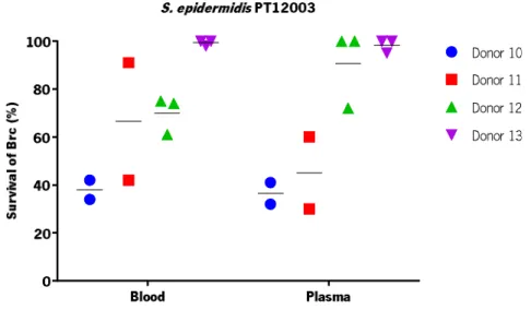
As each strain has unique characteristics and, as a consequence, variability between strains may occur (França et al. 2013), it is advisable to confirm the previous observations in other S. epidermidis strains This will allow to demonstrate if the observable phenomena are relevant in multiple clinical isolates. Therefore, Brc culturability of strains S. epidermidis IE186 and PT12003 were evaluated. Regarding to the strain S. epidermidis IE186 (Figure 12), a higher variance was observed between donors where a more pronounced reduction was observed for the donors 8 and 9, either in the presence of blood or plasma. However, no meaningful reduction was observed for the donor 7. As can be seen in the Figure 13, for the strain S. epidermidis PT12003, different responses between the donors were observed whether after 2 hours of exposure to human blood or plasma. However, no reduction of Brc was observed for donor 13, either in blood or plasma. However, the strains S. epidermidis IE186 and PT12003 showed a different behaviour comparing to the strain S. epidermidis 9142 used as a model. Both strains, IE186 and PT12003, showed similar percentages, with no meaningful differences, of survival either in human blood or plasma for each donor, which does not occur in the strain 9142. This might be relevant as the results showed each strain has its own response after being incubated with the host immune system and do not follow the pattern observed in the strain used as model.
Additionally, as the whole human blood varies from person to person as well as the immune system, each strain showed the capacity to adapt to each case, and as a consequence to remain in the host. Also, the
27
time of day when blood samples were collected might cause sample variation between individuals (Radich
et al. 2004).
Figure 1 2 | Percentage of survival of S. epidermidis IE1 8 6 Brc after incubation with whole
human blood and plasma (comparing to T
0h with a concentration of 1 × 1 0
9CFU/ mL).
The horizontal bars represent the mean of the replicates.
Figure 1 3 | Percentage of survival of S. epidermidis PT1 2 0 0 3 Brc after incubation with
whole human blood and plasma (comparing to T
0h with a concentration of 1 × 1 0
9CFU/ mL).
28
Brc viability of strains S. epidermidis 9142, IE186 and PT12003 was evaluated by imaging assays that measure membrane permeability using the Live/ Dead technique. Accurate determination of live, dead, and total bacteria is important in many microbiology applications (Shapiro 2000; Nebe-von-Caron et al. 2000). Traditionally, viability in bacteria is synonymous with the ability to form colonies in suitable growth medium. Live cells (Figure 14) have intact membranes and are impermeable to dyes such as propidium iodide (PI), which only leaks into cells with compromised membranes (Barbesti et al. 2000). In contrast, the SYTO® 9 stain labels all bacteria, those with intact membranes and those with damage membranes. Thus a combination of these two dyes provides a rapid and reliable method for discriminating live and dead bacteria (Berney et al. 2007).
Figure 1 4 | Image composition of S. epidermidis 9 1 4 2 Brc exposed to blood, acquired with
a CCD color camera DP7 1 (Olympus).
Cells in red represent dead bacteria while cells in green represent live bacteria.
Briefly, after S. epidermidis Brc were incubated with whole human blood and plasma for 2 hours, the suspensions were stained, using Brc before incubation as both positive and negative controls. The quantification of live and dead cells is shown in Figure 15. No significant differences were observed
29
between the strains in both conditions, however, the percentage of live cells present in the strains were higher in the plasma than those present in blood. These findings showed a significant number of live cells (green cells) over the damage cells (red cells) and also, indicates that whole human blood is more capable of reducing the number of Brc than plasma, with punctual exceptions.
Figure 1 5 | Viable Brc after incubation with whole human blood or plasma.
The bars represent the mean with standard deviation of three independent assays, each in triplicate.
One of our main interests in this study was to analyse the key genes of S. epidermidis Brc involved in immune blood evasion, therefore the transcription levels of genes with a central role in biofilm development/ maturation such as lrgB (Bayles 2007) and in immune evasion such as fmtC (Gill et al. 2005) and sepA (Lai, Amer E Villaruz, et al. 2007) were measured. Brc of the strains S. epidermidis 9142, IE186 and PT12003 were exposed to human blood and plasma for a period of 2 hours, following RNA extraction, cDNA synthesis and qPCR of the selected genes was performed.
Over the past several years, qPCR has become one of the leading tools in molecular biology for the detection and quantification of mRNA. It is the most sensitive method for the detection and quantification of gene expression levels, as it allows the detection of small changes in mRNA transcripts expressions levels. The advantages of qPCR include: i) ability to monitor the progress of the PCR reaction as it occurs in real time; ii) high specificity, sensitivity and reproducibility; iii) accurate measurement of the amount of amplicon at each cycle; iv) wide dynamic range of detection (Wong & Medrano 2005; Bustin 2000). However, the disadvantages include the amplification of unspecific products and primer dimers when