from Proteasomal Degradation and Regulates
TAp63
c
-Dependent Growth Arrest
Oshrat Hershkovitz Rokah1, Ofer Shpilberg1,2, Galit Granot1*
1Felsenstein Medical Research Center, Beilinson Hospital, Sackler School of Medicine, Tel Aviv University, Petah-Tikva, Israel,2Institute of Hematology, Beilinson Hospital, Rabin Medical Center, Sackler School of Medicine, Tel Aviv University, Petah-Tikva, Israel
Abstract
Background: p63 is a member of the p53 transcription factor family. p63 is expressed from two promoters resulting in proteins with opposite functions: the transcriptionally active TAp63 and the dominant-negativeDNp63. Similar to p53, the TAp63 isoforms induce cell cycle arrest and apoptosis. TheDNp63 isoforms are dominant-negative variants opposing the activities of p53, TAp63 and TAp73. To avoid unnecessary cell death accompanied by proper response to stress, the expression of the p53 family members must be tightly regulated. NAD(P)H quinone oxidoreductase (NQO1) has recently been shown to interact with and inhibit the degradation of p53. Due to the structural similarities between p53 and p63, we were interested in studying the ability of wild-type and polymorphic, inactive NQO1 to interact with and stabilize p63. We focused on TAp63c, as it is the most potent transcription activator and it is expected to have a role in tumor suppression.
Principal Findings:We show that TAp63ccan be degraded by the 20S proteasomes. Wild-type but not polymorphic, inactive NQO1 physically interacts with TAp63c, stabilizes it and protects it from this degradation. NQO1-mediated TAp63c
stabilization was especially prominent under stress. Accordingly, we found that downregulation of NQO1 inhibits TAp63c -dependant p21 upregulation and TAp63c-induced growth arrest stimulated by doxorubicin.
Conclusions/Significance:Our report is the first to identify this new mechanism demonstrating a physical and functional relationship between NQO1 and the most potent p63 isoform, TAp63c. These findings appoint a direct role for NQO1 in the regulation of TAp63c expression, especially following stress and may therefore have clinical implications for tumor development and therapy.
Citation:Hershkovitz Rokah O, Shpilberg O, Granot G (2010) NAD(P)H Quinone Oxidoreductase Protects TAp63cfrom Proteasomal Degradation and Regulates TAp63c-Dependent Growth Arrest. PLoS ONE 5(6): e11401. doi:10.1371/journal.pone.0011401
Editor:Michael Polymenis, Texas A&M University, United States of America
ReceivedJanuary 25, 2010;AcceptedJune 9, 2010;PublishedJune 30, 2010
Copyright:ß2010 Hershkovitz Rokah et al. This is an open-access article distributed under the terms of the Creative Commons Attribution License, which permits unrestricted use, distribution, and reproduction in any medium, provided the original author and source are credited.
Funding:The authors have no support or funding to report.
Competing Interests:The authors have declared that no competing interests exist.
* E-mail: galitg@clalit.org.il
Introduction
p63, a p53 family member, is expressed from two different promoters generating two classes of proteins: TAp63, containing the N-terminal transactivation (TA) domain, andDNp63, lacking this domain. In addition, alternative splicing generates three different C-termini: a, b and c. Given that the transactivation activity resides in the protein’s N-terminus, TAp63 isoforms function as transcription factors inducing cell cycle arrest and apoptosis. TAp63cis the most potent transcription activator [1,2]. This isoform mimics p53 in culture and is capable of rescuing the growth inhibitory function of p53, in p53-deficient cells [3,4]. These observations suggest that TAp63c has tumor-suppressive properties analogous to p53. Supporting this notion is the fact that p63 maps to a chromosome region often deleted in cancers [5]. Likewise, loss of TAp63 expression has been detected in several cancers and has been associated with increased metastatic potential [6–8]. In opposition to the TA isoforms,DNp63 protects from apoptosis by directly competing for TAp63 target promoters [1,9]. Over-expression of DNp63 isoforms observed in epithelial
cancers suggests that p63 can also act as an oncogene [10–12]. However, the predominant physiological role of p63 is in epithelial development, as demonstrated by lack of epidermis and other epithelia in p63-deficient mice [13].
degradation [16]. In addition to the regulation of p63’s degradation, crosstalk between p63 and proteins such as ASPP1/2 [17], PML [18], Sp1/3 [19] and p300 [20] has been shown to lead to increased transcriptional activity and stability of TAp63.
NAD(P)H quinone oxidoreductase (NQO1) is a cytosolic enzyme that catalyzes two-electron reduction of quinones, with NADH/ NADPH as electron donors. NQO1 expression is induced in response to a variety of signals including oxidants and ionizing radiation. A C609T substitution, encoding for a Pro187Ser amino acid change is the major NQO1 polymorphism described. This polymorphic variant possesses less than 4% of the wild-type enzymatic activity and is associated with increased risk of developing different types of tumors [21–23]. Recently, ornithine decarboxylase, p33ING1b, p53 and p73 were found to be degraded by 20S proteasomes and NQO1 was shown to inhibit this degradation [24–29]. The finding that NQO1 supports the accumulation of p53 attributes to NQO1 a role as a tumor suppressor.
Due to the structural similarities between p53 and p63, it seemed plausible that NQO1 also regulates p63 expression. Our study tests this idea. We have chosen to focus on TAp63c, as it is the most potent transcription activator and it is expected to have a role in tumor suppression. Our data demonstrate that wild-type but not C609T NQO1 binds to, stabilizes and inhibits 20S proteasomal degradation of TAp63c. We further show that downregulation of NQO1 inhibits TAp63c-dependant p21 upregulation and TAp63c -induced growth arrest stimulated by doxorubicin. These findings provide insight into the contribution of NQO1 to p63 stability.
Materials and Methods
Cell lines
HCT116 (kindly provided by Prof. Yossi Shaul, Weizmann Institute of Science, Israel), HCT1162/2(kindly provided by Prof. Moshe Oren, Weizmann Institute of Science, Israel), HEK293 cells (kindly provided by Prof. Yehiel Zick, Weizmann Institute of Science, Israel) were maintained in DMEM supplemented with 10% serum, 2mM glutamine, penicillin/streptomycin and cul-tured at 37uC in a humidified incubator with 5% CO2.
Compounds
Doxorubicin (DOX) (Sigma) was dissolved in H2O; 1mM was added to the cells, unless stated otherwise. The proteasome inhibitor, MG132 (Sigma), was dissolved in DMSO. Cyclohex-amide (Sigma), 10mg/ml was added to the cells.
Plasmids and cloning
Wild-type NQO1 (GenBank accession no. J03934) was cloned into an HA-pCDNA3 expression vector. C609T NQO1 was generated using the QuikChange site-directed mutagenesis kit according to the manufacturer’s instructions (Stratagene) with appropriate primers:
Sense: 59 -GTGGCTTCCAAGTCTTAGAATCTCAACTGA-CATATAGC-39.
Anti-sense: 59 -GCTATATGTCAGTTGAGATTCTAAGAC-TTGGAAGCCAC-39.
Flag-tagged NQO1 expression vector was kindly provided by Prof. Yosef Shaul, Weizmann Institute of Science, Israel. TAp63c
expression vector was kindly provided by Prof. Kurt Engeland, University of Leipzig, Germany.
siRNA transfections
siRNA oligonucleotides targeting NQO1 or scrambled oligo-nucleotides (Ambion) were transfected using siPORT NeoFX (Ambion) following manufacturer’s guidelines. After 24h, cells
were transfected with TAp63cas described below, and incubated for an additional 24h before being harvested.
Transfections
Transfections of TAp63cand NQO1 expression plasmids were performed using jetPEI transfection reagent (Polyplus-transfec-tion), following manufacturer’s guidelines. Briefly: Cells were seeded in 6-well plates 24h before transfection. JetPEI was mixed with each plasmid and complex formation was allowed to take place for 20 min at room temperature before being added to the wells. Cells were harvested 48h later, as described below. Stable transfection of HA-NQO1 in 293 cells was performed in the same manner followed by neomycin-selection. Neomycin-resistant colonies expressing HA-NQO1 were identified by immunoblot analysis with anti-HA antibody.
Western blot
Cells were harvested using lysis buffer (50mM Tris-HCl pH 7.4, 150mM NaCl, 1mM EDTA, 1% TRITON X-100 and protease inhibitor cocktail). Equal amounts of protein were separated by 10% SDS-PAGE and blotted onto PVDF membranes. Mem-branes were blocked over night at 4uC and probed with the appropriate primary for 1h at room temperature and then with the appropriate fluorescently-labeled secondary antibody (Li-Cor Biosciences). Membranes were scanned using ODYSSEY Infrared Imaging System (Li-Cor Biosciences). Primary antibodies used: NQO1, p63 (4A4), GAPDH (Santa Cruz Biotechnology); HA (Covance); FLAG (Sigma).
For stabilization experiments, cells were transfected with TAp63c and NQO1 expression plasmids as described above. Twenty-four hours after the transfections, the cells were treated with 10mg/ml cyclohexamide for 4h. Cells were then collected and analyzed by Western blot analysis as described.
Real-time PCR
RNA was isolated using RNAqueous-4PCR kit (Ambion) and reverse transcribed using the high capacity cDNA RT kit (Ambion). Real-time PCR was then performed using the SDS 7000 machine (Applied Biosystems) in a 20ml reaction containing 40ng RNA, 10ml TaqMan master mix (Ambion), 1ml of target gene or 18S rRNA control primers and a FAM dye-labeled TaqMan probe (Ambion). Amplification conditions were: 50uC for 2 min, followed by 95uC for 10 min, then 40 cycles of 95uC for 15 sec and 60uC for 1 min. The DDCt method was used to calculate
relative expression levels.
Reverse transcription (RT)-PCR
RNA was extracted using Nucleospin RNA II kit (Macherey-Nagel) and reverse transcribed with RevertAid M-MuLV (Fermen-tas). PCR reactions were performed using TAp63 primers and gapdh control primers. TAp63 PCR products were separated on agarose gels and their intensity was calculated relative to the gapdh PCR products, using ImageJ. Primers used: TAp63 forward 59 -TCGTAGAAACCCCAGCTCAT-39; reverse 59 -TTGTTTGTC-GCACCATCTTC-39. gapdh forward 59 -ACCACAGTCCAT-GCCATCAC-39; reverse: 59-CCACCACCCTGTTGCTGTA-39.
Cell cycle analysis
were collected and fixed in 70% ethanol. Nuclei of fixed cells were prepared for analysis using a detergent-trypsin method followed by staining with propidium-iodide [30]. DNA content was analyzed by FACSCALIBUR (Becton Dickinson), using ModFitLT cell cycle analysis software (Verity Software House Inc.).
Co–immunoprecipitation
Immunoprecipitation (IP) was carried out using the ExactaCruz product (Santa Cruz Biotechnology) following manufacturer’s guidelines. Briefly, Cells were transfected with TAp63c and NQO1 expression plasmids, as described above. Following 48h, cells were lysed with 500ml lysis buffer (50mM Tris-HCl pH 7.4, 150mM NaCl, 1mM EDTA, 1% TRITON X-100 and protease inhibitor cocktail) and immunoprecipitated with Exactacruz IP-matrix that was previously conjugated to anti–HA or anti-p63 antibodies. Immunoprecipitated samples were Western blot analyzed using appropriate primary antibodies and the secondary antibodies supplied by the ExactaCruz product.
In-vitro translation
TAp63cand NQO1 expression plasmids were transcribed and translated using the TnTH-T7 Coupled Reticulocyte Lysate System (Promega) and the TranscendH Non-Radioactive Translation Detection Systems (Promega) for incorporating biotinylated lysine residues into proteins during translation, as described by the supplier. Oneml TranscendHtRNA and 1mg of plasmid DNA were routinely used in a 50ml assay. Reactions were incubated at 30uC for 90 min.
In-vitro protein degradation assays
Degradation of in-vitro translated, biotin-labeled p63 with 1mg of purified 20S proteasome (ABR-Affinity BioReagents) was carried out in degradation buffer (100mM Tris-HCL (pH 7.5), 150mM NaCl, 5mM MgCl, 2mM DTT), with or without in-vitro translated, biotin-labeled NQO1 and 1mM NADH, at 37uC for 4h. Samples were then electrophoresed on SDS-PAGE and transferred to PVDF membranes. Membranes were incubated with fluorescently-labeled streptavidin for 30 min at room temperature and scanned using ODYSSEY Infrared Imaging System (Li-Cor Biosciences).
Statistics
Experiments were done in triplicates. Student’st-test was used where indicated. p,0.05 was considered significant.
Results
Wild-type but not C609T NQO1 stabilizes TAp63c expression
The observation that NQO1 stabilizes p53 raised the question whether NQO1 also participates in dictating p63 expression level. To clarify this issue, we cotransfected FLAG-tagged NQO1 and TAp63c expression plasmids into HCT116 and 293 cells. Both these cell lines do not express detectable levels of endogenous TAp63c(Fig. 1A). As predicted, both HCT116 and 293 cells over-expressing NQO1 showed an increase in TAp63c protein level (Fig. 1A). In order to test whether TAp63cstabilization by NQO1 is dependent on p53, these transfections were also conducted in p53 null, HCT1162/2cells. These cells do not express detectable levels of endogenous TAp63ceither (Fig. 1A). Once again, NQO1 over-expression resulted in elevated TAp63c protein levels, suggesting that this effect is independent of the cell’s p53 status (Fig. 1A). In order to confirm that NQO1 indeed prolongs TAp63c stability, the degradation rate of TAp63c in HCT116 cells treated with cyclohexamide was compared to that of TAp63c
in the presence of NQO1 in HCT116 cells treated with cyclohexamide. TAp63c levels decreased significantly following cyclohexamide treatment and by 4 hours were nearly down to 20% compared with non-treated cells (Fig. 1B compare lanes 1, 2). In contrast, the level of TAp63cin the presence of NQO1 was only moderately decreased, to 60%, following cyclohexamide treatment compared with non-treated cells (Fig. 1B compare lanes 3, 4). Similar results were documented in HCT1162/2cells, once again implying that this effect is independent of the cell’s p53 status (data not shown). No upregulation was observed in TAp63c
mRNA level under these conditions in 293 and HCT1162/2cells. A slight upregulation in TAp63cmRNA expression was detected in HCT116 cells (Fig. 1C). To determine whether the enzymatic activity of NQO1 is required for p63 stability, we transfected an HA-tagged C609T NQO1 expression plasmid into all 3 cell lines. Unlike wild-type NQO1, C609T NQO1 did not stabilize cotransfected TAp63c (Fig. 1D). Of note, the expression of transfected C609T NQO1 in these cells was lower than that of transfected wild-type NQO1 (Fig. 1A and 1D, middle panels). Continuous attempts to increase C609T NQO1 expression level, in transient and even in stably transfected lines, failed. This is possibly due to the fact that the mutant protein is considered to be unstable [31]. In a paper published by Traver et al [32], the specific activity of C609T NQO1 purified protein from E. coli cells was 2% of the specific activity of the wild-type recombinant protein probably due to diminished ability to bind FAD. The authors state however that according to their data an additional possible reason for the lack of enzymatic activity could be to be due to very poor expression of the C609T NQO1 protein. These data therefore indicate that TAp63cis stabilized by NQO1 and that NQO1 enzymatic activity may be required for this.
Effect of NQO1 silencing on TAp63cstability
Dicumarol is a common NQO1 inhibitor. However, dicumarol was shown to be nonspecific, inhibiting several quinone reductases and having many ancillary effects [33,34]. Consequently, we decided to inhibit NQO1 by using NQO1 specific siRNA molecules. HCT116 and 293 cells were transfected with siNQO1 oligonucleotides or with scrambled oligonucleotides and with a TAp63cexpression plasmid. Expression of NQO1 mRNA and protein, in the presence of siNQO1, was reduced by an average of 99% and.60% as detected by real-time PCR and Western blot, respectively (data not shown and Fig. 1E). In both cell lines, reduction in NQO1 caused a decrease in TAp63c expression (Fig. 1E). A similar outcome of NQO1 silencing on p63 expression level was observed in HCT1162/2 cells, suggesting once again, that this effect is independent of the cell’s p53 status (Fig. 1E). These data indicate that decreased NQO1 expression leads to downregulation of TAp63c.
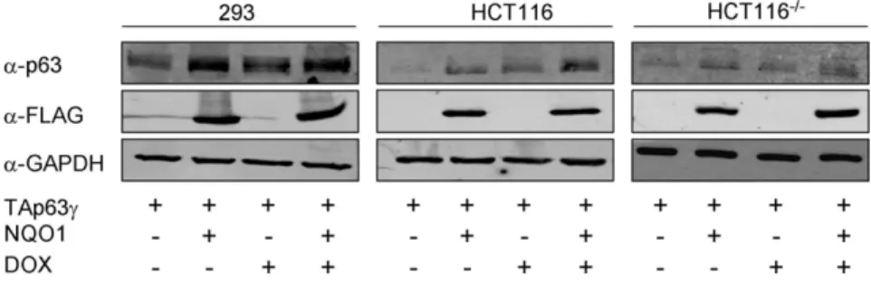
TAp63c stabilization by NQO1 is enhanced under stress
Under normal growth conditions, TAp63c protein level is elevated in cells over-expressing NQO1 (Fig. 1A). p63 expression is known to be induced following stress [35]. At this point, we were interested in determining the effect of NQO1 on TAp63c
following stress. To this end, cells co-transfected with TAp63c
Wild-type but not C609T NQO1 physically interacts with TAp63c
To detect a potential physical association between TAp63c
and NQO1, 293 cells stably expressing HA-NQO1 were transfected with a TAp63cexpression plasmid. Fig. 3A demon-strates that immunoprecipitation of HA-NQO1 pulled down TAp63c (lane 1–2). In agreement, immunoprecipitation of TAp63c pulled down HA-NQO1 (lane 5–6). Conversely, immunoprecipitation of C609T NQO1 did not pull down this p63 isoform under the same conditions (Fig. 3A, lanes 3–4). These data confirm that TAp63c and NQO1 do interact physically and that this interaction may be dependent on the catalytic activity of NQO1.
NQO1 protects TAp63c from 20S proteasomal degradation
To study whether NQO1 protects TAp63cfrom degradation, an in-vitro degradation assay was performed. In this assay, in-vitro translated TAp63cwas used as a substrate for degradation by 20S
proteasomes in the absence or presence of in-vitro translated NQO1. As has previously been shown for p53 and p73 [26–29], TAp63cwas degraded by 20S proteasomes. This degradation was inhibited in the presence of excess NQO1 and its cofactor, NADH, suggesting that NQO1 directly protects TAp63cfrom degradation by 20S proteasomes (Fig. 3B). Unlike wild-type NQO1, C609T NQO1 did not inhibit this degradation (data not shown). It should be noted that the expression level of C609T NQO1 in the presence of 20S proteasomes was very low compared to wild-type NQO1. It is impossible to establish, at this point, whether the absence of inhibition of TAp63cdegradation by C609T NQO1 was due to the incompetence of C609T NQO1 or was it merely a result of its low expression level.
Downregulation of NQO1 inhibits TAp63c-mediated growth arrest
We next determined whether NQO1-dependant TAp63c
accumulation could initiate growth arrest or apoptosis. In order to neutralize the effect of p53, p53 null HCT1162/2 cells were
Figure 1. TAp63cprotein is stabilized by NQO1.(A) 293, HCT116 and HCT1162/2cells expressing endogenous NQO1 or over-expressing
wild-type FLAG-NQO1 were transfected with a plasmid expressing TAp63c. Forty-eight hours post-transfection cell lysates were prepared and resolved by SDS-PAGE. TAp63c, NQO1 and GAPDH levels were detected by Western blot analysis using anti-p63 (4A4), anti-FLAG and anti-GAPDH (loading control) antibodies, respectively. (B) HCT116 cells expressing endogenous NQO1 or over-expressing wild-type FLAG-NQO1 were transfected with a plasmid expressing TAp63c. Twenty-four hours post transfection, 10mg/ml cyclohexamide (CHX) was added for 4 hours. Cell lysates were prepared
and resolved by SDS-PAGE. TAp63c, NQO1 and GAPDH levels were detected by Western blot analysis using anti-p63 (4A4), anti-FLAG and anti-GAPDH (loading control) antibodies, respectively. (C) RNA was prepared from these same cells, reverse transcribed and RT-PCR was performed using primers specific for TAp63 and for gapdh. Data is represented as relative levels of TAp63cnormalized to gapdh. (D) 293, HCT116 and HCT1162/2cells were
co-transfected with plasmids expressing TAp63cand HA-C609T NQO1. Forty-eight hours post-transfection cell lysates were prepared and resolved by SDS-PAGE. TAp63c, NQO1 and GAPDH levels were detected by Western blot analysis using anti-p63 (4A4), anti-HA and anti-GAPDH (loading control) antibodies, respectively. (E) 293, HCT116 and HCT1162/2cells were transfected with scrambled oligonucleotides or siNQO1 oligonucleotides.
Twenty-four hours post-transfection cell were transfected with a plasmid expressing TAp63c. Twenty-four hours post-transfection cell lysates were prepared and resolved by SDS-PAGE. TAp63c, NQO1 and GAPDH levels were detected by Western blot analysis using p63 (4A4), NQO1 and anti-GAPDH (loading control) antibodies, respectively.
used. Since HCT116 cells tend to undergo growth arrest rather than apoptosis we tested the effect of NQO1 on TAp63c-mediated growth arrest. Exposure of HCT1162/2cells to DOX resulted in G2 arrest (Fig. 4C). The presence of TAp63c lead to a slight increase in G2 arrested cells (Fig. 4D). A 5-fold increase in p21 mRNA expression was also detected in these TAp63c over-expressing cells exposed to DOX in comparison to cell exposed to DOX but not expressing TAp63c(Fig. 4I). NQO1 inhibition in DOX treated, TAp63ctransfected cells, resulted in a reduction of G2 arrested cells to the level observed following DOX in cells not expressing TAp63c (Fig. 3H). Accordingly, TAp63cand DOX-mediated increase in p21 level was almost completely reversed by siNQO1 (Fig. 3I). Expression of TAp63c, without an additional stress signal, did not lead to cell cycle arrest in our setting. These results are consistent with our NQO1 overexpression and silencing data and suggest that NQO1 is an important regulator of TAp63c
and consequently of p21. Our data indicate that downregulation
of NQO1 inhibits TAp63c-mediated p21 upregulation and TAp63c-induced G2 arrest in DOX treated HCT1162/2cells.
Discussion
Similar to p53, TAp63cprotein level increases upon treatment with DNA damaging agents resulting in transactivation of p53-responsive genes assigning p63 as an important component of the cell’s apoptotic machinery [35–37]. This p63 upregulation is not the result of transcriptional activation, but is most likely due to an increase at the protein level [36,37]. To date, little is known of the molecular mechanisms regulating p63 protein level. We provide in-vitro evidence for a physical and functional relationship between TAp63c and NQO1 supporting a new pathway regulating p63 stability.
In different types of unstressed cells, wild-type NQO1 bound to and stabilized TAp63c. In contrast, we show that C609T NQO1
Figure 2. TAp63cexpression is stabilized by NQO1 in response to genotoxic stress.293, HCT116 and HCT1162/2 cells expressing
endogenous NQO1 or over-expressing wild-type FLAG-NQO1 were transfected with a plasmid expressing TAp63c. Twenty-four hours post-transfection cells were treated with 1mM DOX for 24h. At this point, cell lysates were prepared and resolved by SDS-PAGE. TAp63c, NQO1 and GAPDH
levels were detected by Western blot analysis using anti-p63 (4A4), anti-FLAG and anti-GAPDH (loading control) antibodies, respectively. doi:10.1371/journal.pone.0011401.g002
Figure 3. NQO1 physically associates with TAp63cand protects it from 20S proteasomal degradation.(A) 293 cells stably expressing
HA-NQO1 or HA-C609T NQO1 were transfected with a plasmid expressing TAp63c. Cellular extracts were prepared and subject to immunoprecipitated (IP) with anti-HA or anti-p63 antibodies. Immunoprecipitated proteins and 5% of input material were detected by Western blot using anti-p63 and anti-HA antibodies. (B) Degradation of in-vitro translated, biotin-labeled TAp63cwith 20S proteasome was carried out in the presence or absence of in-vitro translated, biotin-labeled NQO1 and 1mM NADH at 37uC for 4h. Biotin-labeled TAp63cand NQO1 were detected with fluorescently-labeled streptavidin.
is unable to associate with TAp63cor to affect its protein level. This observation emphasizes the requirement of proper NQO1 enzymatic activity for p63 stabilization. It remains to be determined whether intact enzymatic activity of NQO1 and its binding to TAp63cis sufficient for p63 stabilization. NQO1 did not alter the expression of GAPDH or retinoblastoma (data not shown for retinoblastoma) implying that this protein degradation regulatory pathway is specific to certain proteins.
p63 has been shown to undergo ITCH mediated 26S proteasome degradation [16]. Our results show that, in addition, TAp63c is subjected to 20S proteasomal degradation and that
NQO1 protects TAp63c from such degradation. This finding implies that p63 stabilization may involve several parallel mechanisms.
Many evidence support the notion that p63 has tumor suppressor functions: 1) p63 specific siRNA enhances the transformation potential of p532/2 MEFs [38]; 2) p63 can mediate chemo-sensitivity independent of p53 status by induction of apoptosis [17,39]; 3) The combined absence of p63 and p73 severely impairs the induction of p53-dependent apoptosis in response to DNA damage [40]. These observations suggest that p63 may complement, and in some circumstances substitute for
Figure 4. Downregulation of NQO1 inhibits TAp63c-dependent growth arrest in DOX treated HCT1162/2cells.HCT1162/2cells were
transfected with scrambled oligonucleotides (siControl) (A–D) or siNQO1 oligonucleotides (E–H) and transfected (B, D, F, H) or not (A, C, E, G) with a plasmid expressing TAp63c. Twenty-four hours post-transfection the cells were treated or not with 0.05mM DOX. 24h after this treatment, DNA
content was analyzed by propidium-iodide staining. The percentage of cells in G1 and G2 is presented. (I) RNA was prepared from these same cells, reverse transcribed and real time PCR was performed using primers specific for p21 and 18S rRNA. Data is represented as relative levels of p21 normalized to 18S rRNA. *p,0.05, **p,0.01.
p53. For these reasons complex molecular mechanisms must regulate the expression of the p63 isoforms. Phosphorylation has been reported to be associated with rapid accumulation of TAp63cupon genotoxic treatment whereasDNp63ais degraded under these same conditions [15]. Coherently, in our settings, NQO1 over-expression accompanied by stress (DOX) resulted in the accumulation of TAp63cand not in the accumulation of the antagonizingDN isoforms or the weak TAp63c(data not shown). Stabilization of TAp63cby NQO1 is of biological relevance since the ability of TAp63cto upregulate p21 and to induce G2 arrest in stressed HCT1162/2cells is almost abolished in the presence of siNQO1.
Our results have biological implications concerning the understanding of tumor development. Cells carrying polymorphic inactive NQO1 that are exposed to carcinogens that are substrate for detoxification by NQO1 will accumulate more damage than cells carrying wild-type NQO1. Our results show that polymor-phic NQO1 does not stabilize TAp63cand low wild-type NQO1 levels (as detected in cells harboring one allele of polymorphic NQO1) decrease TAp63c-induced growth arrest. These cells, already harboring damaged DNA, would therefore also exhibit imperfect apoptotic or growth arrest responses, providing them with a growth advantage that could potentially lead to tumor development. Indeed, p63 is not frequently mutated in cancer but rather alterations of p63 expression have been widely reported [10–12,41]. Of note, C609T NQO1 is known to enhance susceptibility to bladder and breast cancers [21–23]. In these same tumor types, TAp63 expression is often misregulated [6–8]. The altered TAp63 expression levels detected in these tumors may be explained by the incompetence of C609T NQO1 to properly stabilize p63. In one study, Fagerholm et al demonstrated that the chemotherapeutic response of C609T homozygous breast
carci-noma cells was impaired in p53-aberrant tumors [21]. This phenomenon could be attributed to the inability of polymorphic NQO1 to stabilize TAp63, whose expression was indeed shown to be significantly reduced in breast cancers. TAp63 reduction could result in the poor chemotherapeutic response characteristic of these tumor cells.
Our report is the first to identify a new mechanism appointing a direct role for NQO1 in the regulation of p63 expression, especially following stress. We propose that NQO1 associates with and stabilizes TAp63c. The increase in TAp63c results in enhanced transcription of p21 consequently leading to growth arrest. Characterization of the interactions between NQO1 and the other p63 family members would be an interesting future study. Such studies will probably elucidate that NQO1 not only regulates the expression of the potent TAp63c but more realistically it regulates the balance of p63 isoforms under normal growth conditions and following stress. It will be interesting to explore how this level of regulation collaborates with others as more regulators of p63 are found. Clearly, comprehension of such regulations of the p63 isoforms will lead to a greater understanding of the role of p63 in tumor suppression.
Acknowledgments
We are grateful to Prof. Yosef Shaul for valuable advice and insightful suggestions.
Author Contributions
Conceived and designed the experiments: OHR GG. Performed the experiments: OHR. Analyzed the data: OHR OS GG. Contributed reagents/materials/analysis tools: OS. Wrote the paper: GG.
References
1. Yang A, Kaghad M, Wang Y, Gillett E, Fleming MD, et al. (1998) p63, a p53 homolog at 3q27–29, encodes multiple products with transactivating, death-inducing, and dominant-negative activities. Mol Cell 2: 305–316.
2. Osada M, Park HL, Nagakawa Y, Yamashita K, Fomenkov A, et al. (2005) Differential recognition of response elements determines target gene specificity for p53 and p63. Mol Cell Biol 25: 6077–6089.
3. Osada M, Ohba M, Kawahara C, Ishioka C, Kanamaru R, et al. (1998) Cloning and functional analysis of human p51, which structurally and functionally resembles p53. Nat Med 4: 839–843.
4. Zeng SX, Dai MS, Keller DM, Lu H (2002) SSRP1 functions as a co-activator of the transcriptional activator p63. EMBO J 21: 5487–5497.
5. Kaelin WG, Jr. (1999) The p53 gene family. Oncogene 18: 7701–7705. 6. Urist MJ, Di Como CJ, Lu ML, Charytonowicz E, Verbel D, et al. (2002) Loss of
p63 expression is associated with tumor progression in bladder cancer. Am J Pathol 161: 1199–1206.
7. Koga F, Kawakami S, Fujii Y, Saito K, Ohtsuka Y, et al. (2003) Impaired p63 expression associates with poor prognosis and uroplakin III expression in invasive urothelial carcinoma of the bladder. Clin Cancer Res 9: 5501–5507. 8. Moll UM, Slade N (2004) p63 and p73: Roles in development and tumor
formation. Mol Cancer Res 2: 371–386.
9. Pietsch EC, Sykes SM, McMahon SB, Murphy ME (2008) The p53 family and programmed cell death. Oncogene 27: 6507–6521.
10. Massion PP, Taflan PM, Jamshedur Rahman SM, Yildiz P, Shyr Y, et al. (2003) Significance of p63 amplification and overexpression in lung cancer develop-ment and prognosis. Cancer Res 63: 7113–7121.
11. Park BJ, Lee SJ, Kim JI, Lee SJ, Lee CH, et al. (2000) Frequent alteration of p63 expression in human primary bladder carcinomas. Cancer Res 60: 3370–3374. 12. Marchini S, Marabese M, Marrazzo E, Mariani P, Cattaneo D, et al. (2008) DeltaNp63 expression is associated with poor survival in ovarian cancer. Ann Oncol 19: 501–507.
13. Mills AA, Zheng B, Wang XJ, Vogel H, Roop DR, et al. (1999) p63 is a p53 homologue required for limb and epidermal morphogenesis. Nature 398: 708–713.
14. Brooks CL, Gu W (2006) p53 ubiquitination: Mdm2 and beyond. Mol Cell 21: 307–315.
15. Papoutsaki M, Moretti F, Lanza M, Marinari B, Sartorelli V, et al. (2005) A p38-dependent pathway regulates DeltaNp63 DNA binding to p53-p38-dependent promoters in UV-induced apoptosis of keratinocytes. Oncogene 24: 6970–6975.
16. Rossi M, Aqeilan RI, Neale M, Candi E, Salomoni P, et al. (2006) The E3 ubiquitin ligase Itch controls the protein stability of p63. Proc Natl Acad Sci USA 103: 12753–12758.
17. Bergamaschi D, Samuels Y, Jin B, Duraisingham S, Crook T, et al. (2004) ASPP1 and ASPP2: common activators of p53 family members. Mol Cell Biol 24: 1341–1350.
18. Bernassola F, Oberst A, Melino G, Pandolfi PP (2005) The promyelocytic leukaemia protein tumour suppressor functions as a transcriptional regulator of p63. Oncogene 24: 6982–6986.
19. Koutsodontis G, Vasilaki E, Chou WC, Papakosta P, Kardassis D (2005) Physical and functional interactions between members of the tumour suppressor p53 and the Sp families of transcription factors: importance for the regulation of genes involved in cell-cycle arrest and apoptosis. Biochem J 389: 443–455. 20. MacPartlin M, Zeng S, Lee H, Stauffer D, Jin Y, et al. (2005) p300 regulates p63
transcriptional activity. J Biol Chem 280: 30604–306010.
21. Fagerholm R, Hofstetter B, Tommiska J, Aaltonen K, Vrtel R, et al. (2008) NAD(P)H:quinone oxidoreductase 1 NQO1*2 genotype (P187S) is a strong prognostic and predictive factor in breast cancer. Nat Genet 40: 844–853. 22. Sanyal S, Ryk C, De Verdier PJ, Steineck G, Larsson P, et al. (2007)
Polymorphisms in NQO1 and the clinical course of urinary bladder neoplasms. Scand J Urol Nephrol 41: 182–190.
23. Chao C, Zhang ZF, Berthiller J, Boffetta P, Hashibe M (2006) NAD(P)H:qui-none oxidoreductase 1 (NQO1) Pro187Ser polymorphism and the risk of lung, bladder, and colorectal cancers: a meta-analysis. Cancer Epidemiol Biomarkers Prev 15: 979–987.
24. Asher G, Bercovich Z, Tsvetkov P, Shaul Y, Kahana C (2005) 20S proteasomal degradation of ornithine decarboxylase is regulated by NQO1. Mol Cell 17: 645–655.
25. Garate M, Wong RP, Campos EI, Wang Y, Li G (2008) NAD(P)H quinone oxidoreductase 1 inhibits the proteasomal degradation of the tumour suppressor p33(ING1b). EMBO Rep 9: 576–581.
26. Asher G, Lotem J, Cohen B, Sachs L, Shaul Y (2001) Regulation of p53 stability and p53-dependent apoptosis by NADH quinone oxidoreductase 1. Proc Natl Acad Sci USA 98: 1188–1193.
27. Asher G, Lotem J, Kama R, Sachs L, Shaul Y (2002) NQO1 stabilizes p53 through a distinct pathway. Proc Natl Acad Sci USA 9: 3099–3104. 28. Asher G, Lotem J, Sachs L, Kahana C, Shaul Y (2002) Mdm-2 and
29. Asher G, Tsvetkov P, Kahana C, Shaul Y (2005) A mechanism of ubiquitin-independent proteasomal degradation of the tumor suppressors p53 and p73. Genes Dev 19: 316–321.
30. Vindelov LL, Christensen IJ, Nissen NI (1983) A detergent-trypsin method for the preparation of nuclei for flow cytometric DNA analysis. Cytometry 3: 323–327.
31. Siegel D, Anwar A, Winski SL, Kepa JK, Zolman KL, et al. (2001) Rapid polyubiquitination and proteasomal degradation of a mutant form of NAD(P)H:quinone oxidoreductase 1. Mol Pharmacol 59: 263–268.
32. Traver RD, Siegel D, Beall HD, Phillips RM, Gibson NW, et al. (1997) Characterization of a polymorphism in NAD(P)H: quinone oxidoreductase (DT-diaphorase). Br J Cancer 75: 69–75.
33. Cross JV, Deak JC, Rich EA, Qian Y, Lewis M, et al. (1999) Quinone reductase inhibitors block SAPK/JNK and NFkappaB pathways and potentiate apoptosis. J Biol Chem 274: 31150–31154.
34. Reigan P, Colucci MA, Siegel D, Chilloux A, Moody CJ, et al. (2007) Development of indolequinone mechanism-based inhibitors of NAD(P)H:qui-none oxidoreductase 1 (NQO1): NQO1 inhibition and growth inhibitory activity in human pancreatic MIA PaCa-2 cancer cells. Biochemistry 46: 5941–5950.
35. Petitjean A, Ruptier C, Tribollet V, Hautefeuille A, Chardon F, et al. (2008) Properties of the six isoforms of p63: p53-like regulation in response to genotoxic stress and cross talk with DeltaNp73. Carcinogenesis 29: 273–281.
36. Katoh I, Aisaki KI, Kurata SI, Ikawa S, Ikawa Y (2000) p51A (TAp63gamma), a p53 homolog, accumulates in response to DNA damage for cell regulation. Oncogene 19: 3126–3130.
37. Okada Y, Osada M, Kurata S, Sato S, Aisaki K, et al. (2002) p53 gene family p51(p63)-encoded, secondary transactivator p51B (TAp63alpha) occurs without forming an immunoprecipitable complex with MDM2, but responds to genotoxic stress by accumulation. Exp Cell Res 276: 194–200.
38. Lang GA, Iwakuma T, Suh YA, Liu G, Rao VA, et al. (2004) Gain of function of a p53 hot spot mutation in a mouse model of Li-Fraumeni syndrome. Cell 119: 861–872.
39. Gressner O, Schilling T, Lorenz K, Schulze Schleithoff E, Koch A, et al. (2005) TAp63alpha induces apoptosis by activating signaling via death receptors and mitochondria. EMBO J 24: 2458–2471.
40. Flores ER, Tsai KY, Crowley D, Sengupta S, Yang A, et al. (2002) p63 and p73 are required for p53-dependent apoptosis in response to DNA damage. Nature 416: 560–564.