Braz J Med Biol Res, March 2010, Volume 43(3) 271-278
Growth inhibitory effect and Chk1-dependent signaling involved
in G /M arrest on human gastric cancer cells induced by diallyl
2disulfide
H. Ling, L. Wen, X.X. Ji, Y.L. Tang, J. He, H. Tan, H. Xia, J.G. Zhou and Q. Su
ISSN 0100-879X
BIOMEDICAL SCIENCES
AND
CLINICAL INVESTIGATION
www.bjournal.com.br
www.bjournal.com.br
Volume 43 (03) 226-324 March 2010
Institutional Sponsors
Growth inhibitory effect and Chk1-dependent
signaling involved in G
2
/M arrest on
human gastric cancer cells induced
by diallyl disulfide
H. Ling, L. Wen, X.X. Ji, Y.L. Tang, J. He, H. Tan, H. Xia, J.G. Zhou and Q. Su
Cancer Research Institute, University of South China, Hengyang, Hunan, China
Abstract
Diallyl disulfide (DADS) inhibits growth and induces cell cycle G2/M arrest in human gastric cancer MGC803 cells. In this study,
15 mg/L DADS exerted similar effects on growth and cell cycle arrest in human gastric cancer BGC823 cells. Due to the im
-portance of cell cycle redistribution in DADS-mediated anti-carcinogenic effects, we investigated the role of checkpoint kinases (Chk1 and Chk2) during DADS-induced cell cycle arrest. We hypothesized that DADS could mediate G2/M phase arrest through
either Chk1 or Chk2 signal transduction pathways. We demonstrated that DADS induced the accumulation of phosphorylated Chk1, but not of Chk2, and that DADS down-regulated Cdc25C and cyclin B1. The expression of mRNA and total protein for Chkl and Chk2 was unchanged. Chk1 is specifically phosphorylated by ATR (ATM-RAD3-related gene). Western blot analysis showed that phospho-ATR was activated by DADS. Taken together, these data suggest that cell cycle G2/M arrest, which
was associated with accumulation of the phosphorylated forms of Chk1, but not of Chk2, was involved in the growth inhibition
induced by DADS in the human gastric cancer cell line BGC823. Furthermore, the DADS-induced G2/M checkpoint response
is mediated by Chk1 signaling through ATR/Chk1/Cdc25C/cyclin B1, and is independent of Chk2.
Key words: Diallyl disulfide; Cell cycle G2/M arrest; Checkpoint kinase-1; Gastric cancer
Introduction
Correspondence: Q. Su, Cancer Research Institute, University of South China, 28 Changshengxi Road, Hengyang, Hunan 421001,
China. Fax: +86-734-828-1547. E-mail: nhdxlh@yahoo.com.cn
Received September 23, 2009. Accepted January 19. 2010. Available online January 29, 2010. Published March 12, 2010.
Gastric cancer is one of the leading causes of death of cancer patients and is the most common type of malig-nancy in China (1). Despite recent advances in surgical and chemotherapeutic procedures, the 5-year survival rate for gastric cancer patients remains very low. Hence, identifying alternative factors that may reduce the initiation and promotion of gastric cancer is important for minimizing the incidence and severity of this disease.
A high intake of garlic is associated with a protective effect against various cancers, including stomach cancer, in humans (2). Several epidemiologic studies have sug-gested that garlic plays an important role in the reduction of cancer-related deaths (3). Garlic oil, or more specifically its diallyl disulfide (DADS) content, has become a quite appealing anti-carcinogenic agent. This is due, in part, to its ability to induce apoptosis in vitro (4) and to inhibit the formation and growth of tumors in vivo (5,6). DADS has been
shown to inhibit the in vitro and in vivo growth of breast,
liver, lung, gastric, colon, and prostate cancers, as well as neuroblastoma and leukemia cell lines(5,7-14). Therefore, DADS may be a useful therapeutic tool for the prevention of environmentally induced cancers.
The antiproliferative property of DADS in cultured hu-man colon tumor cells (HCT-116 and COLO 205) and the prostate cancer cell line (PC-3) is related to its ability to decrease the proportion of cells in the G1 phase and to
in-crease the proportion of cells in the G2/M phase (12,15,16).
272 H. Ling et al.
et al. (18) demonstrated that elevated phosphorylation of checkpoint kinase 1 (Chk1), decreased phosphorylation of Chk2, and decreased levels of Cdc25C, 14-3-3, and cyclin B1 were the critical changes associated with the abrogation of G2/M checkpoint control during transformation of Syrian
hamster embryo cells by Malachite green.
We have previously reported that DADS inhibited the growth of gastric cancer in vitro and in vivo (9,10), and that it induced cell cycle G2/M arrest in human gastric cancer
MGC803 cells (19). A decreased Cdc25C expression plays a crucial role in G2/M arrest after treatment with DADS (19).
In the current study, DADS was shown to induce phospho -rylation of ATR (ATM-RAD3-related gene) and Chk, while suppressing expression of Cdc25C and cyclin B1 in the human gastric cancer cell line BGC823. These observa-tions contribute to our understanding of the mechanisms of the anti-tumor effect of DADS in gastric cancer cells, and furthermore they indicate the potential of DADS for clinical development as a therapeutic drug for combating gastric cancer.
Material and Methods
Material
DADS (MW 146.28) and dimethyl thiazolyl tetrazolium bromide (MTT) were purchased from Fluka Chemika (Ronkonkoma, USA). The kit for bicinchoninic acid protein quantification was manufactured by Pierce (USA). The cell cycle protein test kit, which included the phospho-Chk1 (Ser345) antibody, was from Cell Signaling Technology (USA). The phospho-Chk2 (Thr68) antibody, ATR, and anti-rabbit IgG (HRP-linked) Chk1 and Chk2 antibodies were from Santa Cruz Company (USA). The Cdc25C antibody was from NeoMarkers (USA). The cyclin B1 and β-actin antibodies were from Boster Company (China). Phospho-ATR (Ser428) and ECL LumiGLO reagent were from Cell Signaling Technology.
Cell culture. The human gastric cancer cell line BGC823 was obtained from the Cell Research Institute of the Chinese Academy of Science (China). The cells were maintained in RPMI-1640 medium (Sigma, USA), supplemented with 10% heat-inactivated fetal bovine serum (FBS; Life Tech -nologies, USA), 100 μg/mL streptomycin, and 100 U/mL penicillin G (Invitrogen, USA) in a humidified atmosphere of 5% CO2 and 95% air at 37°C. Cells were suspended at
a final concentration of 104 cells/mL in RPMI-1640 medium
containing 10% FBS in culture flasks. Reagents were added to each flask in various combinations and incubated at 37°C in 5% CO2.
Cell viability assay
The MTT assay was performed using the method of Yuan et al. (19). Briefly, a 200-μL suspension containing approximately 2 x 10⁴ cells was added to each well of a
96-well culture plate and incubated for 24 h at 37°C in a
humidified atmosphere of 95% air and 5% CO2. DADS was
dissolved in dimethyl sulfoxide (DMSO), mixed with culture medium, and then added to the cells on 96-well plates at four different concentrations (5, 10, 15, and 20 mg/L). The concentration of DMSO added to the medium in all con-centrations of DADS was 0.01%. The 96-well culture plate was divided into seven sections, with one section for the control, while the other sections were treated with 100-µL culture media containing 5, 10, 15, and 20 mg/L DADS. The cultures were re-incubated as described above. After 96 h, 100 µL 1 mg/mL MTT solution was added to each well, the cultures were further incubated for 4 h and 100 µL 20% SDS in 50% DMSO was added. The formed crystals were gently dissolved by slowly pipetting two to three times. A microplate reader was used to measure absorbance at 570 nm for each well. The growth inhibition rate was calculated as follows: growth inhibition = [(A570/nm of treated cells) / (A570/nm of control cells)] x 100%.
Cell cycle analysis
Cells were incubated in culture media alone, or in cul-ture media containing 15 mg/LDADS, at 37°C for 12, 24, 36, and 48 h. Cells were harvested in cold PBS, fixed in 700 mL/L ethanol, and stored at 4°C for subsequent cell cycle analysis. Fixed cells were washed with PBS once and suspended in 1 mL propidium iodide-staining reagents (20 mg/L ribonuclease and 50 mg/L propidium iodide). Samples were incubated in the dark for 30 min before cell cycle analysis and the distribution of cells in the various phases of the cell cycle was measured with a flow cytometer (Coulter EPICS-XL, Beckerman, USA). The percentage of cells in the G1, S, and G2/M phases was then calculated
by the CellQuest software on the flow cytometer (Coulter EPICS-XL, Beckerman, USA).
RNA extraction and RT-PCR
comparison, samples were used to compare the density ratio of the objective gene to that of β-actin mRNA.
Western blotting
Human gastric cancer cells were cultured with or without DADS at the indicated concentrations for various periods of time, washed once with ice-cold PBS, and lysed in a buffer consisting of 20 mM Tris/HCl, pH 8.0, 137 mM NaCl, 1.5 mM MgCl2, 1 mM EGTA, 10% glycerol, 100 mM NaF, and
1% Triton X-100. Protein concentration in the lysates was measured with the Protein BCA Assay Kit (Bio-Rad, USA). Thirty micrograms of protein lysate was subjected to 10% SDS-polyacrylamide gel electrophoresis and the proteins separated were transferred to polyvinylidene difluoride membranes (Millipore, USA). To block nonspecific binding, membranes were incubated at room temperature for 1 h with 5% skim milk powder, followed by a 12-h incubation at 4°C with an antiserum containing antibodies against Chk1, Chk2, ATR, Phospho-Chk1 (Ser345), Phospho-Chk2 (Thr68), Phospho-ATR (Ser428), Cdc25C, and cyclin B1. A peroxidase-conjugated secondary antibody (1:5000 di-lution) and ECL Western blotting detection reagents were used to visualize the target proteins (ECL New England Biolabs, USA), which were quantified with a Bio Image Intelligent Quantifier 1-D (Version 2.2.1, Nihon-BioImage Ltd., Japan).
Immunoprecipitation and Western blotting
Human gastric cancer cells were cultured with or without DADS at the indicated concentrations for various periods of time, and then characterized with the Seize (R) Classic Mammalian Immunoprecipitation kit (Piece Technology, USA). The cells were carefully removed from the culture medium, washed once with PBS (0.1 M phosphate, 0.15 M NaCl, pH 7.2), harvested and lysed with the lysis buffer of the M-PER reagent. Lysates were collected, transferred to a microcentrifuge tube and centrifuged at 13,000 g for 5-10 min to separate the cell debris. The supernatants were transferred to another tube for further analysis. Purified Chk1 or Chk2 antibody was added to the sample, followed by overnight incubation at 4°C. The immune complex was
added to the spin cup containing equilibrated protein G beads. Then, 190 μL elution buffer was added to the spin cup and the samples were eluted. The levels of immunopre-cipitated protein were determined by Western blotting with the anti-Cdc25C antibody and the ECL detection system.
Statistical analysis
Data are reported as means ± SD. One-way ANOVA
was used to compare difference among groups and P values of less than 0.05 were considered to be statistically significant.
Results
Cell viability
DADS inhibits the growth of BGC823 cells for 96 h, in a dose-dependent manner. The exposure of BGC823 cells to 5 mg/L DADS for 96 h inhibited growth by 34.2%/96 h, whereas exposure to 15 mg/L DADS increased the inhibition to 55.7%/96 h, as assessed by the MTT test (P < 0.05; Table 1). From these results, the IC50 value of DADS for 96 h was
calculated as ≈15 mg/L DADS. Hence, for further studies, we selected 15 mg/L DADS IC50 to maximize the effects of
dose levels so that the mechanism of action of DADS could be identified. The results indicated that the proliferation of BGC823 cells was significantly reduced by treatment with DADS for 96 h in a dose-dependent manner.
Cell cycle arrest of BGC823 cells
DADS inhibited the proliferation of BGC823 cells, as also shown by FACS analysis. As described previously, the inhibitory effects of DADS on proliferation are often due to cell cycle arrest (15). Consistent with its effect on cell growth inhibition, DADS induced a significant cell cycle arrest at the G2/M phase in BGC823 cells. As shown in
Table 2, approximately 48.3 (24 h) and 18.6% (24 h) of the untreated cells were in the G0/G1 and G2/M phases,
respectively, and 33.1% (24 h) of the cells were in the S phase. In contrast, after treatment with 15 mg/LDADS, the proportion of cells in the G2/M phase was 58.1 (24 h) and
50.2% (36 h), or more than three times and two times,
re-Table 1. Inhibitory effects on human gastric cancer BGC823 cells exposed to various concentrations of diallyl disul
-fide (DADS) for 96 h.
BGC823 cells DADS (mg/L)
5 10 15 20
Absorption value 0.79 ± 0.049 0.52 ± 0.033* 0.48 ± 0.018* 0.35 ± 0.023* 0.24 ± 0.032*
Inhibition rate (%/96 h) 34.2 39.2 55.7 69.6
The number of BGC823 cells is reported as 2 x 10⁵/mL. The initial concentration of cells was 1 x 10⁵/mL. Data are
274 H. Ling et al.
spectively, compared to untreated cells. Taken together, these data suggest that DADS reduces the proliferation of BGC823 cells by increasing the proportion of cells in the G2/M stage of the cell cycle.
Effect of DADS on Chk1 and Chk2 mRNA expression in BGC823 cells
The expression of Chk1 and Chk2 mRNA associated with the cell cycle arrest of BGC823 cells after treatment with 15 mg/LDADS for 1 or 2 days was revealed by RT-PCR. As shown in Figure 1, the expression of Chk1 and Chk2 mRNA was unchanged in treated cells compared to untreated cells.
Expression of Chk1 and phospho-Chk1 in BGC823 cells induced by DADS
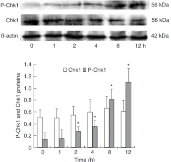
Using antibodies specific for Chk1 phosphopeptides, we analyzed Chk1 phosphorylation at serine 345, following exposure of BGC823 cells to DADS. As illustrated in Figure 2, after stimulation with 15 mg/LDADS for 2 h, phospho-Chk1 showed a significant increase and this expression continued to increase gradually with time (P < 0.05). On the other hand, the levels of total Chk1 protein and β-actin, an inner control, did not change significantly.
Expression of Chk2 and phospho-Chk2 induced by DADSin BGC823 cells
Figure 3 shows the effects of DADS on the expres-sion of phospho-Chk2 in BGC823 cells at different
Figure 1. Expression of Chk1 and Chk2 mRNA induced by diallyl disulfide (DADS).BGC823
cells (1 x 10⁶/mL)were treated with 15 mg/LDADS and expression of Chk1 and Chk2 mRNA
was analyzed by RT-PCR. The relative amount of each mRNA was compared to β-actin.The
data are reported as means ± SD (N = 3) on the right side of the figure. Each result is the aver
-age of two determinations. Left side of the figure: Lane 1, marker; lane 2, BGC823 cells; lanes 3
and 4, BGC823 cells treated with 15 mg/L DADS for 1 or 2 days. Right side of the figure: column
1 = BGC823 cells; column 2 = BGC823 cells treated with 15 mg/L DADS for 1 day; column 3 = BGC823 cells treated with 15 mg/L DADS for 2 days. There were no significant differences (t -test).
Table 2. Effect of diallyl disulfide (DADS) on the percent of cells
in the cell cycle.
Group G1 S G2/M
Control
12 h 49.1 ± 1.25 32.7 ± 0.83 18.2 ± 0.91
24 h 48.3 ± 1.14 33.1 ± 1.09 18.6 ± 1.82
36 h 50.1 ± 2.40 30.7 ± 1.52 19.2 ± 1.34
48 h 50.2 ± 2.21 29.4 ± 1.11 20.4 ± 1.42
DADS (15 mg/L)
12 h 38.4 ± 1.07 31.5 ± 0.31 30.1 ± 1.20*
24 h 15.1 ± 0.19 26.8 ± 0.23 58.1 ± 1.45*
36 h 35.6 ± 0.62 14.2 ± 0.09 50.2 ± 1.71* 48 h 47.7 ± 0.96 16.1 ± 0.18 36.2 ± 1.36*
The concentration of BGC823 cells was 1 x 10⁴/mL. Data are
reported as means ± SD for N = 3. *P < 0.05 compared with
Figure 2. Activation of Chk1 phosphorylation by diallyl disul
-fide (DADS). BGC823 cells (1 x 10⁶/mL) were treated with 15 mg/LDADS. Expression and phosphorylation of Chk1 (P-Chk1) were analyzed by Western blot analysis. The relative amount of
each protein was compared to β-actin. The data are reported as means ± SD (N = 3). Each result is the average of two determina -tions. *P < 0.05 vs control (t-test).
Figure 3. Activation of Chk2 phosphorylation by diallyl disulfide
(DADS). BGC823 cells (1 x 10⁶/mL) were treated with 15 mg/L
DADS. Expression and phosphorylation of Chk2 (P-Chk2) were
analyzed by Western blot analysis. The relative amount of each
protein was compared to β-actin.Thedata are reported as means
± SD (N = 3). Each result is the average of two determinations. There were no significant differences (t-test).
Figure 4. Expression of Cdc25C by diallyl disulfide (DADS).
BGC823 cells (1 x 10⁶/mL) were treated with 15 mg/LDADS and
expression of Cdc25C was analyzed by Western blot.The
rela-tive amount of each protein was compared to β-actin. The data are reported as mean ± SD (N = 3). Each result is the average of two determinations. *P < 0.05 vs control (t-test).
times. The expression of phospho-Chk2 was weak and even weaker after stimulation with DADS, but the change was not statistically significant (P > 0.05). Chk2 and β-actin, the inner control, showed no significant changes.
Expression of Cdc25C and cyclin B1, downstream molecules of Chk, induced by DADS in BGC823 cells
The results in Figure 4 show that, after 12 h, 15 mg/LDADS inhibited the expression of the cell cycle-associated phosphatase Cdc25C in BGC823 cells, which was decreased by 81% after 48 h (P < 0.05). As shown in Figure 5, the expression of cyclin B1 increased after 12 h of DADS treatment, and then decreased after 36 h, being reduced by 87.5% after 48 h (P < 0.05).
Role of ATR, as an upstream molecule of Chk, in BGC823 cells treated with DADS
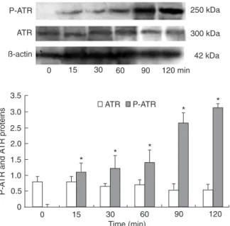
ATR mediates checkpoint signaling through its downstream effect or Chk1 (20). As shown in Figure 6, treatment of BCC823 cells for 15 min to 2 h resulted in an increase in phospho-ATR expression, whereas no change was found in ATR expression.
Chk1 or Chk2 interacts with Cdc25C
-276 H. Ling et al.
ments. The Chk1 Ab increasingly precipitated Cdc25C (Fig-ure 7A). In contrast, Chk2 Ab failed to precipitate Cdc25C in BGC823 cells (Figure 7B). Unrelated rabbit immunoglobulin (IgG) was used as a negative control.
Discussion
DADS, an oil-soluble organosulfur compound and al-lyl mercaptan, is formed in individuals who eat raw garlic. Moreover, DADS is the major component of cooked garlic, which is reduced to allyl mercaptan in blood (21). DADS is reported to make up about 60% of garlic oil (22), thus, indicating that it is the most appropriate compound to use in the study of the possible effects of raw and cooked garlic in humans. Studies have shown that DADS suppresses the proliferation of cancer cells such as MGC803, SW480, and HL-60-cultured cells (9,23,24). A previous study also showed that DADS inhibits the proliferation of prostate cancer cells through cell cycle arrest at the G2/M transition (12). In the
present study, an apparent and dose-dependent growth inhibition characteristic was observed in the BGC823 cell line treated with DADS. At a high concentration of 15 mg/L, DADS displayed a time-dependent and steady inhibition that was not demonstrated at lower concentrations. Cell cycle arrest at the G2/M transition was shown to be involved in
the growth inhibition induced by DADS in the human gastric cancer cell line.
Anticancer insights derived from cell cycle research have given rise to the idea that abrogation of the cell cycle G2
checkpoint can be viewed as a potential cancer cell-specific therapy. This idea is based on the discovery that many cancer cells have a defective G1 checkpoint that results in
a dependence on the G2 checkpoint during cell replication
(25,26). The cell cycle has recently become a new, more appealing target at which anti-carcinogenic agents can be directed. This represents a new mechanism for therapeutic drugs as regulators of Chk in tumor cells (27). In the present study, treatment with DADSproduced cell cycle arrest in
Figure 5. Expression of cyclin B1 by diallyl disulfide (DADS).
BGC823 cells(1 x 10⁶/mL)were treated with 15 mg/LDADS and
expression of cyclin B1 was analyzed by Western blot.The
rela-tive amount of each protein was compared to β-actin. Thedata
are reported as means ± SD (N = 3). Each result is the average of
two determinations. *P < 0.05 vs control (t-test).
Figure 6. Activation of ATR phosphorylation by diallyl disulfide
(DADS). BGC823 cells(1 x 10⁶/mL) were treated with 15 mg/L
DADS and the expression and phosphorylation of ATR (P-ATR)
were analyzed by Western blot.The relative amount of each
pro-tein was compared to β-actin. Thedata are reported as means ±
SD (N = 3). Each result is the average of two determinations. *P
< 0.05 vs control (t-test).
Figure 7. Expression of Chk1 and Chk2 in BGC823 cells by
immunoprecipitation (IP) and Western blotting. A, B: Lane 1,
BGC823 cells; lane 2, BGC823 cells treated with 15 mg/Ldiallyl
disulfide (DADS); lane 3, negative control. Thedata are reported
References
1. Sun XD, Mu R, Zhou YS, Dai XD, Zhang SW, Huangfu XM,
et al. [Analysis of mortality rate of stomach cancer and its trend in twenty years in China]. Zhonghua Zhong Liu Za Zhi
2004; 26: 4-9.
2. Fleischauer AT, Arab L. Garlic and cancer: a critical review
of the epidemiologic literature. J Nutr 2001; 131:
1032S-1040S.
3. Ryzhenkov VE, Makarov VG. [Biologically active substances
in garlic (Allium sativum L.) and their application in nutrition
for humans]. Vopr Pitan 2003; 72: 42-46.
4. Milner JA. A historical perspective on garlic and cancer. J Nutr 2001; 131: 1027S-1031S.
5. Nakagawa H, Tsuta K, Kiuchi K, Senzaki H, Tanaka K,
Hioki K, et al. Growth inhibitory effects of diallyl disulfide on
human breast cancer cell lines. Carcinogenesis 2001; 22:
891-897.
6. Kalra N, Arora A, Shukla Y. Involvement of multiple signaling pathways in diallyl sulfide mediated apoptosis in mouse skin
tumors. Asian Pac J Cancer Prev 2006; 7: 556-562.
7. De Martino A, Filomeni G, Aquilano K, Ciriolo MR, Rotilio G. Effects of water garlic extracts on cell cycle and viability
of HepG2 hepatoma cells. J Nutr Biochem 2006; 17:
742-749.
8. Wu XJ, Kassie F, Mersch-Sundermann V. The role of reac
-tive oxygen species (ROS) production on diallyl disulfide (DADS) induced apoptosis and cell cycle arrest in human A549 lung carcinoma cells. Mutat Res 2005; 579: 115-124.
9. Ling H, Zhang LY, Su Q, Song Y, Luo ZY, Zhou XT, et al. Erk is involved in the differentiation induced by diallyl disulfide
in the human gastric cancer cell line MGC803. Cell Mol Biol Lett 2006; 11: 408-423.
10. Xiang SL, Xiao XL, Ling H, Liao QJ, Zhou XT, Dong L, et al.
[Antitumor effect of diallyl disulfide on human gastric cancer MGC803 cells xenograft in nude mice]. Ai Zheng 2005; 24:
940-944.
11. Jakubikova J, Sedlak J. Garlic-derived organosulfides in
-duce cytotoxicity, apoptosis, cell cycle arrest and oxidative
stress in human colon carcinoma cell lines. Neoplasma
2006; 53: 191-199.
12. Arunkumar A, Vijayababu MR, Srinivasan N, Aruldhas MM, Arunakaran J. Garlic compound, diallyl disulfide induces
cell cycle arrest in prostate cancer cell line PC-3. Mol Cell
the G2 phase,which was associated with accumulation of
the phosphorylated formsof Chk1, but not of Chk2. Human Cdc25C is one of the central targets and regulators of the G2/M checkpoint mechanisms activated
in response to DNA injury (28). Cdc25C is thought to be the major effector of the G2/M DNA damage checkpoint
kinase Chk1 or/and Chk2 (29,30), which triggers the cyclin B1/CDK1 complex (30). The present study demonstrated that phospho-Chk1 upregulation, Cdc25C and cyclin B1 down-regulation in human gastric cancer cells enhanced the sensitivity to DADS, whereas phospho-Chk2 had no ef-fect. These results havepotentially vital implications for the mechanism of actionof DADS and its future clinical use.
The subsequent degradation of Cdc25C in response to DADSwas mediated by Chk1. Neither Chk1 nor Chk2 were directly responsiblefor the inhibition of DNA synthesis induced by DADS;however, Chk1 negatively regulated the entry of DADS-treated cells into mitosis. These findings suggest that DADS stimulates Chk1to initiate a G2-M cell
cycle checkpoint. Furthermore, it would appear that Chk1 acts to coordinate the cell cycle withDNA synthesis, thus preventing premature mitotic entry in DADS-treatedcells. Co-immunoprecipitated Chk1 or Chk2 was detected by anti-Chk1 or anti-Chk2 immunoprecipitation followed by anti-Cdc25C immunoblotting. DADS treatment enhanced the binding activity of Chk1 with Cdc25C in BGC823 cells; however, it did not influence the binding activity of Chk2 with Cdc25C. This confirms that DADS induces G2/M arrest by
the interaction of Chk1 and Cdc25C in BGC823 cells. There are two possible scenarios explaining how DNA synthesis mightbe arrested. Firstly, arrest of DNAsynthesis could be directly mediated by checkpoint activation.Alterna
-tively, arrest might occur by other mechanisms that activate other checkpoints, for example, chain termination.
ATR is capable of specifically phosphorylating Chk1 (31). Another important finding of the present study was that ATR isrequired for maximal phosphorylation of Chk1 checkpointproteins following DADS treatment. However, ATR only phosphorylates Chk1 (Ser345) and not Chk2. This substratespecificity may reflect the initial oxidative DNA strandbreaks.
Ourhypothesis is that the substrate specificity of these kinases iscoordinated by the unique binding and activation of ATR with auxiliary proteins at the sites of DNA lesions. Hypothetically, the activation of these kinases toward selective substratesmay be critical for activating specific DNA repair mechanisms. ATRs are very large proteins whose undefinedamino-terminal domains could interact with numerous proteinsinvolved in determining substrate specificities toward checkpoint control. Future studies should evaluate how DADS stimulates selective activation of phosphoinositide 3-kinase-related protein kinases such as ATR and DNA-dependent protein kinase. This would further clarify how these substrates regulate the cellular response to DADS.
Acknowledgments
278 H. Ling et al.
Biochem 2006; 288: 107-113.
13. Aquilano K, Filomeni G, Baldelli S, Piccirillo S, De Martino A, Rotilio G, et al. Neuronal nitric oxide synthase protects neuroblastoma cells from oxidative stress mediated by garlic
derivatives. J Neurochem 2007; 101: 1327-1337.
14. Yang JS, Kok LF, Lin YH, Kuo TC, Yang JL, Lin CC, et al. Diallyl disulfide inhibits WEHI-3 leukemia cells in vivo. Anti-cancer Res 2006; 26: 219-225.
15. Song JD, Lee SK, Kim KM, Park SE, Park SJ, Kim KH, et al. Molecular mechanism of diallyl disulfide in cell cycle arrest and apoptosis in HCT-116 colon cancer cells. J Biochem Mol Toxicol 2009; 23: 71-79.
16. Yang JS, Chen GW, Hsia TC, Ho HC, Ho CC, Lin MW, et al. Diallyl disulfide induces apoptosis in human colon can
-cer cell line (COLO 205) through the induction of reactive oxygen species, endoplasmic reticulum stress, caspases
casade and mitochondrial-dependent pathways. Food Chem Toxicol 2009; 47: 171-179.
17. Knowles LM, Milner JA. Diallyl disulfide inhibits p34(cdc2) kinase activity through changes in complex formation and
phosphorylation. Carcinogenesis 2000; 21: 1129-1134.
18. Ashra H, Rao KV. Elevated phosphorylation of Chk1 and
decreased phosphorylation of Chk2 are associated with abrogation of G2/M checkpoint control during transformation
of Syrian hamster embryo (SHE) cells by Malachite green.
Cancer Lett 2006; 237: 188-198.
19. Yuan JP, Ling H, Zhang MX, Liu Y, Song Y, Su Q. [Diallyl
disulfide-induced G2/M arrest of human gastric cancer MGC803 cells involves activation of p38 MAP kinase path
-ways]. Ai Zheng 2004; 23: 169-172.
20. Jamil S, Mojtabavi S, Hojabrpour P, Cheah S, Duronio V. An essential role for MCL-1 in ATR-mediated CHK1 phospho -rylation. Mol Biol Cell 2008; 19: 3212-3220.
21. Lawson LD, Wang ZJ. Pre-hepatic fate of the organosulfur
compounds derived from garlic [Allium sativum]. Planta Med
1993; 59: A688-A689 (Abstract).
22. Dausch JG, Nixon DW. Garlic: a review of its relationship to
malignant disease. Prev Med 1990; 19: 346-361.
23. Liao QJ, Su J, Zhou XT, Tang HL, Song Y, Su Q. [Inhibitory
effect of diallyl disulfide on proliferation of human colon cancer cell line SW480 in nude mice]. Ai Zheng 2007; 26:
828-832.
24. Yi L, Zeng X, Tan H, Ge L, Ji XX, Lin M, et al. Proteomics
analysis of apoptosis initiation induced by diallyl disulfide in human leukemia HL-60 cells. Chin J Cancer 2009; 28:
33-37.
25. Levine AJ. p53, the cellular gatekeeper for growth and divi -sion. Cell 1997; 88: 323-331.
26. Suganuma M, Kawabe T, Hori H, Funabiki T, Okamoto T. Sensitization of cancer cells to DNA damage-induced cell death by specific cell cycle G2 checkpoint abrogation. Can-cer Res 1999; 59: 5887-5891.
27. Uto K, Inoue D, Shimuta K, Nakajo N, Sagata N. Chk1, but not Chk2, inhibits Cdc25 phosphatases by a novel common mechanism. EMBO J 2004; 23: 3386-3396.
28. Aressy B, Ducommun B. Cell cycle control by the CDC25
phosphatases. Anticancer Agents Med Chem 2008; 8:
818-824.
29. Guo Z, Kumagai A, Wang SX, Dunphy WG. Requirement for Atr in phosphorylation of Chk1 and cell cycle regulation in response to DNA replication blocks and UV-damaged DNA
in Xenopus egg extracts. Genes Dev 2000; 14: 2745-2756.
30. Bartek J, Lukas J. Chk1 and Chk2 kinases in checkpoint control and cancer. Cancer Cell 2003; 3: 421-429.
31. Flatten K, Dai NT, Vroman BT, Loegering D, Erlichman C,
Karnitz LM, et al. The role of checkpoint kinase 1 in sensi-tivity to topoisomerase I poisons. J Biol Chem 2005; 280: