A
rti
c
le
0103 - 5053 $6.00+0.00
*e-mail: [email protected]
Biological Activity of Metal-edds (ethylenediaminedisuccinate)
Complexes in K562 and PBMC Cells
Natália J. S. Costa,
aSzulim B. Zyngier,
bCíntia R. Bombardieri,
bJuliana S. Kuribayashi,
bMaristela M. de Camargo
band Breno P. Espósito
*,aaInstituto de Química, Universidade de São Paulo, Av. Lineu Prestes 748 sala 1265, 05508-000 São Paulo-SP, Brazil
bInstituto de Ciências Biomédicas, Universidade de São Paulo, Av. Lineu Prestes 2415, 05508-000 São Paulo-SP, Brazil
O efeito do ácido S,S-etilenodiaminodi-succínico (edds) na supressão da oxidação do ácido ascórbico catalisada por metais (Mn, Fe, Co, Ni, Cu, Zn) foi testado in vitro através da oxidação da sonda fluorescente cloreto de 1,2,3-diidrorodamina. A atividade pró-oxidante do ferro não foi totalmente suprimida, mesmo sob excesso molar de quatro vezes do ligante. O efeito do meio de cultura na toxicidade dos complexos para células mononucleares do sangue periférico (PBMC) e da linhagem K562 foi estudado. A citotoxicidade de Fe-edds foi abolida na presença de Trolox ou de proteínas do soro. Os prováveis mecanismos de toxicidade celular foram investigados através do bloqueio de transportadores de monocarboxilatos (MCT) e estudos de ciclo celular por citometria de fluxo. Células tratadas com os complexos metálicos e com o ácido -ciano-4-hidroxicinâmico, um conhecido bloqueador de MCT, mostraram recuperação de viabilidade, sugerindo que esses transportadores possam estar envolvidos na internalização dos complexos metal-edds. O ácido livre promoveu a parada do ciclo celular na fase G0/G1 (PBMC) ou S (K562), sugerindo dano direto ao DNA ou interferência na sua duplicação.
The effect of S,S-ethylenediaminedisuccinic acid (edds) on the quenching of metal-catalyzed (metal = Mn, Fe, Co, Ni, Cu, Zn) oxidation of ascorbic acid was tested in vitro via oxidation of the fluorescent probe 1,2,3-dihydrorhodamine dihydrochloride. The pro-oxidant activity of iron was not fully suppressed, even at a four-fold molar excess of the ligand. The effect of serum on the toxicity to peripheral blood mononuclear cells (PBMC) and K562 cells was investigated. The cytotoxic effect of Fe-edds was abrogated in the presence of Trolox or serum proteins. The probable pathways of cell toxicity were investigated through blocking of the monocarboxylate transporters (MCT) in association with cell cycle studies by flow cytometry. Cells treated with metal comple-xes and -cyano-4-hydroxycinnamic acid, a known MCT inhibitor, showed recovery of viability, suggesting that MCT proteins may be involved in the internalization of metal-edds complexes. The free acid induced cell cycle arrest in G0/G1 (PBMC) and S (K562) phases, suggesting direct DNA damage or interference in DNA replication.
Keywords: K562, edds, antitumor, pro-oxidant, cell cycle, PBMC
Introduction
Ethylenediamminedisuccinic acid (edds) was the first natural aminopolycarboxylic acid discovered, isolated from
a culture of Amycolatopsis orientalis. It was first detected
due to its ability to inhibit Zn2+-dependent phospholipase
C activity. Edds has two chiral carbon atoms, resulting in
three possible stereoisomers [S,S], [R,S] and [R,R].1,2
Technical applications of edds usually involve reme-diation of soils contaminated with heavy metals, either in
extraction columns,3 induced in situ phytoextraction4,5 or
soil washing in permeable barriers.5,6 Comparative tests
involving [S,S]-edds and edta (ethylenediaminetetraacetic
acid) were performed to determine their relative efficiencies
in inducing phytoextraction of heavy metals; [S,S]-edds was
more efficient for Cu2+removal, while edta removed Pb2+
preferentially. However, since edds is more biodegradable than edta, the former has been recommended as a potential
replacement for the latter in environmental applications.7,8
The S,S stereoisomer was found to give rise to slightly
more stable complexes (around 0.3 – 0.5 log units) than the
coordination.9 [S,S]-edds is also efficient for radiochemical
decontamination.10
There are relatively few studies addressing the
biologi-cal activity of this chelator. It is toxic to algae11 and inhibits
the formation of biofilms by Xylella fastidiosa.12 Its antiviral
activity in vitro and in vivo, which has been demonstrated
against cytomegaloviruses, is probably due to inhibition of
ribonucleotide reductase activity.13
Metal complexes of [S,S]-edds are also highly
biode-gradable,14 but there are no reports of their behavior when
in biological medium. Their stability is comparable to that of edta-containing analogues; therefore, they might be involved in altered metal mobilization and availability when present in the environment. An understanding of the biological activity of metal-edds complexes is important both for decontamination applications and for the develop-ment of metallopharmaceuticals.
In this paper, we report the biological and pro-oxidant
activities of several M-[S,S]-edds complexes (M = Fe3+,
Co2+, Ni2+, Zn2+, Cu2+, Mn2+) in K562 and PBMC cells. Also,
in this study the protective effect of serum proteins against toxicity was verified. We found that the oxidant activity of iron was not fully inhibited by edds, but Fe-edds cell toxicity was halted by the presence of serum and Trolox, which acted as antioxidants. In the absence of medium, blocking of membrane monocarboxylate transporters decreased K562 mortality caused by Fe- and Co-edds complexes, which implies that this transport route may be used for the internalization of these complexes. Free edds induced cell cycle arrest in G0/G1 (PBMC) and S (K562) phases, indicating DNA damage or interference with DNA replication.
Experimental
Abbreviations: edds, S,S-ethylenediamminedisuccinic acid; PBMC, Peripheral Blood Mononuclear Cells; 4-HCA,
-cyano-4-hydroxycinnamic acid.
Reagents
The following reagents were used without further
purification: MnSO4H2O, FeSO47H2O, CuSO45H2O,
NiSO46H2O, ZnSO47H2O, CoSO47H2O, edta disodium
salt, nitrilotriacetic acid (nta), L-ascorbic acid, NaCl
(Cromoline, Diadema, Brazil); Octaquest E30 ([S,S]-edds
trisodium salt; The Associated Octel Co., UK); Trolox, Chelex , -cyano-4-hydroxycinnamic acid (4-HCA;
Sigma-Aldrich, St. Louis, MO); N-2-hydroxyethylpiperazine-N’
-2-ethanesulfonic acid (HEPES; Vetec, Rio de Janeiro, Brazil); 1,2,3-dihydrorhodamine dihydrochloride (DHR;
Biotium, Hayward, CA). Ascorbic acid and DHR were
prepared as concentrated stocks of 8 mmol L-1 in water
or 50 mmol L-1 in dimethyl sulfoxide, respectively, kept
frozen as aliquots and thawed immediately before use. HEPES-Buffered Saline (HBS) was prepared with 20 mmol
L-1 HEPES, 150 mmol L-1 NaCl, pH 7.4. Phosphate Buffer
Saline (PBS) was prepared with 0.14 mol L-1 NaCl; 2.6
mmol L-1 NaH
2PO4H2O and 7.4 mmol L
-1 Na
2HPO47H2O,
pH 7.4. When required, metal-free HBS was prepared by
treating the solution with Chelex resin (10 mg mL-1).
Instruments
Spectra were recorded using a Shimadzu UV-1650PC (UV-Visible) or a Bomem MB-100 spectrophotometer in KBr pellets (infrared). Fluorescence measurements were performed in a Tecan GENios microplate reader (Tecan,
Austria; exc= 485 nm; em= 535 nm) in 96-well microplates
(TPP, Switzerland). Flow cytometry measurements were carried out using a FACS Calibur (Becton Dickinson, CA) with laser excitation at 488 nm.
Complexes
M-edds (M = Mn2+, Fe2+, Co2+, Ni2+, Cu2+ or Zn2+)
com-plexes were prepared once a week through the dissolution
of the appropriate mass of metal salt in a 0.5 mol L-1 edds
aqueous solution to give a 1:1 metal:edds final stoichiom-etry. Stock solutions were not kept for longer than one week due to the development of mold.
Pro-oxidant activity
The ability of edds to prevent the metal-catalyzed oxida-tion of ascorbic acid was assessed by a slight modificaoxida-tion
of a previously reported method.15 Metal-nta stock solutions
were prepared by mixing 100 mmol L-1 nta (titrated to pH
7.0 with NaOH) with the appropriate mass of metal salt to produce a metal:nta molar ratio of 1:10. The objective of this procedure was to prevent metal hydrolysis by protect-ing the metal ion in a well-defined chelate. The nitrilotria-cetic acid was chosen as the protective ligand because the formation constants of its metal complexes are lower than
those of metal-edds analogues,16 and thus the metal can be
transferred to edds in the reaction medium.
The metal-nta solutions were further diluted in water to
give a final metal concentration of 5 mol L-1. 20 L
ali-quots of these solutions were then transferred, in duplicate, to a microplate and mixed with 20 L aliquots of different
edds solutions ([edds]final = 0, 2.5, 5, 10 or 20 mol L-1). The
a fluorogenic solution consisting of 40 mol L-1 ascorbic
acid and 50 mol L-1 DHR in metal-free HBS. Fluorescence
reading was performed at 37ºC for 40 minutes.
Cell studies
In vitro studies were performed using K562
hu-man leukemia cells cultured in complete RPMI-1640
medium (RPMI, 2% glutamax, 40 mg L-1 gentamicine,
10% heat-inactivated fetal calf serum) at 37 °C and 5%
CO2. Cell viability was determined through Trypan Blue
counting. Peripheral blood mononuclear cells (PBMC) were obtained from healthy donors by centrifugation in a Ficoll-Paque™PLUS gradient (GE Biosciences). Briefly, blood samples were diluted 1:1 with PBS and layered over
Ficoll-Paque™PLUS. After centrifugation at 900 gfor 30
minutes at 20 C, PBMC were harvested at the interface, washed in PBS and immediately processed.
To assess the toxicity of the metal-edds complexes and the effect of serum proteins, triplicates of 1.0 mL of a cell
suspension in either complete RPMI medium or PBS (ca.
106 cells mL-1) were transferred into a 24-well plate and
treated with M-edds solutions at final concentrations of 0,
10, 100 and 1000 mol L-1 of M-edds. After 24 hours of
incubation, cells were centrifuged at 500 g for 3 minutes
and resuspended in 50% Trypan Blue in PBS. Live and dead cells were counted.
The inhibition of cell monocarboxylate transporters (MCT) was assessed in a similar setup, except that cells
were kept in PBS and treated with 10 mmol L-1 4-HCA
for 24 h before the addition of 100 mol L-1 M-edds
com-plexes (M = Fe, Ni, Co). Controls with no 4-HCA were kept under the same conditions. To assess the role of pro-oxidant activity in cell viability, cells were kept in PBS
and treated with 100 mol L-1 of the antioxidant Trolox
immediately before the addition of Fe-edds (final
concen-trations: 100 and 1000 mol L-1). Controls had no Trolox
added. Analysis of variance (ANOVA, p < 0.05) followed
by group comparison using Fisher’s LSD test (Protected
t-Test) were performed with GB-STAT® software (version
9.0; Dynamic Microsystems).
For the flow cytometry experiments, the cells (1.0 mL
aliquots; 1.0 106 mL-1) were incubated in complete RPMI
for 1 h at 37 ºC and 5% CO2 in the presence of 10 mol L-1
M-edds or edds alone. Cells were then centrifuged and stained with propidium iodide (PI) in hypotonic fluorochrome solution
(50 g mL-1 PI in 0.1% sodium citrate, 0.1% Triton X-100)
and PI fluorescence was measured by flow cytometry (FACS Calibur, Becton Dickinson, San Jose, CA). Data were acquired using CellQuest (BD, CA) and analyzed with FlowJo (TreeStar, CA) software. The percentage distribution of the cell population among the different phases of the cell cycle was calculated using the Watson Pragmatic method (G2 defined as 2 G1).
Results and Discussion
Complexes
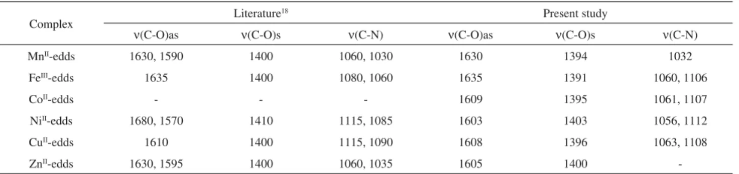
The spectroscopic data indicated that edds forms stable, hexadentate complexes with most transition metals. The infrared analysis of the crystals obtained by recrystalliza-tion (Table 1) showed the characteristic displacement of the asymmetric carboxylate stretching to lower frequencies
for the complexes, as previously established,17,18 implying
that this group is involved in the coordination to the metal. UV-visible measurements of aqueous solutions of
[M(H2O)6]2+ and M-edds (M = Co, Ni, Cu; [M] = [edds] =
5 mol L-1) were also consistent with the formation of
complexes, with the displacement of the absorption maxima
( abs) to 492 nm (Co-edds19), 369 and 583 nm (Ni-edds20)
and 670 nm (Cu-edds21). Upon oxidation to FeIII during
complex formation, Fe-edds solutions turn
yellowish-brown and display a small shoulder at ca. 490 nm.
Pro-oxidant activity
The DHR probe (non-fluorescent in its reduced form) undergoes unspecific oxidation and develops fluorescence
Table 1. Main infrared absorptions (cm-1) for metal-edds complexes
Complex Literature
18 Present study
(C-O)as (C-O)s (C-N) (C-O)as (C-O)s (C-N)
MnII-edds 1630, 1590 1400 1060, 1030 1630 1394 1032
FeIII-edds 1635 1400 1080, 1060 1635 1391 1060, 1106
CoII-edds - - - 1609 1395 1061, 1107
NiII-edds 1680, 1570 1410 1115, 1085 1603 1403 1056, 1112
CuII-edds 1610 1400 1115, 1090 1608 1396 1063, 1108
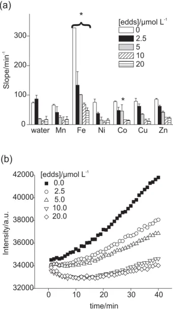
-in an oxidant concentration-dependent manner. In our experimental setting, the slope of a kinetic fluorescence curve is directly dependent on the amount of free radicals produced during the metal-catalyzed oxidation of ascorbic
acid in the reaction media. Metal ions, such as Fe3+and
Cu2+, are known to catalyze this reaction22,23 when in their
free form, but a plethora of chelators might decrease their activity provided that they can bind to the metal through
available coordination sites.24 Thus, we aimed to verify
whether edds might behave as an antioxidant in this metal-promoted oxidation by removing the metal through the formation of a stable chelate.
We observed that, except for Fe, all tested metals be-haved similarly to the control with respect to the decrease
in the rate of ascorbate oxidation versus [edds] (Figure 1).
Traces of iron present in the buffer may be responsible for the apparent “redox activity” of water alone, which subsided with increasing [edds]. However, this should not exclude the occurrence of important redox reactions promoted by other cations, since the experimental setting
was optimized for clinical evaluation of redox-active iron.15
As expected, the Zn-edds complex did not show any redox activity.
“Free” iron displays high redox activity (Figure 1). Interestingly, iron-catalyzed ascorbate oxidation was not totally suppressed, even at a four-fold molar excess of edds. Two important conclusions might be drawn from this
ob-servation. First, edds is not an antioxidant per se. Second,
iron chelates such as Fe-edta, which present at least one free coordination site, are known to promote the formation
of reactive oxygen species.24 Indeed, Fe-edta is an excellent
catalyst of ascorbate oxidation.25 Our observations indicate
that this is also the case for Fe-edds, since Fe3+ tends to
form less stable bonds with relatively soft bases like the N atoms of edds, which implies that these groups are read-ily replaced by solvent molecules, the metal remaining
redox-active. On the other hand, Kovaleva et al.18identified
a peak at 1725 cm-1 attributed to the (C=O)
as of a free
car-boxylic group in Fe-edds. They suggested that the nitrogen atoms, three oxygen atoms from the carboxyl groups and
one water molecule form the coordination sphere of Fe3+
in Fe-edds. We did not identify this particular stretching frequency in our experiments (Table 1). Kovaleva’s model also explains the persistence of available positions (the water molecule) through which ascorbate could interact with Fe and initiate a cascade of oxidation. It should be noted that complete blocking of all available coordination
sites, in order to prevent unwanted redox side-reactions in
vivo,is one of the requisites for molecules employed in
iron chelation therapy.15
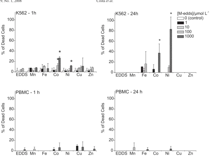
Cell studies
Initially, we compared the effect of the incubation time of M-edds complexes on K562 and PBMC viability (Figure 2). The tumor line was less resistant to the toxic effects of some of the complexes (Fe-edds, Co-edds and Ni-edds) after 24 h when compared to PBMC. In fact, virtually no toxic effects were observed for non-tumoral ex-vivo cells even at high M-edds concentrations and prolonged
expo-sure. Edds is known to capture Zn2+ in biological media,2
therefore it can compete with enzymes for this metal and induce loss of enzymatic functions. Curiously, free edds did
not show high toxicity to the cells (Figure 2), indicating that the toxicity of M-edds complexes, when present, depends mainly on the nature of the metal being transported into the cell. Iron, cobalt and nickel complexes were the most toxic to K562 cells, albeit at the relatively high concentrations of
100 and 1000 mol L-1. This observation supports previous
claims that edds is a useful chelator to reduce metal
con-tamination risks,1 but at the same time indicates that some
of the M-edds complexes could be effective metallodrugs for tumor chemotherapy.
Since the toxicity of metallodrugs in vivo is critically
determined by the outcome of their interactions with plasma
proteins,26 and serum deprivation may stress cells leading
to higher susceptibility to toxicants and apoptosis, we conducted a set of experiments to assess the toxicity of M-edds complexes to K562 in the presence and absence of serum (Figure 3).
As anticipated, all control groups displayed higher mortality when treated with PBS only (compare right up-per panel of Figure 2 with Figure 3). Also, Zn-edds was more toxic under this condition than in the presence of the culture medium. Interestingly, Fe-edds displayed a greatly
increased rate of mortality in the absence of serum. Pro-teins such as albumin have extensive antioxidative effects in vivo, and we previously noted (Figure 1) that Fe-edds is
Figure 2. Cytotoxicity of M-edds complexes to K562 and PBMC cells after 1 and 24 h incubation (mean ± S.D.) in complete RPMI medium. Asterisks indicate significant differences from the control according to analysis of variance (ANOVA, p < 0.05) and comparison of the groups using Fisher’s LSD test (Protected t-Test). The Trypan Blue exclusion test was used to evaluate cell viability.
octanol-water partition (data not shown). A previous report accounted for the facilitated transport of aluminum citrate by MCT1, suggesting that the transporter would recognize
a free citrate carboxylate.33 However, it should be noted
that potential metal ligands, such as acetoacetate, which lack this free carboxylic acid motif, are recognized MCT
substrates.32 To our knowledge, the present work is the first
report of complexes lacking a free carboxylic function in the chelating agent being transported by MCTs.
The mechanisms by which metal ions cause cell cycle arrest are not fully understood, since cell cycle regulation is a complex process with several steps, which is prone to interference by contaminants. Induction of apoptosis in tumor cells is an important strategy for the development of improved platinum-based metallodrugs. However, the biological response to these drugs seems to be dependent
on both the cell line and the metal complex structure.34
For instance, vanadium compounds have been shown to
induce both apoptosis and S phase arrest35 depending on
the experimental model. Exogenous chelators also interfere with the cell cycle, the best known examples coming from studies of siderophores employed in iron chelation therapy, which may exert useful antitumoral effects by deactivating the metalloenzyme ribonucleotide reductase and inducing
G1/S arrest.36
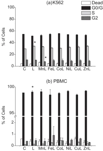
As with the previous results (Figure 2), we observed that cell mortality (apoptosis) was higher for K562 cells irrespective of the treatment (Figure 6 and Electronic Supplementary Information), which indicates that at least some tumor cell lines are less capable than normal cells of coping with the stress presented by the metal complexes. an effective pro-oxidant of ascorbate (and, presumably, of
other biological substrates). Therefore, we hypothesized that the observed decreased viability of cells treated with Fe-edds in PBS might be the result of an oxidative aggres-sion towards the cells, as observed by other researchers for
other iron complexes.27,28 To test this point, we conducted a
comparative study on the viability of cells challenged with Fe-edds, using cells which had been treated or not treated with the antioxidant Trolox in the absence of serum (Figure 4). The presence of the antioxidant suppressed toxicity even at the highest concentrations of Fe-edds.
To gain some insight into the mechanisms of biologi-cal action, the cell toxicity studies in the absence of serum were repeated in the presence and absence of 4-HCA, a known inhibitor of membrane monocarboxylate transporter
(MCT) proteins.29-31 This family of proteins is involved in
the transport of short chain carboxylates (pyruvate, lactate), ketone bodies and hormones through several biological
interfaces.32 This test was not performed with cells grown in
complete RPMI due to the fact that, as previously observed, cell toxicity in this case was difficult to observe.
We observed (Figure 5) that 4-HCA-treated cells re-sisted Fe- and Co-edds toxicity. Ni-edds did not show a similar recovery, probably because the concentration was too high to verify the role of MCT blocking. This indicates that membrane monocarboxylate transporters may be required for the internalization of these metal complexes. Our results do not rule out other internalization routes such as passive diffusion. However, for the anionic M-edds complexes, passive diffusion through cell membranes is unlikely, since none of these complexes showed a detectable
Figure 4. Effect of 100 mol L-1 Trolox on K562 cell viability after treat-ment with 100 mol L-1 and 1000 mol L-1 (mean ± S.D.) of Fe-edds in PBS. Asterisks indicate significant differences from the control according to analysis of variance (ANOVA, p < 0.05) and comparison of the groups using Fisher’s LSD test (Protected t-Test). The Trypan Blue exclusion test was used to evaluate cell viability.
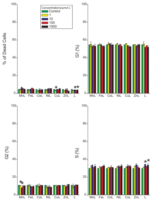
Figure 6. Cell cycle arrest induced by different M-edds complexes of K562 and PBMC cells after 1h treatments (mean ± S.D.) in complete RPMI medium. C = control; L = edds ; [edds] = [M-edds] = 10 mol L-1. Asterisks indicate significant differences from the control according to analysis of variance (ANOVA, p < 0.05) and comparison of the groups using Fisher’s LSD test (Protected t-Test).
In both K562 and PBMC, the free acid edds induced a significant phase-specific arrest (G0/G1 in PBMC and S in K562). K562 cells treated with Mn-edds showed a decreased population in the G2 phase as compared to the control, which may be due to the slightly increased number of dead cells and/or cells in other phases.
Leukemic cell lines such as K562 are susceptible to a number of inducers of apoptosis, and a fraction of terminally differentiated cells undergo apoptosis during
maturation as well.37 Our data corroborate these findings,
in the sense that K562 cells were more sensitive to M-edds toxicity than PBMC cells in all cases.
The G1 checkpoint controls the progression through the cell cycle by checking for cell size, presence of growth factors, presence of nutrients and DNA damage. Progres-sion to the S phase occurs after proteins of the Rb family are phosphorylated and unbound from E2F transcription factors in the DNA molecule. Cells with damaged DNA are prevented from progressing to the S phase by the p53
protein which, among other roles, blocks Rb
phosphory-lation.38 Our results suggest that edds alone, and possibly
Mn-edds, might either induce direct DNA damage or block p53 activity in K562 leukemic cells. Further experiments,
e.g. an evaluation of nucleotide excision fragments or
im-paired expression of the p53 protein, and the role of edds in relation to zinc-finger transcription factors, should be performed in order to uncover the precise mechanism of action of these metal complexes.
Conclusions
Biodegradable ethylenediamminedisuccinic acid (edds) has been proposed as an environmentally friendly alterna-tive to persistent edta for heavy metal remediation and decontamination. The biological activities studied here
suggest that this acid is not toxic per se to human cells,
and the toxicity of M-edds complexes is dependent upon the nature of M. The toxicity of Fe-edds may, in part, be due to its ability to induce redox reactions, although other models of oxidative damage (lipid peroxidation, DNA damage) should be investigated. Monocarboxylate trans-porters may be involved in the internalization of M-edds complexes. In our study, M-edds complexes also displayed some anti-proliferative activity, which should be explored further for the development of new metallodrugs for cancer chemotherapy.
Acknowledgements
This research was funded in part by CNPq (Brazilian Council for Scientific and Technological Development) and FAPESP (São Paulo State Council for Scientific Research). Octel Performance Chemicals kindly provided us with a sample of edds. Dr. Heraldo Possolo de Souza (School of Medicine, University of São Paulo) kindly permitted the use of the fluorescence microplate reader.
Supplementary Information
Cell cycle arrest induced by different M-edds complexes of K562 and PBMC cells after 1 h treatments (mean ± S.D.) in complete RPMI medium. C = control; L = edds ;
[edds] = [M-edds] = 1, 100 and 1000 mol L-1. Supplementary
data are available free of charge at http://jbcs.sbq.org.br as PDF file.
References
1. Schowanek, D.; Feijtel, T. C. J.; Perkins, C.M.; Hartman, F. A.;
2. Bucheli-Witschel, M.; Egli, T.; FEMS Microbiol. Rev.2001,
25, 69.
3. Hauser, L.; Tandy, S.; Schulin, R.; Nowack, B.; Environ. Sci.
Technol.2005,39, 6819.
4. Kos, B.; Lestan, D.; Environ. Pollut.2004,132, 333.
5. Kos, B.; Lestan, D.; Environ. Sci. Technol.2003,37, 624.
6. Finzgar, N.; Kos, B.; Lestan, D.; Chemosphere2004,57, 655.
7. Meers, E.; Ruttens, A.; Hopgood, M. J.; Samson, D.; Tack, F.
M. G.; Chemosphere2005,58, 1011.
8. Luo, C. L.; Shen, Z.G.; Li, X. D.; Chemosphere2005,59, 1.
9. Orama, M.; Hyvonen, H.; Saarinen, H.; Aksela, R.; J. Chem.
Soc., Dalton Trans.2002, 4644.
10. Jones, P. W.; Williams, D.R.; Appl. Radiat. Isot.2001,54,
587.
11. Jaworska, J. S.; Schowanek, D.; Feijtel, T. C. J.; Chemosphere
1999,38, 3597.
12. Toney, J. H.; Koh, M. L.; JALA2006,11, 30.
13. Vogel, J. U.; Michaelis, M.; Neyts, J.; Blaheta, R. A.; Snoeck, R.; Andrei, G.; De Clercq, E.; Rabenau, H. F.; Kreuter, J.; Cinatl,
J.; Doerr, H.W.; Antiviral Res.2002,55, 179.
14. Van Devivere, P. C.; Saveyn, H.; Verstraete, W.; Feijtel, T. C.
J.; Schowanek, D. R.; Environ. Sci. Technol.2001,35, 1765.
15. Esposito, B. P.; Breuer, W.; Sirankapracha, P.; Pootrakul, P.;
Hershko, C.; Cabantchik, Z. I.; Blood2003,102, 2670.
16. Martell, A. E.; Smith, R. M.; Critical Stability Constants – vol.
1,Plenum Press: New York, 1974.
17. Nakamoto, K.; Infrared and Raman Spectra of Inorganic and
Coordination Compounds Part B: Applications in
Coordina-tion, Organometallic, and Bioinorganic Chemistry,
Wiley-Interscience: New York, 1986.
18. Kovaleva, I.; Mitrofanova, N.; Martynenko, L.; Russ. J. Inorg.
Chem.1992,37, 41.
19. Vasilev, V.; Zaitseva, G.; Sapronova, N.; Russ. J. Inorg. Chem.
1991,36, 1225.
20. Samsonov, A.; Gorelov, I.; Russ. J. Inorg. Chem.1972,17,
1148.
21. Babich, V.; Gorelov, I.; J. Anal. Chem. USSR1971,26, 1736.
22. Halliwell, B.; Gutteridge, J.; Free radicals in biology and
medicine, 3rd. ed., Oxford Science Publishers: Oxford, 1999.
23. Halliwell, B.; Gutteridge, Meth. Enzymol.1990,180, 1.
24. Graf, E.; Mahoney, J. R.; Bryant, R. G.; Eaton, J. W.; J. Biol.
Chem.1984,259, 3620.
25. Buettner, G.; Jurkiewicz, B. In: Analysis of Free Radicals in
Biological Systems; Favier, A., ed., Birkhäuser Verlag: Basel,
1995.
26. Esposito, B. P.; Najjar, R. Coord. Chem. Rev.2002,232, 137.
27. Yao, D. C.; Shi, W. B.; Gou, Y. L.; Zhou, X. R.; Aw, T. Y.; Zhou,
Y. K.; Liu, Z. X.; Free Rad. Biol. Med.2005,39, 1385.
28. Zodl, B.; Zeiner, M.; Paukovits, P.; Steffan, I.; Marktl, W.;
Ekmekcioglu, C.; Microchem. J.2005,79, 393.
29. Bui, B. V.; Kalloniatis, M.; Vingrys, A. J.; Invest. Ophth. Vis.
Sci.2004,45, 584.
30. Lin, R. Y.; Vera, J. C.; Chaganti, R. S. K.; Golde, D. W.; J. Biol.
Chem.1998,273, 28959.
31. Wang, X.; Levi, A. J.; Halestrap, A. P.; Am. J. Physiol.-Heart
C.1996,39, H476.
32. Galic, S.; Schneider, H. P.; Broer, A.; Deitmer, J. W.; Broer, S.;
Biochem. J.2003,376, 413.
33. Yokel, R. A.; Allen, D. D.; Ackley, D. C.; J. Inorg. Biochem.
1999,76, 127.
34. Valentini, A.; Pucci, D.; Crispini, A.; Federici, G.; Bernardini, S.;Chem.-Biol. Interact.2006,161, 241.
35. Leonard, S. S.; Bower, J. J.; Shi, X. L.; Mol. Cell. Biochem.
2004,255, 3.
36. Kalinowski, D. S.; Richardson, D. R.; Pharmacol. Rev.2005,
57, 547.
37. Tsiftsoglou, A. S.; Pappas, I. S.; Vizirianakis, I. S.; Pharmacol.
Ther.2003,100, 257.
38. Macaluso, M.; Montanari, M.; Giordano, A.; Oncogene2006,
25, 5263.
Received: December 21, 2006
Published on the web: January 28, 2008
S
u
p
p
le
m
e
nta
ry
Inf
o
rm
a
ti
o
n
J. Braz. Chem. Soc., Vol. 19, No. 1, S1-S2, 2008. Printed in Brazil - ©2008 Sociedade Brasileira de Química 0103 - 5053 $6.00+0.00
*e-mail: [email protected]
Biological Activity of Metal-edds (ethylenediaminedisuccinate)
Complexes in K562 and PBMC Cells
Natália J. S. Costa,
aSzulim B. Zyngier,
bCíntia R. Bombardieri,
bJuliana S. Kuribayashi,
bMaristela M. de Camargo
band Breno P. Espósito
a,*aInstituto de Química, Universidade de São Paulo, Av. Lineu Prestes 748 sala 1265, 05508-000, São Paulo-SP, Brazil
bInstituto de Ciências Biomédicas, Universidade de São Paulo, Av. Lineu Prestes 2415, 05508-000, São Paulo-SP, Brazil
Figure S1. K562, 1h – Cell cycle arrest induced by different M-edds complexes on K562 cells after 1-h treatments (mean ± S.D.) in complete RPMI medium. L = edds ; [edds] = [M-edds] = 1 - 1000 µmol L-1. Asterisks indicate significant differences from the control according to analysis of variance (ANOVA, p < 0.05) and comparison of the groups using Fisher’s LSD test (Protected t-Test).
Biological Activity of Metal-edds (ethylenediaminedisuccinate) J. Braz. Chem. Soc. S2
Figure S2. PBMC, 1h – Cell cycle arrest induced by different M-edds complexes on PBMC cells after 1-h treatments (mean ± S.D.) in complete RPMI medium. L = edds ; [edds] = [M-edds] = 1 - 1000 µmol L-1. Asterisks indicate significant differences from the control according to analysis of variance (ANOVA, p < 0.05) and comparison of the groups using Fisher’s LSD test (Protected t-Test).
1
2
3
4
5
6
7
8
9
10
11
12
13
14
15
16
17
18
19
20
21
22
23
24
25
26
27
28
29
30
31
32
33
34
35
36
37
38
39
40
41
42
43
44
45
46
47
48
49
50
51
52
53
54
55
56
57
58
59
60
61
62
63
64
65
66
67
68
69
70
71
72
73
74
75
76
77
78
79
80
81
82
83
84
85
86
87
88
89
90
91
92
93
94
95
96
97
98
99
100
101
102