TRAIL/DR5 Death Receptor Dependent Apoptosis
Richa Tiwary1., Weiping Yu1., Jing Li1
, Sook-Kyung Park1, Bob G. Sanders1, Kimberly Kline2*
1School of Biological Sciences/C0900, University of Texas at Austin, Austin, Texas, United States of America,2Department of Nutritional Sciences/A2703, University of Texas at Austin, Austin, Texas, United States of America
Abstract
Background:a-TEA (RRR-a-tocopherol ether-linked acetic acid analog), a derivative of RRR-a-tocopherol (vitamin E) exhibits anticancer actionsin vitroandin vivoin variety of cancer types. The objective of this study was to obtain additional insights
into the mechanisms involved ina-TEA induced apoptosis in human breast cancer cells.
Methodology/Principal Findings:a-TEA induces endoplasmic reticulum (ER) stress as indicated by increased expression of CCAAT/enhancer binding protein homologous protein (CHOP) as well as by enhanced expression or activation of specific markers of ER stress such as glucose regulated protein (GRP78), phosphorylated alpha subunit of eukaryotic initiation factor 2 (peIF-2a), and spliced XBP-1 mRNA. Knockdown studies using siRNAs to TRAIL, DR5, JNK and CHOP as well as chemical inhibitors of ER stress and caspase-8 showed that: i)a-TEA activation of DR5/caspase-8 induces an ER stress mediated JNK/ CHOP/DR5 positive amplification loop; ii)a-TEA downregulation of c-FLIP (L) protein levels is mediated by JNK/CHOP/DR5 loop via a JNK dependent Itch E3 ligase ubiquitination that further serves to enhance the JNK/CHOP/DR5 amplification loop by preventing c-FLIP’s inhibition of caspase-8; and (iii) a-TEA downregulation of Bcl-2 is mediated by the ER stress dependent JNK/CHOP/DR5 signaling.
Conclusion:Taken together, ER stress plays an important role ina-TEA induced apoptosis by enhancing DR5/caspase-8 pro-apoptotic signaling and suppressing anti-pro-apoptotic factors c-FLIP and Bcl-2 via ER stress mediated JNK/CHOP/DR5/caspase-8 signaling.
Citation:Tiwary R, Yu W, Li J, Park S-K, Sanders BG, et al. (2010) Role of Endoplasmic Reticulum Stress ina-TEA Mediated TRAIL/DR5 Death Receptor Dependent Apoptosis. PLoS ONE 5(7): e11865. doi:10.1371/journal.pone.0011865
Editor:Mikhail V. Blagosklonny, Roswell Park Cancer Institute, United States of America
ReceivedMay 24, 2010;AcceptedJuly 7, 2010;PublishedJuly 29, 2010
Copyright:ß2010 Tiwary et al. This is an open-access article distributed under the terms of the Creative Commons Attribution License, which permits unrestricted use, distribution, and reproduction in any medium, provided the original author and source are credited.
Funding:The Clayton Foundation for Research, the National Institute of Environmental Health Sciences Center Grant ES007784, the Center for Molecular and Cellular Toxicology at the University of Texas at Austin and a NIEHS/NIH Toxicology Training Grant T32 ES07247. The funders had no role in study design, data collection and analysis, decision to publish, or preparation of the manuscript.
Competing Interests:US and international patents ona-TEA are held by the Research Development Foundation. Kimberly Kline and Bob G. Sanders are listed as inventors. No commercial applications or financial gain have been realized.
* E-mail: [email protected]
.These authors contributed equally to this work.
Introduction
Targeting cell surface death receptors, especially tumor necrosis factor-related apoptosis-inducing ligand (TRAIL/Apo2L) binding receptors, holds promise for cancer treatment [1,2]. TRAIL selectively induces apoptosis in a wide variety of cancer cells with little or no toxicity towards normal cells [1,2]. Thus, agents that can enhance TRAIL death receptor (R/DR4 or TRAIL-R2/DR5) signaling or sensitize TRAIL resistant cells to TRAIL induced apoptosis are of interest [2,3]. TRAIL/DR4/DR5 apoptotic signaling includes: interaction of TRAIL with DR4 or DR5, receptor clustering, recruitment of the adaptor molecule FADD, and activation of initiator caspases-8 or -10, leading to cleavage of downstream effector caspases (mitochondrial-indepen-dent apoptosis) or cleavage of Bid, a pro-apoptotic Bcl-2 family member, leading to mitochondrial-dependent apoptosis [4].
Evading apoptosis is a hallmark of cancer [5]. One way tumor cells can escape death signals is by expression of anti-apoptotic pro-survival proteins [6]. Therefore, targeting anti-apoptotic proteins also holds promise for killing cancer cells and sensitizing
them to different therapeutics [7]. c-FLIP (cellular FADD-like IL-1a-converting enzyme inhibitory protein), is a death effector domain containing protein that regulates extrinsic death receptor signaling from the tumor necrosis factor-a(TNF-a) family of cell surface death receptors, including DR4/DR5, Fas (CD95/APO-1), and TNFR [8]. c-FLIP is a catalytically inactive caspase-8/10 homolog and typically functions as a caspase-8 inhibitor resulting in chemotherapeutic drug resistance [8].
suppression of c-FLIP-L, survivin and phospho-Akt (pAkt), leading to death receptor mediated caspase-8 activation and mitochondria dependent apoptosis [17,18,19,20,21,22,23].
These data are the first to show thata-TEA induces ER stress dependent increases in death mediators JNK/CHOP/DR5 and decreases in survival mediators c-FLIP-L and Bcl-2 in human breast cancer cells. These ER stress mediated events function downstream of a-TEA triggered TRAIL/DR5/caspase-8 signal-ing, leading to up-regulation of JNK, CHOP and DR5 and downregulation of c-FLIP and Bcl-2.
Materials and Methods
Chemicals
a-TEA was made in house as previously described [9]. ER stress inhibitor salubrinal was purchased from Calbiochem (La Jolla, CA). Caspase-8 inhibitor Z-IETD-FMK was purchased from BioVision (Mountain View, CA).
Cell Culture
MDA-MB-231 estrogen-receptor negative human breast cancer cells were purchased from the American Type Culture Collection (Manassas, VA). MCF-7 estrogen-responsive human breast cancer cells were originally provided by Dr. Suzanne Fuqua (Baylor College of Medicine, Houston, TX). Both cell lines were cultured in MEM media with 10% FBS. For experiments, FBS was reduced to 2% and cells were allowed to attach overnight before treatments. a-TEA (40 mM) was dissolved in ethanol as stock solution. Equivalent level of ethanol (0.1%) was used as vehicle control (VEH) fora-TEA treatment (40mM).
Quantification of apoptosis
Apoptosis was quantified by Annexin V-FITC/PI assay following the manufacturer’s instructions (Invitrogen).
Western Blot Analyses
Whole cell protein lysates were prepared and western blot analyses were conducted as described previously [24]. Antibodies to the following proteins were used: poly (ADP-ribose) polymerase (PARP), c-FLIP, CHOP, GRP-78, Bcl-2, total JNK, TRAIL and phospho-JNK (pJNK) (Santa Cruz Biotechnology, Santa Cruz, CA) and Bid (Pharmigen, Rockville, MD), phospho-eIF-2a (peIF-2a), eIF-2a (eIF-2a), caspase-8, caspase-9, DR5 and glyceraldehyde-3-phosphate dehydrogenase (GAPDH) (Cell Sig-naling Technology, Beverly, MA).
RT-PCR detection of DR5, Bcl-2 and XBP-1 mRNA expression
Total RNA was extracted using RNA isolation kit (Qiagen Inc. Valencia, CA). Semi-quantitative analyses were conducted to detect DR5, Bcl-2 and XBP-1mRNA form by reverse transcriptase -polymerase chain reaction (RT-PCR) using the housekeeping gene b-actin as control. 5mg total RNA was reverse transcribed to cDNA
using 1ml Superscript RTase (250 U, Invitrogen) following the
manufacture’s instructions. 1ml cDNA was used per PCR reaction
with 15ml Taq PCR Master Mix Kit (Qiagen Inc) plus 10mM
oligonucleotide primer pairs (Invitrogen). Primer sequences are available upon request.
RNA Interference
A scrambled RNA duplex purchased from Ambion (Austin, TX) that does not target any of the known genes was used as the nonspecific negative control for RNAi (referred to as control
siRNA). Transfection of MCF-7 or MDA-MB-231 cells with siRNA to DR5, TRAIL, CHOP, JNK, Itch or control (Ambion, Austin, TX) was performed in 100 mm cell culture dishes at a density of 26106cells/dish using Lipofectamine 2000 (Invitrogen) and siRNA duplex, resulting in a final siRNA concentration of 30 nM following the company’s instructions. After one day exposure to transfection conditions, the cells were re-cultured in 100 mm dish at 26106 cells/dish and incubated for one day followed by treatments.
Chromatin immunoprecipiation (ChIP) assay
MDA-MB-231 cells were treated with 40mM of a-TEA or
vehicle (0.1% ethanol) for 15 hours. Protein to DNA cross-linking was conducted by adding 1% formaldehyde to the cell culture medium for 12 min followed by the addition of glycine (0.125M) to stop the cross-linking. Cells were collected for ChIP assay as described by Nelsonet al.[25]. The CHOP antibody used for the Western blot analysis was used for immunoprecipitation. Normal mouse IgG1purchased from Santa Cruz Biotechnology was used as an isotype control. Polymerase chain reaction (PCR) was conducted using the primers as described by Adbelrahimet al.[26] to detect CHOP binding sites in the DR5 promoter region.
Ectopic Expression of c-FLIP (L)
Transient transfection of MCF-7 cells with wildtype His-tagged FLIP expression construct, pcDNA3.1-His-c-FLIP (L), which was kindly provided by Dr. John C. Reed (The Burnham Inst. La Jolla, CA) [27] was performed following the procedure published before [23]. Transfected cells were cultured overnight before a-TEA treatment.
Statistical Analyses
Apoptosis data were analyzed using a two-tailed studentt-test to determine statistical differences among treatments. Differences were considered statistically significant atp,0.05.
Results
a-TEA induces TRAIL/DR5 and CHOP dependent apoptosis
a-TEA mediated increases in DR5 and CHOP protein levels are JNK dependent and CHOP influences JNK phosphorylation status
Since prior studies showed that JNK activation is a critical component ina-TEA induced apoptosis and other studies have shown that JNK is involved in upregulation of CHOP protein expression [29] the possible involvement of JNK in a-TEA mediated increases in CHOP/DR5 protein expression was investigated in MCF-7 cells. siRNA to JNK reduced total JNK and reduced the ability of a-TEA to increase phosphorylated JNK2/1 (pJNK), increase CHOP and DR5 (L/S) protein levels and to induce apoptosis as measured by PARP cleavage (Fig 2A), suggesting that JNK is a critical contributor to increases in CHOP and DR5 (L/S) protein level. Interestingly, siRNA to CHOP reduced a-TEA mediated increases in
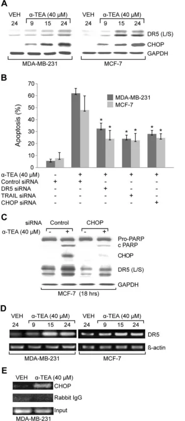
Figure 1. a-TEA induces TRAIL/DR5 and CHOP dependent
apoptosis.A. MDA-MB-231 and MCF-7 cells were treated with 40mM a-TEA for 9, 15, and 24 hrs. Western blot analyses were performed to evaluate DR5 (L/S) and CHOP protein levels using GAPDH as loading control. B. MDA-MB-231 and MCF-7 cells were transiently transfected with siRNAs to DR5, TRAIL or CHOP using non-specific siRNA as negative control followed by treatment with 40mMa-TEA for 18 hrs. Apoptosis was determined by Annexin V/FACS. C. Samples (from B) were analyzed by western blot for PARP cleavage, CHOP and DR5 (L/S) protein levels
using GAPDH as loading control. D. mRNA levels of DR5 were determined by RT-PCR. E. The binding activity of CHOP on DR5 promoter was determined by CHIP assay. Data from A, C, D and E are representative of two or more independent experiments. Data from B are the mean6S.D. of three independent experiments. * p,0.05 = sig-nificantly different from control siRNA determined byt-test.
doi:10.1371/journal.pone.0011865.g001
Figure 2.a-TEA induced increased CHOP and DR5 (L/S) protein
levels are JNK dependent.In turn, CHOP regulates pJNK2/1 protein levels. A. MCF-7 cells were transiently transfected with siRNA to JNK using non-specific siRNA as negative control followed by treatment with 40mMa-TEA for 18 hrs. Western blot analyses were performed to evaluate PARP cleavage, pJNK2/1, total JNK2/1, CHOP, and DR5 (L/S) protein levels using GAPDH as loading control. B. MCF-7 cells were transiently transfected with siRNA to CHOP using non-specific siRNA as negative control followed by treatment with 40mMa-TEA for 18 hrs. Western blot analyses were performed to evaluate pJNK2/1 protein levels using GAPDH as loading control. Data from A and B are representative of two or more independent experiments.
pJNK2/1 protein levels (Fig 2B), suggesting that a-TEA mediated increases in JNK phosphorylation are dependent on CHOP expression.
Endoplasmic reticulum stress involvement ina-TEA induced apoptosis contributes to JNK/CHOP/DR5 upregulation
a-TEA treatment of MDA-MB-231 and MCF-7 cells increased levels of endoplasmic reticulum stress (ER-stress) indicators: phosphorylated eukaryotic initiation factor 2-alpha (peIF-2a) and glucose-regulated protein of 78kDA (GRP78) also known as BiP (Fig 3A), as well as spliced mRNA forms of X-box binding protein-1 (XBP-protein-1) (Fig 3B) in a time-dependent manner, suggesting thata -TEA treatment induced ER stress in both cell lines. Furthermore, ER-stress inhibitor salubrinal significantly reduced the ability ofa -TEA to induce apoptosis detected by Annexin V/FACS (Fig 3C). Since JNK/CHOP/DR5 pathway can be activated in response to ER stress [30], studies were conducted to examine if a-TEA mediated JNK/CHOP/DR5 (L/S) signaling events via ER stress. Data show that ER-stress inhibitor salubrinal significantly reduced the ability ofa-TEA to upregulate pJNK2/1, CHOP, and DR5 (L/S), as well as to induce PARP cleavage in both cell lines (Fig. 3D). Salubrinal significantly reduced the ability ofa-TEA to upregulate ER stress markers GRP78 and peIF2a, confirming the efficiency of ER-stress inhibitor. These data demonstrate thata -TEA induction of JNK/CHOP/DR5 and apoptosis is mediated via ER stress.
a-TEA induction of ER stress-dependent JNK/CHOP/DR5 signaling is mediated by TRAIL/DR5 pathway
Since both JNK and DR5 can be regulated by TRAIL/DR5 signaling [30,31], studies were conducted to determine ifa-TEA mediated ER stress events are downstream events of TRAIL/DR5 using siRNA knockdown procedures. MCF-7 and MDA-MB-231 cells transiently transfected with siRNAs to DR5 or TRAIL exhibited reduced levels of DR5 and TRAIL, respectively, and showed blockage ofa-TEA’s ability to produce PARP cleavage and to increase levels of pJNK2/1, CHOP, DR5 (L/S), as well as blockeda-TEA’s ability to upregulate ER stress markers GRP78 and peIF-2a (Fig 4 A, B & C), demonstrating that a-TEA induction of ER stress-dependent JNK/CHOP/DR5 signaling is mediated by TRAIL/DR5 pathway.
ER stress dependent JNK/CHOP/DR5 upregulation occurs both upstream and downstream of caspase-8 activation
In an effort to determine if ER stress proceeds or followsa-TEA activation of DR5 signaling, caspase-8 inhibitor (Z-IETD-FMK) was employed. Data show that caspase-8 inhibitor significantly reduceda-TEA abilities to induce apoptosis (Fig 5A), and cleavage of Bid, caspase-9 and PARP, as well as increase protein levels of pJNK2/1, CHOP, DR5, and ER stress markers GRP78 and peIF-2a in both cell types (Fig 5B). These data suggest that a-TEA activation of caspase-8 is upstream of ER-stress mediated JNK/ CHOP/DR5 signaling. siRNA knockdown of JNK, CHOP and DR5 in MCF-7 cells reduced the ability of a-TEA to cleave caspases 8 and 9 (Fig 5 C), suggesting that not only DR5 but also JNK and CHOP are involved in activation of caspases-8 and -9. Furthermore, treatment of MDA-MB-231 and MCF-7 cells with ER inhibitor salubrinal blocked a-TEA’s ability to induce caspases-8 and -9 cleavage (Fig 5 D), providing additional evidence for caspase 8 acting prior to and aftera-TEA induced ER stress.
Figure 3. ER stress is involved ina-TEA induced apoptosis and
a-TEA down regulation of c-FLIP (L) protein levels is in part, mediated by caspase-8/ER stress/JNK/CHOP/DR5 signaling via Itch E3 ligase ubiquitination
a-TEA downregulates c-FLIP (L) protein levels in both MDA-MB-231 and MCF-7 breast cancer cells in a time dependent manner (Fig 6A). MCF-7 cells transiently transfected with siRNA to CHOP, JNK or DR5, and both MDA-MB-231 and MCF-7 cells cultured with ER-stress inhibitor salubrinal or caspase 8 inhibitor Z-IETD-FMK prevented the ability ofa-TEA to decrease c-FLIP (L) protein levels (Fig 6B and C), providing evidence that a-TEA downregulation of c-FLIP is mediated by caspase-8/ER stress/
JNK/CHOP/DR5 signaling. Since c-FLIP has been reported to be degraded via JNK-dependent E3 ubiquitin ligase Itch [32], studies were conducted to determine if Itch E3 ligase mediated ubiquitina-tion was involved ina-TEA down-regulation of c-FLIP. Knock-down of Itch E3 ligase with siRNA to Itch reduceda-TEA’s ability to downregulate c-FLIP (L) protein levels in MCF-7 cells (Fig. 6D). siRNA to Itch significantly reduced the ability ofa-TEA to induce apoptosis in MDA-MB-231 and MCF-7 cells (Fig 6E). These data suggest that a-TEA induced downregulation of c-FLIP (L) is mediated by a caspase-8/ER stress/JNK/CHOP/DR5 pathway via Itch ligase ubiquitination.
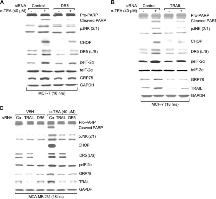
Figure 4. ER stress dependent JNK/CHOP/DR5 upregulation is mediated by TRAIL/DR5 pathway ina-TEA induced apoptosis.A & B.
MCF-7 cells were transiently transfected with siRNA to DR5 or TRAIL, using non-specific siRNA as negative control followed by treatment with 40mM a-TEA for 18 hrs. Western blot analyses were performed to evaluate PARP cleavage, pJNK2/1, CHOP, DR5 (L/S), peIF-2atotal eIF-2aand GRP78 protein levels, using GAPDH as loading control for (A & B) and TRAIL protein levels for siRNA knockdown efficiency (B). C. MDA-MB-231 cells were transiently transfected with siRNA to DR5 or TRAIL, using non-specific siRNA as negative controls followed by treatment with 40mMa-TEA for 18 hrs. Western blot analyses were performed to evaluate PARP cleavage, pJNK (2/1), CHOP, DR5 (L/S), peIF-2a, GRP78, and TRAIL protein levels, using GAPDH as loading control. Data from A, B and C are representative of at least 2 individual experiments.
a-TEA downregulation of c-FLIP mediates ER stress-dependent JNK/CHOP/DR5 signaling via activation of caspase-8
Since c-FLIP is an inhibitor of caspase-8 activation, we hypothesized thata-TEA downregulation of c-FLIP may contrib-ute to activation of caspase-8 and subsequent ER stress-dependent JNK/CHOP/DR5 signaling. To test this hypothesis, MCF-7 cells were transiently transfected with wildtype c-FLIP (L) plasmid or Itch siRNA followed by treatment witha-TEA (40mM) for 18 hrs.
Over-expression of c-FLIP enhanced c-FLIP (L) levels (Fig 6G) and siRNA to itch blocked a-TEA’s ability to reduce c-FLIP protein expression (Fig 6D). Both overexpression of c-FLIP (L) protein and restoration of c-FLIP protein expression by silencing itch ina-TEA treatment reduced the ability ofa-TEA to cleave PARP and caspase-8, and to increase protein levels of pJNK2/1, CHOP, DR5, and GRP 78 (Fig 6G and H). These data suggest thata-TEA’s ability to downregulate c-FLIP (L) is important for caspase-8 activation and subsequent activation of the ER stress/ JNK/CHOP/DR5 signaling.
a-TEA downregulation of Bcl-2 protein and mRNA expression is mediated by ER stress
a-TEA treatment of MDA-MB-231 and MCF-7 cells reduced both Bcl-2 protein and mRNA levels in a time dependent manner (Fig 6A and F). siRNA to JNK, CHOP, and DR5 (Fig 6B) as well as caspase-8 and ER stress inhibitors (Fig 6C) blocked the ability of a-TEA to decrease Bcl-2 protein expression. These data suggest that a-TEA induced down-regulation of Bcl-2 is mediated by caspase-8, ER stress, JNK, CHOP and DR5.
Discussion
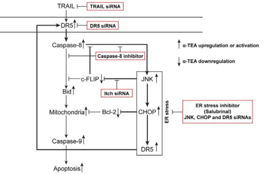
The underlying mechanisms of how a-TEA, a potential chemotherapeutic drug, enhances DR5 death signaling path-ways were investigated. For the first time, studies demonstrate that: (i) a-TEA induces ER stress dependent apoptosis, (ii) a -TEA induced ER stress regulates JNK/CHOP/DR5 signaling and is mediated by TRAIL/DR5/caspase-8, forming a positive loop, (iii) a-TEA downregulation of c-FLIP is mediated by ER stress-dependent JNK/CHOP/DR5 signaling via JNK activa-tion of Itch E3 ligase ubiquitinaactiva-tion and involved in activaactiva-tion of the ER-stress-dependent events via reducing the inhibitory effect of c-FLIP on caspase-8, and (iv)a-TEA downregulation of Bcl-2 protein levels is mediated by the ER stress-dependent events. Taken together, these data show that a-TEA induces TRAIL/DR5/caspase-8 dependent ER stress that triggers JNK/CHOP/DR5 signaling enhancing DR5/caspase-8 medi-ated mitochondria-dependent apoptotic signaling and produces a downregulation of two key anti-apoptotic factors; c-FLIP and Bcl-2. Based on these data, we provide a schematic diagram depicting a series of events including a positive-acting feedback loop, which is operative in a-TEA induced DR5 dependent apoptosis of human breast cancer cells in culture (Fig 7). DR5
signaling triggers caspase-8 activation, leading to two a-TEA initiated pathways: (i) a classic mitochondria-dependent apo-ptotic cascade initiated by cleavage of Bid and (ii) an ER-stress dependent activation of JNK/CHOP/DR5 sequence leading to further activation of caspase-8 and downregulation of anti-apoptotic factors; c-FLIP and Bcl-2. Importantly, down-regulation of c-FLIP serves to further enhance caspase-8 activation, which sustains the ER-stress/JNK/CHOP/DR5 amplifying loop.
TRAIL has emerged as a potent anticancer agent based on its ability to induce apoptosis in tumor cells but not in most normal cells [1,2,3]. Thus, targeting TRAIL/DR4/DR5 signaling pathways holds promise for inducing pro-apoptotic signaling in many cancer types while sparing normal cells and tissues [33,34]. Although considerable effort has been made in investigating the biological ramifications of TRAIL death receptor signaling, a complete understanding of the multiple pathways and events involved remain unclear. Typically, signaling pathways that initiate apoptosis have been broadly classified as (i) extrinsic death receptor initiated pathways or (ii) intrinsic pathways initiated by mitochondrial events [35]. These two pathways are not mutually exclusive, and JNK and caspase-8 have been observed to play central roles in both [4,36]. Our findings that TRAIL/DR5/casapse-8 triggers an ER stress dependent JNK/CHOP/DR5 amplifying loop and a JNK/c-FLIP/caspase-8 amplifying loop in a-TEA induced apoptosis provides new insights into TRAIL/DR5-mediated apoptotic signaling.
DR5 upregulation has been reported to be mediated by NF-kB in TRAIL- induced apoptosis in MCF-7 and MDA-MB-231 cells [37]. However, inhibition of NF-kB using IkB phosphorylation inhibitor BAY-11-7085 [38] enhancesa-TEA induced upregula-tion of DR5 and apoptosis in MCF-7 cells (data not shown), suggesting that NF-kB plays an anti-apoptotic role in a -TEA-induced apoptosis. Data reported here showing thata-TEA up-regulates DR5 via CHOP binding to the DR5 promoter is supported by published reports showing that CHOP can directly regulate DR5 expression through a CHOP binding site in the 59 -flanking region of the DR5 promoter [28,39].
JNK is a stress responsive kinase that has been reported to induce CHOP expression via an AP-1 binding site in the CHOP promoter [40,41]. JNK has been shown to be activated and is required fora-TEA-induced apoptosis of human breast, ovarian and prostate cancer cells [17,18,19,20]. Data presented here show that siRNA to JNK blocksa-TEA induced CHOP as well as DR5, indicating that CHOP/DR5 upregulation is mediated via JNK in a-TEA induced apoptosis. These data are in agreement with other studies showing that CHOP is an ER stress-regulated protein [42], JNK activation can contribute to CHOP expression during ER stress [43], and coupling of JNK/CHOP/DR5 are involved in ER stress-mediated apoptosis [29].
Present study show that a-TEA induces increased levels of peIF-2aand GRP78 proteins and spliced XBP-1, all of which are Figure 5. ER stress-dependent JNK/CHOP/DR5 upregulation is both upstream and downstream events of caspase-8.A. MDA-MB-231 and MCF-7 cells were cultured with caspase-8 inhibitor (Z-IETD-FMK) or DMSO vehicle control plus 40mMa-TEA for 18 hrs. Apoptosis was determined by Annexin V/FACS. B. Samples (from A) were analyzed by western blot to evaluate caspase-8 cleavage of Bid to tBid, cleaved caspase 9 and PARP, pJNK2/1, CHOP, DR5 (L/S), peIF-2aand GRP78 protein levels using GAPDH as loading control. C. MCF-7 cells were transiently transfected with siRNA to JNK, CHOP and DR5, using non-specific siRNA as negative control followed by treatment with 40mMa-TEA for 18 hrs. Western blot analyses were performed to evaluate caspase-8 and -9 cleavages, GAPDH served as lane controls. D. MDA-MB-231 and MCF-7 cells were cultured with ER-stress inhibitor salubrinal at 40mM plus 40mMa-TEA for 18 hrs. Western blot was conducted to evaluate caspase-8 and -9 cleavages, GAPDH served as lane controls. Data from B, C and D are representative of two or more individual experiments. Data from A are presented as the mean6S.D. of three independent experiments. *P,0.05 = caspase-8 inhibitor (Z-IETD-FMK) at 2mM plus 40mMa-TEA treatment is significantly different from 40mMa
-TEA determined byt-test.
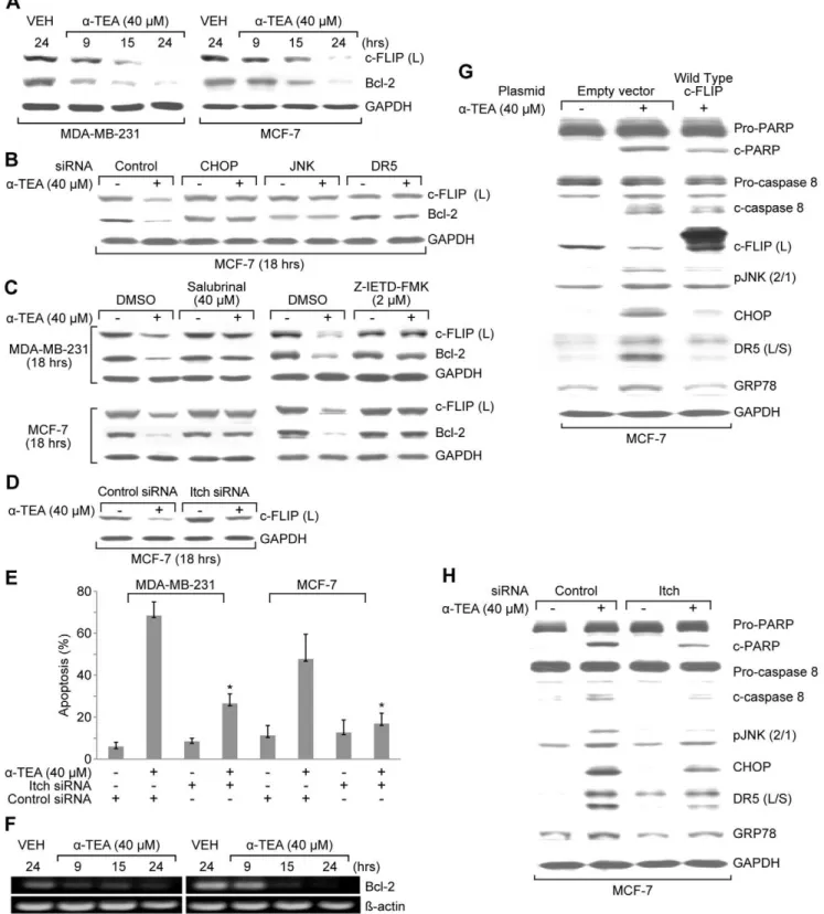
Figure 6. a-TEA decreased Bcl-2 and c-FLIP (L) protein levels via caspase-8 dependent ER stress-mediated JNK/CHOP/DR5
recognized as ER stress markers [44,45] and that salubrinal, a selective inhibitor of eIF-2a dephosphorylation (protects cells from ER stress) [46] blockeda-TEA induced apoptosis and the ability ofa-TEA to activate JNK/CHOP/DR5 and upregulate ER stress markers, confirming that ER stress is induced by a -TEA and is involved in both apoptosis and activation of JNK/ CHOP/DR5.
Present data show that a-TEA induction of the ER stress dependent JNK/CHOP/DR5 pathway is a downstream conse-quence of TRAIL/DR5 signaling. Furthermore, a caspase-8 inhibitor not only blocks a-TEA induced mitochondria-depen-dent apoptotic cascade as indicated by blockage of Bid and pro-caspase-9 cleavage, but also blocks ER stress signaling events, indicating that TRAIL/DR5 downstream effector caspase-8 contributes to a-TEA induced ER stress-dependent events. To our knowledge, this is the first report that ER stress can be triggered by a TRAIL/DR5/caspase-8 pathway and that ER stress induces a JNK/CHOP/DR5 amplifying loop that can further enhance TRAIL/DR5-mediated apoptosis. The mecha-nism(s) whereby activated caspase-8 induces ER stress were not investigated in this study. However, studies using hepatoblastoma HepG2 cells suggest that activated caspase-8 may promote and amplify the ER stress response by cleaving BAP31, an integral membrane protein of the ER, in glycochenodeoxycholic acid-induced apoptosis [47].
As a caspase-8 inhibitor, c-FLIP plays an important role in death receptor dependent apoptosis. Published data showed that down-regulation of c-FLIP is involved in a-TEA induced
apoptosis in human ovarian and prostate cancer cells [18,22,23]. However, howa-TEA regulates c-FLIP is not known. Data reported here show that down-regulation of c-FLIP is mediated by the caspase-8 dependent ER stress JNK/CHOP/ DR5 loop via JNK-mediated phosphorylation and activation of the E3 ubiquitin ligase Itch. JNK-mediated protein degradation of c-FLIP has been reported for TNF-induced cell death [48]. However, it is the first report that JNK-mediated c-FLIP degradation is ER stress dependent.
Bcl-2 is a well established anti-apoptotic factor, which inhibits mitochondrial dependent apoptosis. It has been reported that ER stress induces downregulation of Bcl-2 via CHOP [49]. Although CHOP is a transcription factor, a CHOP responsive site in the Bcl-2 promoter has not been reported; suggesting that if CHOP mediates transcriptional regulation of Bcl-2 expression it must form a complex with other proteins [49]. Here, for the first time, we demonstrate that a-TEA downregulates Bcl-2 at both protein and mRNA levels, which is regulated by ER-stress mediated JNK/CHOP/ DR5 signaling.
In conclusion, data show thata-TEA induces a TRAIL/DR5/ caspase-8 dependent ER stress-mediated JNK/CHOP/DR5 loop, which amplifies caspase-8-mediated mitochondrial-dependent apoptosis via downregulation of c-FLIP and Bcl-2. These data are significant in that they provide a better understanding of the pro-apoptotic actions of the anti-cancer agenta-TEA, and provide new insights into DR5-mediated apoptotic signaling in human breast cancer cells.
treatment with 40mMa-TEA for 18 hrs. Western blot analyses were performed to evaluate over expression of c-FLIP on ability ofa-TEA to cleave PARP and caspase-8, and down-regulate c-FLIP (L), pJNK2/1, CHOP, DR5 (L/S), peIF-2aand GRP78 protein expression using GAPDH as loading control. H. MCF-7 cells were transiently transfected with siRNA to Itch, using non-specific siRNA as negative control followed by treatment with 40mMa-TEA for 18 hrs. Western blot analyses were performed to evaluate elevated levels of c-FLIP (L) on ability ofa-TEA to cleave PARP and caspase-8 and to down-regulate pJNK2/1, CHOP, DR5 (L/S) and GRP78 protein levels using GAPDH as loading control. Data for A, B, C, D, F, G and H are representative of two or more individual experiments. Data from E are the mean 6 S.D. of three independent experiments. * p,0.05 = significantly different from control siRNA determined byt-test.
doi:10.1371/journal.pone.0011865.g006
Figure 7. Schematic diagram showinga-TEA to induce breast cancer cells to undergo apoptosis via caspase-8 mediated ER stress
Acknowledgments
Authors thank Dr. John C. Reed for the kind gift of c-FLIP plasmids.
Author Contributions
Conceived and designed the experiments: RT WY BGS KK. Performed the experiments: RT JL SKP. Analyzed the data: RT WY BGS KK. Wrote the paper: RT WY BGS KK.
References
1. Ashkenazi A (2008) Directing cancer cells to self-destruct with pro-apoptotic receptor agonists. Nat Rev Drug Discov 7: 1001–12.
2. Kruyt FA (2008) TRAIL and cancer therapy. Cancer Letters 263: 14–25. 3. Seok-Hyun KM, Ricci S, El-Deiry WS (2008) Mcl-1- A Gateway to TRAIL
Sensitization. Cancer Research 68: 2062–4.
4. Jin Z, El-Deiry WS (2005) Overview of cell death signaling pathways. Cancer Biol Ther 4: 139–63.
5. Hanahan D, Weinberg RA (2000) The hallmarks of cancer. Cell 100: 57–70. 6. Spencer SL, Gaudet S, Albeck JG, Burke JM, Sorger PK (2009) Non-genetic
origins of cell-to-cell variability in TRAIL-induced apoptosis. Nature 459: 428–32.
7. Tuma RS (2009) Agents targeting apoptosis show promise in early trials. J Natl Cancer Inst 101: 138–9.
8. Safa AR, Day TW, Wu CH (2008) Cellular FLICE-like inhibitory protein (c-FLIP): a novel target for cancer therapy. Curr Cancer Drug Targets 8(1): 37–46. 9. Lawson KA, Anderson K, Menchaca M, Atkinson J, Sun L, et al. (2003) Novel vitamin E analogue decreases syngeneic mouse mammary tumor burden and reduces lung metastasis. Mol Cancer Ther 2: 437–44.
10. Lawson KA, Anderson K, Simmons-Menchaca M, Atkinson J, Sun L, et al. (2004) Comparison of vitamin E derivatives a-TEA and VES in reduction of mouse mammary tumor burden and metastasis. Exp Biol Med 229: 954–63. 11. Anderson K, Lawson KA, Simmons-Menchaca M, Sun L, Sanders BG, et al.
(2004) a-TEA plus cisplatin reduces human cisplatin-resistant ovarian cancer cell tumor burden and metastasis. Exp Biol Med 229: 1169–76.
12. Hahn T, Szabo L, Gold M, Ramanathapuram L, Hurley LH, et al. (2006) Dietary administration of the proapoptotic vitamin E analogue a-tocopherylox-yacetic acid inhibits metastatic murine breast cancer. Cancer Res 66: 9374–78. 13. Jia L, Yu W, Wang P, Sanders BG, Kline K (2008) In vivo and in vitro studies of anticancer actions of a-TEA for human prostate cancer cell. Prostate 68: 849–60.
14. Yu W, Shun MC, Anderson K, Chen H, Sanders BG, et al. (2006) alpha-TEA inhibits survival and enhances death pathways in cisplatin sensitive and resistant human ovarian cancer cells. Apoptosis 11(10): 1813–23.
15. Kline K, Lawson KA, Yu W, Sanders BG (2007) Vitamin E and cancer. Vitam Horm 76: 435–61.
16. THahn K, Fried LH, Hurley LH, Akporiaye ET (2009) Orally active a-tocopheryloxyacetic acid suppresses tumor growth and multiplicity of sponta-neous murine breast cancer. Mol Cancer Ther 8: 1570–8.
17. Shun MC, Yu W, Gapor A, Parsons R, Atkinson J, et al. (2004) Pro-apoptotic mechanisms of action of a novel vitamin E analog (a-TEA) and a naturally occurring form of vitamin E (a-tocotrienol) in MDA-MB-435 human breast cancer cells. Nutr Cancer 48: 95–105.
18. Yu W, Shun MC, Anderson K, Chen H, Sanders BG, et al. (2006) alpha-TEA inhibits survival and enhances death pathways in cisplatin sensitive and resistant human ovarian cancer cells. Apoptosis 11: 1813–23.
19. Jia L, Yu W, Wang P, Li J, Sanders BG, et al. (2008) Critical roles for JNK, c-Jun, and Fas/FasL-Signaling in vitamin E analog-induced apoptosis in human prostate cancer cells. Prostate 68: 427–41.
20. Wang P, Yu W, Hu Z, Jia L, Iyer VR, et al. (2008) Involvement of JNK/p73/ NOXA in vitamin E analog-induced apoptosis of human breast cancer cells. Mol Carcinogenesis 7: 436–45.
21. Yu W, Jia L, Park SK, Li J, Gopalan A, et al. (2009) Anticancer actions of natural and synthetic vitamin E forms: RRR-alpha-tocopherol blocks the anticancer actions of gamma-tocopherol. Mol Nutr Food Res 12: 1573–81. 22. Shun MC, Yu W, Park SK, Sanders BG, Kline K (2010) Downregulation of
Epidermal Growth Factor Receptor Expression Contributes to alpha-TEA’s Proapoptotic Effects in Human Ovarian Cancer Cell Lines. J Onco 824571. 23. Jia L, Yu W, Wang P, Sanders BG, Kline K (2008) In vivo and in vitro studies of
anticancer actions of alpha-TEA for human prostate cancer cells. Prostate 68: 849–60.
24. Yu W, Sanders BG, Kline K (2003) RRR-a-tocopheryl succinate-induced apoptosis of human breast cancer cells involves Bax translocation to mitochondria. Cancer Res 63: 2483–91.
25. Nelson JD, Denisenko O, Bomsztyk K (2006) Protocol for the fast chromatin immunoprecipitation (ChIP) method. Nat Protoc 1: 179–85.
26. Abdelrahim M, Newman K, Vanderlaag K, Samudio I, Safe S (2006) 3, 30-Diindolylmethane (DIM) and its derivatives induce apoptosis in pancreatic
cancer cells through endoplasmic reticulum stress-dependent upregulation of DR5. Carcinogenesis 27: 717–28.
27. Naito M, Katayama R, Ishioka T, Suga A, Takubo K, et al. (2004) Cellular FLIP inhibitsb-catenin ubiquitylation and enhances Wnt signaling. Mol Cell Biol 24: 8418–27.
28. Yamaguchi H, Wang HG (2004) CHOP is involved in endoplasmic reticulum stress-induced apoptosis by enhancing DR5 expression in human carcinoma cells. J Biol Chem 279: 45495–502.
29. Zou W, Yue P, Khuri FR, Sun SY (2008) Coupling of endoplasmic reticulum stress to CDDO-Me-induced up-regulation of death receptor 5 via a CHOP-dependent mechanism involving JNK activation. Cancer Res 688: 7484–92. 30. Zou W, Liu X, Yue P, Zhou Z, Sporn MB, et al. (2004) c-Jun NH2-terminal
kinase-mediated up-regulation of death receptor 5 contributes to induction of apoptosis by the novel synthetic triterpenoid methyl-2-cyano-3,12-dioxooleana-1, 9-dien-28-oate in human lung cancer cells. Cancer Res 64: 7570–8. 31. Shetty S, Gladden JB, Henson ES, Hu X, Villanueva J, et al. (2002) Tumor
necrosis factor-related apoptosis inducing ligand (TRAIL) up-regulates death receptor 5 (DR5) mediated by NFkappaB activation in epithelial derived cell lines. Apoptosis 7: 413–20.
32. Chang L, Kamata H, Solinas G, Luo JL, Maeda S, et al. (2006) The E3 ubiquitin ligase itch couples JNK activation to TNFalpha-induced cell death by inducing c-FLIP (L) turnover. Cell 124: 601–13.
33. Ashkenazi A, Pai RC, Fong S, Leung S, Lawrence DA, et al. (1999) Safety and antitumor activity of recombinant soluble Apo2 ligand. J Clin Invest 104: 155–62.
34. Elrod HA, Sun SY (2008) Modulation of death receptors by cancer therapeutic agents. Cancer Biol Ther 7: 163–73.
35. Elmore S (2007) Apoptosis: A Review of Programmed Cell Death. Toxicol Pathol 35: 495–516.
36. Dhanasekaran DN, Reddy EP (2008) JNK signaling in apoptosis. Oncogene 27: 6245–51.
37. Shetty S, Gladden JB, Henson ES, Hu X, Villanueva J, et al. (2002) Tumor necrosis factor-related apoptosis inducing ligand (TRAIL) up-regulates death receptor 5 (DR5) mediated by NF kappaB activation in epithelial derived cell lines. Apoptosis 7: 413–20.
38. Herna´ndez-Gutierrez S, Garcı´a-Pela´ez I, Zentella-Dehesa A, Ramos-Kuri M, Herna´ndez-Franco P, et al. (2006) NF-kB signaling blockade by Bay 11-7085 during early cardiac morphogenesis induces alterations of the outflow tract in chicken heart. Apoptosis 11: 1101–9.
39. Yoshida T, Shiraishi T, Nakata S, Horinaka M, Wakada M, et al. (2005) Proteasome inhibitor MG132 induces death receptor 5 through CCAAT/ enhancer-binding protein homologous protein. Cancer Res 65: 5662–7. 40. Guyton KZ, Xu Q, Holbrook NJ (1996) Induction of the mammalian stress
response gene GADD153 by oxidative stress: role of AP-1 element. Biochem 314: 547–54.
41. Ubeda M, Vallejo M, Habener JF (1999) CHOP enhancement of gene transcription by interactions with Jun/Fos AP-1 complex proteins. Mol Cell Biol 9: 7589–99.
42. Oyadomari S, Mori M (2004) Roles of CHOP/GADD153 in endoplasmic reticulum stress. Cell Death Differ 11: 381–9.
43. Li J, Holbrook NJ (2004) Elevated gadd153/chop expression and enhanced c-Jun N-terminal protein kinase activation sensitizes aged cells to ER stress. Exp Gerontol 39: 735–44.
44. Ron D, Walter P (2007) Signal integration in the endoplasmic reticulum unfolded protein response. Nat Rev Mol Cell Biol 8: 519–29.
45. Szegezdi E, Logue SE, Gorman AM, Samali A (2006) Mediators of endoplasmic reticulum stress-induced apoptosis. EMBO Rep 7: 880–5.
46. Boyce M, Bryant KF, Jousse C, Long K, Harding HP, et al. (2005) A selective inhibitor of eIF2alpha dephosphorylation protects cells from ER stress. Science 307: 935–9.
47. Lizaka T, Tsuji M, Oyamada H, Morio Y, Oguchi K (2007) Interaction between caspase-8 activation and endoplasmic reticulum stress in glycochenodeoxycholic acid-induced apoptotic HepG2 cells. Toxicology 241: 146–156.
48. Chang L, Kamata H, Solinas G, Luo JL, Maeda S, et al. (2006) The E3 ubiquitin ligase itch couples JNK activation to TNF alpha-induced cell death by inducing c-FLIP(L) turnover. Cell 124: 601–13.