Affect the Asymmetry of the LPP Component
Jing Zhang4, Renlai Zhou1,2,3*
1Beijing Key Lab of Applied Experimental Psychology, School of Psychology, Beijing Normal University, Beijing, China,2State Key Laboratory of Cognitive Neuroscience and Learning, Beijing Normal University, Beijing, China,3Center for Collaboration and Innovation in Brain and Learning Sciences, Beijing, China,4Department of Psychology, Renmin University of China, Beijing, China
Abstract
The main goal of this study was to investigate how automatic emotion regulation altered the hemispheric asymmetry of ERPs elicited by emotion processing. We examined the effect of individual differences in automatic emotion regulation on the late positive potential (LPP) when participants were viewing blocks of positive high arousal, positive low arousal, negative high arousal and negative low arousal pictures from International affect picture system (IAPS). Two participant groups were categorized by the Emotion Regulation-Implicit Association Test which has been used in previous research to identify two groups of participants with automatic emotion control and with automatic emotion express. The main finding was that automatic emotion express group showed a right dominance of the LPP component at posterior electrodes, especially in high arousal conditions. But no right dominance of the LPP component was observed for automatic emotion control group. We also found the group with automatic emotion control showed no differences in the right posterior LPP amplitude between high- and low-arousal emotion conditions, while the participants with automatic emotion express showed larger LPP amplitude in the right posterior in high-arousal conditions compared to low-arousal conditions. This result suggested that AER (Automatic emotion regulation) modulated the hemispheric asymmetry of LPP on posterior electrodes and supported the right hemisphere hypothesis.
Citation:Zhang J, Zhou R (2014) Individual Differences in Automatic Emotion Regulation Affect the Asymmetry of the LPP Component. PLoS ONE 9(2): e88261. doi:10.1371/journal.pone.0088261
Editor:Philip Allen, University of Akron, United States of America
ReceivedJune 7, 2013;AcceptedJanuary 7, 2014;PublishedFebruary 11, 2014
Copyright:ß2014 Zhang, Zhou. This is an open-access article distributed under the terms of the Creative Commons Attribution License, which permits unrestricted use, distribution, and reproduction in any medium, provided the original author and source are credited.
Funding:This research was supported by the Major Project of the National Social Science Fund (11&ZD187), the evaluation and training to the people with drug addiction in Beijing, the National Natural Science Foundation of China (Grant No. 31200845) and the Basic research funds in Renmin University of China from the central government (code: 12XNLF10). The funders had no role in study design, data collection and analysis, decision to publish, or preparation of the manuscript.
Competing Interests:The authors have declared that no competing interests exist.
* E-mail: [email protected]
Introduction
The question of how the two separate hemispheres of the brain process emotion has been the focus of many neuropsychological studies employing event-related potentials (ERPs). Some ERPs studies assessed hemispheric asymmetry within a number of tasks, including the determination of emotional content from a picture showing a baby’s expression [1], detecting an emotion from sound [2], and memorizing emotional expressions [3]. However, recent studies have documented hemispheric asymmetry in passive viewing tasks, in which participants only view pictures [4–12].
Several competing hypotheses have been proposed to explain the neuropsychological mechanisms of ERPs hemispheric asym-metry. The right hemisphere hypothesis suggests that the right hemisphere is dominant over the left for emotional perception and the experience of either positive or negative emotions [4–10]. The valence hypothesis refers to anterior hemispheric asymmetry for the production and perception of emotions depending on the valence of that emotion, with the right hemisphere being dominant for negative emotions and the left dominant for positive emotions [13–17]. Davidson et al. [18] extended this approach in the approach–withdrawal model suggesting that left sided anterior neural activity is involved in approach related emotions whereas right sided anterior activity is involved in withdrawal related emotions.
ERPs studies focusing on the late positive potential (LPP), a positive slow modulation of the ERPs with a posterior midline scalp distribution and an onset around 500 ms after stimulus presentation, found that the LPP was larger at the right electrodes than at the left electrodes in emotion perception [19]. The LPP component is a sustained positive deflection in the event related potential, which is related to attentional processing and evaluation processing to affective stimuli and that is larger following emotional compared to neutral visual stimuli [20]. A previous study measured the LPP to determine whether the asymmetry of LPP would be modulated by affective evaluative categorization [19]. Participants were asked to evaluatively categorize each food as either positive vs. nonpositive, or nonevaluatively categorize each food as either vegetable vs. nonvegetable. Results revealed that affective evaluative categorizations evoked larger LPP over the right than the left electrodes compared with the LPP evoked by nonevaluative categorizations [19].
emotion hemispheric asymmetry, participants passively viewed pleasant, unpleasant and neutral pictures with no response. Participants with different automatic emotion regulation tenden-cies would decrease or enhance the feelings of the pictures simultaneously with no explicit goal.
It has been shown that automatic emotion regulation can modulate the arousal of emotion not only by reducing the self-report arousal or maladaptive cardiovascular changes in anger situation, but also by attenuating the amplitude of the LPP component in emotion conditions [22,23]. Concerning the LPP component, previous studies have documented the amplitude of the LPP decreases as a function of implicit emotion regulation [23]. Hajcak, Moser, & Simons [22] demonstrated that the LPP amplitude decreased when participants were not paying attention to the emotional aspects of the stimuli in the implicit regulation condition. Mocaiber et al. [23] also examined whether the implicit reappraisal strategy could modulate the LPP. Mocaiber et al. [23] invited participants to perform the bar-orientation task under the unpleasant (or neutral) picture distracters in two contexts in which a prior description presented them as taken from either movie scenes (fictitious, provided as a manipulation of implicit reap-praisal strategy) or real scenes. They found the LPP amplitude was attenuated under the fictitious context but not in the real context and concluded that responses to affective stimuli could be modulated by an implicit emotion regulation [23]. These findings have led to the view that the LPP is an index of automatic emotion regulation. However, it remains largely unknown, whether automatic emotion regulation affects the asymmetry of the LPP component in emotion conditions. In the present study, we examined whether automatic emotion regulation modulated the hemispheric asymmetry of LPP component, in response to emotion combined with the effects of emotion arousal and valence. We also intended to replicate the findings that emotion arousal would be changed by automatic emotion regulation [21], especially at the level of ERPs.
How to manipulate automatic emotion regulation is an important question. Two kinds of methods have been used in previous studies: one is aimed at seeking the individual differences in automatic emotion regulation by emotion regulation-Implicit Association Test (ER-IAT) [24], resting prefrontal asymmetric activation measurement [25] or emotion regulation questionnaire [26]; the other method attempts to activate different regulation tendency by taking advantage of priming techniques [27,28]. The present study followed Mauss e. al. [21]’s ER-IAT to separate participants into automatic emotion control and automatic emotion express groups.
In summary, we hypothesized that the asymmetry of the LPP component would be modulated by automatic emotion regulation during the passive viewing of emotion pictures. We expected that the automatic emotion express group, but not the automatic emotion control group, would show right dominance in high arousal conditions. We also expected that automatic emotion control would diminish the difference of right posterior LPP amplitude in high arousal emotion and low arousal emotion.
Materials and Methods
Ethics Statement
The protocol of this study was approved by the Institutional Review Board of the State Key Laboratory of Cognitive Neurosciences and Learning of Beijing Normal University. Informed written consent was obtained from each subject before the experiment.
Participants
Sixty female students (mean age: 23.262.53 years, range: 18–28 years) from Beijing Normal University voluntarily participated in the Emotion Regulation-Implicit Association Test (ER-IAT) study. All participants were right-handed. Data from all participants were included in the analysis. Fifteen participants, whose scores in the emotion control category of ER-IAT were in the top 27% of the 60 participants, were recruited into the high-automatic emotion regulation group of the ERP study. Fifteen participants, whose scores in the emotion control category were in the last 27% of the total participants, were recruited into the low-automatic emotion regulation group of the ERP study. One participant was excluded due to technical problems, and four participants did not finish the experiment. All participants had normal or corrected-to-normal vision, reported no medication use at the time of the study and had no history of neurological disorders or drug abuse. Each participant received 15 Chinese yuan for participating in the ER-IAT study and 30 Chinese yuan for participating in the ERP study.
Stimuli
We adapted the ER-IAT to assess individual differences in the implicit evaluation of emotion regulation [29]. This paradigm has previously been used by Mauss et al. [29] in the assessment of automatic emotion regulation. The present study applied the same paradigm. Twenty items of the ER-IAT were translated into Chinese by a graduate student who majored in psychology. A lecturer who majored in English and psychology translated the twenty Chinese items into English. We changed the Chinese wording if the translation did not match its English equivalent until all twenty Chinese items matched the corresponding English items. We then transferred the Chinese items to the ER-IAT.
The visual stimuli consisted of a picture of a gray square (16612 cm, RGB, 144, 144, 144) and four sets of 36 emotional pictures drawn from the IAPS, defined as high negative arousal (NH, badly mutilated bodies, threatening animals and events), low negative arousal (NL, gloomy faces, scenes and events), high positive arousal (PH, delicious food, warm groups of people, sweet girls, money and exciting sport scenes, but without sexual pictures) and low positive arousal (PL, cute animals, smiling faces, less exciting sports). The allocation of pictures to these categories had been confirmed in a pilot study, in which 291 Chinese participants (Mean age 21.51 years; SD = 2.45) rated the valence, arousal and dominance of all 704 IAPS pictures on a 9-point Self-Assessment Manikin (SAM; [30,31]. In the pilot study, participants were instructed to rate what the degrees of the positive or negative feelings are (valence) and how strong the feelings were (arousal) when they watch the picture. The means and standard deviations of the valence and arousal ratings for the four picture sets were as follows: NH: Valence: M = 2.47, SD = 0.66, Arousal: M = 5.80, SD = 0.50; NL, Valence: M = 3.24, SD = 0.95, Arousal: M = 4.79, SD = 0.59; PH, Valence: M = 6.34, SD = 0.77, Arousal: M = 6.09, SD = 0.62; PL, Valence: M = 6.68, SD = 0.83, Arousal: M = 5.40, SD = 0.43. A 2 (Arousal)62 (Valence) repeated measures ANOVA was conducted on the normative ratings of arousal and valence. The NH and NL blocks differed in arousal (F(1, 35)= 79.35,
p,.001) but not in valence (p..05). The same result also emerged for the PH and PL pictures (F(1, 35)= 40.65, p,.001; p..05). The
NH and PH blocks were significantly different in valence (F(1, 35)=
770.63, p,.001) but not in arousal (p..05).The same held for the NL and PL pictures (F(1, 35)= 206.06, p,.001; p..05). The gray
In our study, the pictures (16612 cm) were presented on a 17-inch CRT monitor positioned in front of the participant. The distance from the participants’ eyes to the screen was 60 cm, with a visual angle of 15uhorizontally and 11uvertically to the picture.
Procedures
ER-IAT. This study applied the IAT paradigm, which has been used in the study by Mauss et al. [29]. Participants were told that they should categorize each stimulus word as quickly as possible and with no errors. The task was administered on a 17-inch CRT monitor using the program Inquisit for Windows XP. In Block 1, the participants practiced categorizing items into the positive versus negative categories. In the critical Block 3, the participants finished categorizing 40 items into emotion regulation and positive categories versus emotion expression and negative categories. In Block 2, the participants practiced for 20 items. In Block 5, the participants finished categorizing 40 items into emotion regulation and negative categories versus emotion expression and positive categories. In Block 4, the participants practiced for 20 items.
The participants also completed the Chinese version of the Beck Depression Inventory (BDI) [33] and the State–Trait Anxiety Inventory (STAI) [34] at the very beginning of the experiment.
ERPs procedure. Two participant groups took part in the ERPs study. One group was the high-automatic emotion regulation group and the other was the low-automatic emotion regulation group. The experiment was conducted individually in a private suite of a laboratory. Each participant sat in a chair for the placement of 64 Ag-AgCl electrodes on scalp sites identified by the extended International 10–20 System. Following electrode place-ment, the participant was seated in a comfortable chair in a dimly lit testing room. To reduce muscle artifacts in the EEG signal, the participant was instructed to assume a comfortable position and to avoid movement and unnecessary eye blinks. The presence of an adequate EEG signal was determined by a visual inspection of the raw signal on the monitor. The participant was then asked to view a series of slides that was divided into five blocks based on the picture type (high arousal pleasant, low arousal pleasant, high arousal unpleasant and low arousal unpleasant color pictures and neutral gray squares). Each block included 72 trials. Each trial was comprised of 1,000 ms of picture presentation and a randomized inter-stimulus interval (ISI) of 2,500–3,000 ms.
The participant first completed 72 trials in the neutral block followed by a rest period of 3 min. Thereafter, the participant completed 72 trials in one of the four emotion blocks. Immediately after the completion of 72 trials the participant was asked to complete a self-rating measure of their emotional state during the picture viewing period in terms of arousal (0 = very calm, 9 = highly aroused) and valence (0 = very unpleasant, 9 = very pleasant). The participant then rested for 3 min before the next emotion block and subsequent self-rating survey. Participants completed all emotion blocks, and the order of presentation was randomized.
EEG Recording
Scalp voltages were recorded by a NeuroSCAN system (according to the 10–20 system) using a 64-channel quick cap with Ag-AgCl electrodes (Neurosoft, Inc. Sterling, USA). Hori-zontal electro-oculography (HEOG) was recorded bi-polarly from the outer canthi of both eyes, and vertical EOG (VEOG) was recorded from above and below the left eye. All electrodes were referred to the left mastoid with a Fpz ground. Electrode impedance was kept below 5 kV. The amplifier bandwidth was .05–100 Hz. EEGs and EOGs were sampled with a digitization
rate of 1000 Hz. All EEG and EOG signals were saved on a computer hard disk for offline analysis.
ER-IAT Analysis
We excluded the trials with latencies greater than 10,000 ms. Response latencies were log-transformed and aggregated sepa-rately for trials with compatible (emotion regulation and positive vs. emotion expression and negative) and incompatible (emotion regulation and negative vs. emotion expression and positive) response assignments. Then, we calculated the SDs across the practice and test trials. The means of the latencies of practice and test trials were divided by the resulting SDs. Finally, we subtracted the means of Block 3 from the means of Block 5. The data of all 60 participants were analyzed. Higher scores indicate more positive implicit evaluation of emotion regulation relative to emotion expression.
ERP Analysis
The EEG data of four participants displaying persistent muscle artifacts or frequent blinking were excluded from analysis. Offline ocular artifacts were corrected with Neuro-Scan EDIT (Version 4.3.1). The trigger threshold for ocular artifacts was set to 10%. The minimum number of sweeps that were required to construct an averaged VEOG artifact was 2. The duration of the average artifact was 400 ms. After correcting for ocular artifacts, the continuous EEG data were segmented into epochs from 100 ms pre-stimulus to 1,000 ms post-stimulus. The 100 ms pre-stimulus epoch served as the baseline. EEGs were de-trended and baseline-corrected. Epochs exceeding the range of250 to 50mV at any channel except HEOG and VEOG were rejected as artifacts. The remaining trials were averaged for each operation separately for each participant. The averaged waveform was filtered with a low pass of 40 Hz (zero phase, 12 dB/octave). The overall average was obtained by averaging each participant’s averages separately for each block.
Results
Behavioral results of ER-IAT
The mean of BDI scores before ER-IAT was 7.00 (SD = 4.19). The mean of STAI-T scores before ER-IAT was 41.50 (SD = 6.71). The score of 60 participants were 1124.15 ms (SD = 566.05) in compatible response assignments and 1210.94 ms (SD = 584.97) in incompatible response assignments. Fifteen participants, whose scores in the emotion control category of ER-IAT were in the first 27% of the 60 participants, were recruited into the high-automatic emotion regulation group. Fifteen participants, whose scores in the emotion control category were in the last 27% of the total participants, were recruited into the low-automatic emotion regulation group. Finally, 24 partic-ipants (mean age = 23.2, SD = 2.5) took part in the ERP experiment. Table 1 shows the means and SD of the two groups. An independent t test showed that these two groups were significantly different in emotion control score t(2,24)= 10.811
(p,.001).
ERP results at anterior lobe electrodes
The ANOVA was performed on the LPP component at anterior lobe electrodes with one between-subject factor: automatic emotion regulation (control group and express group), and with three within-subject factors: arousal (high arousal and low arousal), valence (positive and negative) and asymmetry (left and right). The results revealed significant main effect of arousal on LPP component (F(1, 23)= 23.240, p,.001, gp2= .503) with larger
mean amplitude in high-arousal emotion than for low-arousal emotion, and significant valence6laterality (F(1, 23)= 4.475, p,.05, gp2= .163) on LPP component. Simple effect analysis revealed no significant difference (p..05). Another significant valence6arousal interaction (F(1, 23)= 14.981, p,.001,gp2= .394) was also found
on the LPP component. Simple effect analysis showed that larger LPP amplitude in high negative arousal emotion than that in low negative arousal emotion (p,.05), and larger LPP amplitude in low negative arousal emotion than that in low positive arousal emotion (p,.05). No other main effects or interaction effects were found (Fig. 1).
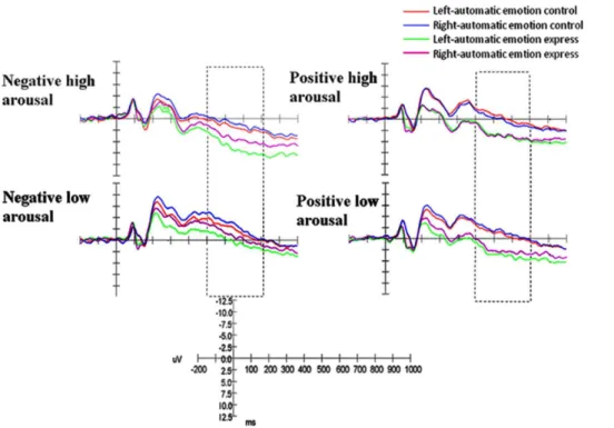
ERP results at posterior lobe electrodes
The ANOVA was performed on the LPP component at posterior lobe electrodes with one between-subject factor: auto-matic emotion regulation (control group and express group), and with three within-subject factors: arousal (high arousal and low arousal), valence (positive and negative) and asymmetry (left and right). Statistically significant interactions were shown on the LPP component: valence6arousal (F(1, 23)= 49.145, p,.001, gp2= .681) and arousal6laterality6group (F(1, 23)= 4.639,
p,.05, gp2= .189). Simple effect analysis revealed that larger
LPP amplitude appeared in negative high arousal emotion than in positive high arousal emotion (p,.001). The three-way simple effect analysis revealed that automatic emotion express group had larger LPP amplitude in the right posterior lobe than the left posterior lobe (p,.05), but automatic emotion control group showed no significant difference (p..05) in high arousal emotion conditions. The result also showed that the automatic emotion express group showed larger LPP amplitudes at right electrodes in high arousal emotion than that in low arousal emotion (p,.05), but no such difference was observed in the automatic emotion control group (p..05) (Fig. 2).
Discussion
The present study examined whether individual difference in automatic emotion regulation (AER) affected emotion-related hemispheric asymmetry for LPP component, with the interaction between valence and arousal. Importantly, we observed a right dominance of LPP component at posterior electrodes in high arousal conditions for automatic emotion express group, but no similar difference was observed for automatic emotion control group. This result suggested that AER modulated the hemispheric asymmetry of LPP on posterior electrodes and supported the right hemisphere hypothesis. We also observed an attenuated difference in the LPP amplitude at right posterior electrodes between high arousal and low arousal emotion for automatic emotion control group compared with automatic emotion express group. This suggested that a declined LPP component on right posterior electrodes would be an indicator of automatic emotion control, especially when compared to automatic emotion express condi-tion.
This result partly supported our hypothesis that AER modulat-ed the right dominance of the mean amplitude of the LPP in posterior electrodes, and also documented the right hemisphere hypothesis, which proposed a dominance of the right lobes in emotion processing. More importantly, our result provided an indication that the right dominance of the LPP component in posterior electrodes happened just for automatic emotion express participants, but not for automatic emotion control participants.
The findings also showed an attenuated difference for automatic emotion control participants in the LPP amplitude at right posterior electrodes between high arousal and low arousal emotion, compared with automatic emotion express participants. Previous researches suggested that the LPP reflected the evaluation of emotion and could be an index of emotion regulation; when emotion regulation happens, the amplitude of LPP would be diminished. For example, Hajcak and Nieuwenhuis found cognitive re-appraisal strategy diminished the amplitude of the LPP and reduced self-reporting negative feelings [38]. The present finding indicated that automatic emotion regulation, including emotion control and emotion express, would regulate the amplitudes of the LPP component in emotion conditions and induce emotion change with no deliberated manipulation.
It would be possible to say that our findings supported the hypothesis that the LPP component would be an index of automatic emotion regulation on the ERPs level. However, some shortcomings in the present study limited our understanding of the LPP component as the index of automatic emotion regulation. Firstly, it is important that deliberated conditions could be manipulated compared with automatic conditions. Previous studies have documented the LPP component as an index of emotion regulation [39]. Thus, future studies should examine the difference of the LPP component in automatic emotion regulation and deliberated emotion regulation. Secondly, although ER-IAT
Table 1.The scores of emotion control category of ER-IAT of automatic emotion control group and automatic emotion express group.
The scores of emotion control category of ER-IAT
M SD
Automatic emotion control group 0.3 0.15
Automatic emotion express group 20.18 0.11
is an effective way to assign participants to automatic emotion control group and automatic emotion express group, as has been evidenced by our findings and Mauss et al.’s study, future studies will be needed to directly speak to the usage of automatic emotion regulation in the experiment [29].
This study provided some indication on the ERPs level that ER-IAT could predict the tendency of automatic emotion regulation,
including automatic emotion control or automatic emotion express. This was consistent with Mauss et al.’s study, in which they examined the relationship of automatic emotion regulation and the feeling of anger, cardiac activity and skin conductivity level and found that a higher score in ER-IAT was related to lower levels of physiological index and self-reported feelings of anger [29].
Figure 1. ERPs for automatic emotion control and automatic emotion express groups during four emotion blocks at anterior electrodes.Left = left electrodes. Right = right electrodes.
doi:10.1371/journal.pone.0088261.g001
Figure 2. ERPs for automatic emotion control and automatic emotion express groups during four emotion blocks at posterior electrodes.Left = left electrodes. Right = right electrodes.
Conclusion
The present findings provide further evidence that automatic emotion regulation affects the asymmetry of the LPP component when participants view affective pictures passively. Our results showed that a right dominance of the LPP component at posterior electrodes appeared for automatic emotion express participants, but not for automatic emotion control ones. In this way, our study provides further support to the right hemisphere hypothesis.
Author Contributions
Conceived and designed the experiments: JZ. Performed the experiments: JZ. Analyzed the data: JZ. Contributed reagents/materials/analysis tools: JZ RLZ. Wrote the paper: JZ. Designed the software used in analysis: JZ RLZ.
References
1. Proverbio AM, Brignone V, Matarazzo S, Zotto MD, Zani A (2006) Gender differences in hemispheric asymmetry for face processing. BMC Neurosci 8: 44– 53.
2. Erhan H, Borod JC, Tenke CE, Bruder GE (1998) Identification of emotion in a dichotic listening task: event-related brain potential and behavioral findings. Brain Cogn 37: 286–307.
3. Dolcos F, Cabeza R (2002) Event-related potentials of emotional memory: Encoding pleasant, unpleasant, and neutral pictures. J Cogn Neurosci 2: 252– 263.
4. Kayser J, Bruder GE, Tenke CE, Stewart JW, Quitkin FM (2000) Event-related potentials (ERPs) to hemifield presentations of emotional stimuli: differences between depressed patients and healthy adults in P3 amplitude and asymmetry. Int J Psychophysiol 36: 211–236.
5. Kayser J, Tenke C, Nordby H, Hammerborg D, Hugdahl K, et al. (1997) Event-related potential (ERP) asymmetries to emotional stimuli in a visual half-field paradigm. Psychophysiology 34: 414–426.
6. Keil A, Mu¨ller MM, Gruber T, Wienbruch C, Stolarova M, et al. (2001) Effects of emotional arousal in the cerebral hemispheres: a study of oscillatory brain activity and event-related potentials. Clin Neurophysiol 112: 2057–2068. 7. Zhang J, Zhou RL, Oei TPS (2011) The Effects of Valence and Arousal on
Hemispheric Asymmetry of Emotion. J Psychophysiol 25: 95–103.
8. Kestenbaum R, Nelson CA (1992) Neural and Behavioral-Correlates of Emotion Recognition in Children and Adults. J Exp Child Psychol 54: 1–18. 9. Laurian S, Bader M, Lanares J, Oros L (1991) Topography of Event-Related
Potentials Elicited by Visual Emotional Stimuli. Int J Psychophysiol 10: 231– 238.
10. Munte TF, Brack M, Grootheer O, Wieringa BM, Matzke M, et al. (1998) Brain potentials reveal the timing of face identity and expression judgments. Neurosci Res 30: 25–34.
11. Vanderploeg RD, Brown WS, Marsh JT (1987) Judgements of emotion in words and faces: ERP correlates. Int J Psychophysiol 5: 193–205.
12. Kayser J, Bruder GE, Tenke CE, Stewart JE, Quitkin FM (2000) Event-related potentials (ERPs) to hemifield presentations of emotional stimuli: differences between depressed patients and healthy adults in P3 amplitude and asymmetry. Int J Psychophysiol 36: 211–236.
13. Coan JA, Allen JJB (2004) Frontal EEG asymmetry as a moderator and mediator of emotion. Biol Psychol 67: 7–49.
14. Davidson RJ (1992) Emotion and Affective Style - Hemispheric Substrates. Psychol Sci 3: 39–43.
15. Davidson RJ (1988) Eeg Measures of Cerebral Asymmetry - Conceptual and Methodological Issues. Int J Neurosci 39: 71–89.
16. Pizzagalli DA, Jahn AL, O’Shea JP (2005) Toward an objective characterization of an anhedonic phenotype: A signal detection approach. Biol Psychiatry 57: 319–327.
17. Tomarken AJ, Davidson RJ, Henriques JB (1990) Resting Frontal Brain Asymmetry Predicts Affective Responses to Films. J Pers Soc Psychol 59: 791– 801.
18. Davidson RJ (1992) Emotion and affective style: Hemispheric substrates. Psychol Sci 3: 39–43.
19. Cacioppo JT, Crites SL, Gardner WL (1996) Attitudes to the right: Evaluative processing is associated with lateralized late positive event-related brain potentials. Pers Soc Psychol Bull 22: 1205–1219.
20. Cuthbert BN, Schupp HT, Bradley MM, Birbaumer N, Lang PJ (2000) Brain potentials in affective picture processing: covariation with autonomic arousal and affective report. Biol Psychol 52: 95–111.
21. Mauss IB, Cook CL, Gross JJ (2007) Automatic emotion regulation during anger provocation. J Exp Soc Psychol 43: 698–711.
22. Hajcak G, Moser JS, Simons RF (2006) Attending to affect: appraisal strategies modulate the electrocortical response to arousing pictures. Emotion 6: 517. 23. Mocaiber I, Pereira MG, Erthal FS, Machado-Pinheiro W, David IA, et al.
(2010) Fact or fiction? An event-related potential study of implicit emotion regulation. Neurosci Lett 476: 84–88.
24. Mauss IB, Evers C, Wilhelm FH, Gross JJ (2006) How to bite your tongue without blowing your top: Implicit evaluation of emotion regulation predicts affective responding to anger provocation. Pers Soc Psychol Bull 32: 589–602. 25. Schmidt B, Hanslmayr S (2009) Resting frontal EEG alpha-asymmetry predicts
the evaluation of affective musical stimuli. Neurosci Lett 460: 237–240. 26. Drabant EM, McRae K, Manuck SB, Hariri AR, Gross JJ (2009) Individual
differences in typical reappraisal use predict amygdala and prefrontal responses. Biol Psychiatry 65: 367–373.
27. Williams LE, Bargh JA, Nocera CC, Gray JR (2009) The unconscious regulation of emotion: nonconscious reappraisal goals modulate emotional reactivity. Emotion 9: 847.
28. Vogt J, Lozo L, Koster EHW, De Houwer J (2011) On the role of goal relevance in emotional attention: Disgust evokes early attention to cleanliness. Cogn Emot 25: 466–477.
29. Mauss IB, Evers C, Wilhelm FH, Gross JJ (2006) How to bite your tongue without blowing your top: implicit evaluation of emotion regulation predicts affective responding to anger provocatio. Pers Soc Psychol Bull 32: 589–602. 30. Bradley MM, Greenwald MK, Petry MC, Lang PJ (1992) Remembering
pictures: pleasure and arousal in memory. J Exp Psychol Learn Mem Cogn 18: 379.
31. Liu X, Xu A, Zhou RL (2009) Native research of international affective picture system: assessment in university students. Chin J Clin Psychol 17: 687–692. 32. Sato W, Aoki S (2006) Right hemispheric dominance in processing of
unconscious negative emotion. Brain Cogn 62: 261–266.
33. Beck AT, Steer RA (1987) Beck Depression Inventory manual. San Antonio, TX: Psychological Corporation.
34. Spielberger CD (1989) State-Trait Anxiety Inventory: a comprehensive bibliography. Palo Alto, CA: Consulting Psychologists Press.
35. Heller W (1993) Neuropsychological Mechanisms of Individual Differences in Emotion, Personality, and Arousal. Neuropsychology 7: 476–489.
36. Keil A, Bradley MM, Hauk O, Rockstroh B, Elbert T, et al. (2002) Large-scale neural correlates of affective picture processing. Psychophysiology 39: 641–649. 37. Olofsson JK, Nordin S, Sequeira H, Polich J (2008) Affective picture processing:
An integrative review of ERP findings. Biol Psychol 77: 247–265.
38. Hajcak G, Nieuwenhuis S (2006) Reappraisal modulates the electrocortical response to unpleasant pictures. Cogn Affect Behav Neurosci 6: 291–297. 39. Dennis TA, Hajcak G (2009) The late positive potential: a neurophysiological