Characterisation of
Pseudomonas aeruginosa
Adaptation
to a Novel Antimicrobial Substance
Peter Cierniak1, Martin Ju¨bner1, Stefan Mu¨ller2, Katja Bender1*
1Institute of Legal Medicine, Medical Faculty, University of Cologne, Cologne, Germany,2Center for Molecular Medicine Cologne, University of Cologne, Cologne, Germany
Abstract
Antibiotic resistance has been reported since the introduction of synthetic antibiotics. Bacteria, such as one of the most common nosocomial pathogensP. aeruginosa, adapt quickly to changing environmental conditions, due to their short generation time. Thus microevolutional changes can be monitoredin situ. In this study, the microevolutional process of
Pseudomonas aeruginosa PAO1 resistance against a recently developed novel antibacterial zinc Schiff-base (ZSB) was investigated at the proteome level. After extended exposure to ZSB the passaged strain differed in tolerance against ZSB, with the adaptedP. aeruginosaPAO1 exhibiting 1.6 times higher minimal inhibitory concentration. Using Two-dimensional Difference Gel Electrophoresis, the changes in the proteome of ZSB adapted P. aeruginosa PAO1 were examined by comparison with the non-adaptedP. aeruginosaPAO1. The proteome of the adaptedP. aeruginosaPAO1 strain differed significantly from the non-adapted in the abundance of two proteins when both strains were grown under stressing conditions. One protein could be identified as the outer membrane protein D that plays a role in uptake of basic amino acids as well as in carbapeneme resistance. The second protein has been identified as alkyl peroxide reductase subunit F. Our data indicated a slight increase in abundance of alkyl peroxide reductase F (AhpF) in the case of ZSB passagedP. aeruginosaPAO1. Higher abundance of Ahp has been discussed in the literature as a promoter of accelerated detoxification of benzene derivatives. The observed up-regulated AhpF thus appears to be connected to an increased tolerance against ZSB. Changes in the abundance of proteins connected to oxidative stress were also found after short-time exposure ofP.
aeruginosaPAO1 to the ZSB. Furthermore, adapted P. aeruginosa PAO1 showed increased tolerance against hydrogen
peroxide and, in addition, showed accelerated degradation of ZSB, as determined by HPLC measurements.
Citation:Cierniak P, Ju¨bner M, Mu¨ller S, Bender K (2013) Insights into Mechanisms and Proteomic Characterisation ofPseudomonas aeruginosaAdaptation to a Novel Antimicrobial Substance. PLoS ONE 8(7): e66862. doi:10.1371/journal.pone.0066862
Editor:Helene F. Rosenberg, NIAID, United States of America
ReceivedDecember 27, 2012;AcceptedMay 11, 2013;PublishedJuly 15, 2013
Copyright:ß2013 Cierniak et al. This is an open-access article distributed under the terms of the Creative Commons Attribution License, which permits unrestricted use, distribution, and reproduction in any medium, provided the original author and source are credited.
Funding:The authors kindly acknowledge funding of this work through the European Commission, (EMBEK1) grant 211436 (http://www.mpip-mainz.mpg.de/ eu-projekte/embek1/index.php). The funders had no role in study design, data collection and analysis, decision to publish, or preparation of the manuscript.
Competing Interests:The authors have declared that no competing interests exist.
* E-mail: katja.bender@uk-koeln.de
Introduction
Microevolution is defined as an adaptive modification in a population over a given time that occurs as a continuous natural process with gradual change [1]. The gradual change is caused, as observed by Charles Darwin and Alfred Russel Wallace, by different principles: 1) All individual variations among organisms are propagated vertically from parental to the next generation. 2) All organisms generate more offspring than necessary to replace themselves. 3) Environmental changes cause a distinct pressure by limiting the survivability of a population, which controls the population size. 4) Individuals who are still able to survive and generate offspring within those changed conditions predominantly take benefit of their individual variations, and are better adapted to the changed conditions than the non-survivors [1–3]. In contrast to many multicellular organisms many bacteria have very short generation times. It is therefore possible to follow the microevolution in a bacterial population described by the named principlesin situ, thus making predictions on population changes possible [4].
An alarming example of adaptation processes is the rapid onset in resistance of bacteria against antibiotics or biocides [5]. Resistances were reported soon after the introduction of synthetic antibiotics in the early twentieth century [6], and these resistances are on the increase [7]. Different modes of acquired and intrinsic resistance are known. One mode is the alteration of the antibiotic drug target; such as changes in the ribosome structure [8]. Another mode of resistance is decreased membrane permeability, thereby preventing the uptake of the drug [9]. Over-expression of efflux systems that lead to removal of the antibacterial drug is another general mode of resistance [10]. A prominent mechanism of resistance to antibiotics is the enzymatic destruction of the drug by enzymes such as beta-lactamases [11].
increasing interest in antimicrobial materials as surface impreg-nations, attention should also be paid to the evolution of resistances against these materials. Besides silver or copper, other metals, such as zinc, show antimicrobial properties (reviewed in [13]). Thus zinc-containing materials are currently under inves-tigation in the production, and for use as, antimicrobial surfaces – both for the protection of medical devices as well as household items. The aim of our study was to elucidate the possibility of resistance development against zinc-containing compounds as an antimicrobial material, and to gain knowledge of the underlying mechanism.
Recently, various Schiff-base derivatives have aroused consid-erable interest [14–17] due to their antibacterial activities [15], offering a source for antibacterial substances. The Schiff-base bis(N-allylsalicylideneiminato)-zinc (ZSB), developed, synthesized and characterized by Poulter and colleagues, demonstrated antibacterial properties, when deposited on surfaces by plasma-enhanced chemical vapour deposition (PECVP) [15]. In our study the same ZSB was used as a prototype of a new antibacterial material, to investigate microevolution.
Pseudomonaeare a ubiquitous genus of gram-negative bacteria that occur in terrestrial as well as in aquatic environments. They are found associated together with plants as well as animals, emphasising the versatility of this genus, both genetically and metabolically (reviewed in [18]). P. aeruginosa is a nosocomial pathogen that accounts for around a third of all intensive care unit infections in Europe [19]. It is an opportunistic pathogen that causes infections in immuno-compromised persons [20], and is associated with cystic fibrosis with a prevalence of more than 80% [21], as well as with burn wound infections [22]. Pseudomonas aeruginosa PAO1 was used as the prototype bacterial strain for microevolution experiments.
In this study we present data on the adaptation ofP. aeruginosa
PAO1 to ZSB for nearly 300 generations. The adapted strain was compared to its non-adapted, ancestral strain by Two-dimensional Difference Gel Electrophoresis (2-D DIGE) and mass spectrosco-py. The results of the proteomic work were verified by comparison of the physiological behaviour of the adapted and the non-adapted
P. aeruginosa. Based on this data, we demonstrate the formation of tolerances by means of natural selection in P. aeruginosa PAO1. Tolerance comes along with accelerated degradation of the antibacterial compound. Except for directed genetic modifications [23] [24] the presented study is, to our knowledge, the only study that has monitored evolutional changes in stressed bacteria at the proteome level against a newly synthesised antimicrobial sub-stance.
Materials and Methods
Bacterial strain and culture conditions
Pseudomonas aeruginosa PAO1 (kindly provided by A. T. A. Jenkins, University of Bath, UK) was routinely grown in Lysogeny Broth [25,26] (Luria/Miller, LB, Carl Roth, Karlsruhe, Germany) at 37uC in an orbital shaking incubator (GFL, Burgwedel, Germany) at 170 rpm, unless otherwise stated. Stock cultures were stored in 20% (v/v) glycerol at280uC.
Antibacterial agents
The zinc Schiff-base complex bis(N-allylsalicylideneiminato)-zinc (ZSB), (MW 771 g mol21
) was synthesized and characterized by Poulter et al. [15]. The compound consists of two zinc atoms chelated by four allylsalicylideneimin Schiff-base (SB) ligands. ZSB and the zinc free ligand SB were provided by Poulter and colleagues, University of Bath, UK. ZSB and SB were dissolved in
.99.99% dimethyl sulfoxide (DMSO) (Aldrich; Sigma-Aldrich Chemie GmbH, Taufkirchen Germany) respectively and diluted in LB medium to a final DMSO concentration of 2% (v/v).
Adaptation ofP. aeruginosaPAO1 in zinc Schiff-base Microevolution was achieved according to a modified protocol of Lenski and colleagues [4]. The experimental setup for the microevolution with ZSB included 3 to 6 bacterial cultures per passage. The cultures were grown under continuous agitation at 170 rpm for 20 to 48 h in glass tubes containing 6 mL LB medium with a distinct concentration of ZSB. Starting concentration for the first passages was 0.5 mM of ZSB, which was increased stepwise by 0.2 mM, and in the final step by 0.4 mM, up to 1.3 mM in total. To monitor the growth of the bacteria, the optical density was measured after 20 to 48 h of cultivation at wavelength l 600 nm (OD600) (Geneys UV10, Thermo Fisher Scientific Germany Ltd. & Co. KG, Braunschweig, Germany). After each incubation period, the culture with the highest OD600
was used for the next passage. Additionally, in order to make comparison with the ancestral cultures possible, at the start of the experiment and at every 5–10th passage, glycerol cultures were prepared and stored at280uC. The ZSB Stock solutions were exchanged every 5thto 10thpassage. The initial viable cell number before incubation of each passage was 107CFU mL21
, and after incubation 109 CFU mL21
. In total, the microevolution was performed over almost 300 generations (45 passages) until significant adaptation was reached.
The purity of the cultures was tested after every 8th –10th passage, in order to detect contamination. LB agar and Cetrimide agar (Sigma-Aldrich Chemie GmbH, Taufkirchen, Germany) plates were inoculated with the respective passaged liquid culture and incubated for 24 h at 37uC. The culture was only utilised for further passages if a single colony type could be identified on LB agar, and no change between the passaged and the non-passaged
P. aeruginosawas found on Cetrimide agar with respect to colony appearance, pigmentation and fluorescence at 254 nm.
To verify whether the ZSB adapted strain was able to grow in the presence of significantly higher concentrations of ZSB, both the non-adapted cultures and the ZSB adapted cultures were grown in LB medium at increasing concentrations of ZSB for 23 to 24 h. Prior to the experiments, in order to reduce the influence of metabolic modifications, the non-adapted culture was incubated for one passage at 0.2 mM of the ZSB. The adapted and the preconditioned non-adapted cultures were diluted with LB medium to OD600= 1. Then 6 mL of LB medium containing
2% (v/v) DMSO and a defined concentration of ZSB was inoculated with 60mL of the prepared bacteria dilutions. After incubation the OD600 was measured. All experiments were
prepared in duplicate on two separate days. LB medium containing 2% (v/v) of DMSO was used as a control.
In order to verify whether the increased tolerance to ZSB was due to amplified metabolic activity or to adaptation, the P. aeruginosaPAO1 P45strain was grown for two passages in ZSB-free
LB medium (P45L). The OD600of the non-adapted P0, the initial
adapted (P45) and the P45L strain were compared after incubation
in LB medium containing 1.3 mM ZSB at 24 h intervals for up to 96 h.
Proteomic analysis of adapted and non-adaptedP. aeruginosaPAO1
from different metabolic activities of the cultures. Cultures were grown in baffled Erlenmeyer flasks in 20 mL ZSB-free LB medium for three to four hours, until the cultures reached an OD600of
0.65, after which ZSB was added to a final concentration of 0.5 mM to both adapted and non-adapted P. aeruginosa PAO1. Once both experimental groups had reached an OD600of 0.9, the
samples were harvested by centrifugation at 5000gfor 10 min at 23uC (Biofuge Primo R, Thermo Fisher Scientific Germany Ltd. & Co. KG, Braunschweig, Germany). Pellets were washed three times with PBS pH 7.6 (Carl Roth, Karlsruhe, Germany) and stored at280uC until use. Each experimental group consisted of four biological replicates.
Comparative proteome analysis of adapted and non-adapted strains: Proteomic analysis was performed, with some minor adjustments (listed below), according to the protocols from A. Go¨rg and colleagues [27] [28]. The protein pellets were thawed on ice and resuspended in 1 mL lysis buffer (7 M urea, 2 M thiourea, 30 mM TRIS (pH 7.5), 4% v/v CHAPS) containing a final concentration of 1 x nuclease mix and 1 x protease inhibitor. The samples were homogenized by ultrasonication (Bandelin electron-ic, Berlin, Germany) on ice at 40% cycle rate and 13% amplitude for 1 min and subsequently incubated for 15 min at room temperature. The samples were centrifuged at 8000gfor 20 min at 4uC to remove insoluble material after incubation. The supernatant was then transferred into 1.5 mL centrifuge tubes and centrifuged again at 25000gfor 45 min at 4uC. Subsequently, proteins were isolated from the crude extracts by the 2-D Clean-Up Kit (GE Healthcare, Clean-Uppsala, Sweden) according to manu-facturer’s instructions and dissolved in lysis buffer (7 M urea, 2 M thiourea, 4% (v/v) CHAPS, 30 mM tris (pH 7.5)). The pH of the dissolved samples was adjusted to 8.5 on ice with 0.1 M NaOH (analytical grade, Carl Roth, Karlsruhe, Germany) and the protein concentration was photometrically determined using the 2-D Quant kit (GE Healthcare, Uppsala, Sweden). The proteins of the samples were then labelled by applying the CyDyeTMDIGE Fluor minimal labelling kit (GE Healthcare, Uppsala, Sweden) according to manufacturer’s instructions. After labelling, the samples were mixed 1:1 with sample buffer (7 M urea, 2 M thiourea, 4% (v/v) CHAPS, 2% (v/v) ampholytes pH 4–7) and pooled. Each pooled sample consisted of an assay of the non-adapted group, an assay of the adapted group and a mixture of all samples (internal standard). The dye order was swapped intentionally from one sample to the next. The samples were then applied cathodically onto an immobilized pH gradient (IPG) 18 cm stripe, pH 4–7 (GE Healthcare, Uppsala, Sweden). Samples were separated in the second dimension (Ettan DALTsix, GE Healthcare, Uppsala, Sweden) for 5 h. A fluorescent scanner equipped with lasers emitting three different wavelength, with associated band pass filters, was used to scan the gels (Typhoon Trio, GE Healthcare, Uppsala, Sweden). Experimental groups were analysed by DeCyderTMV6 (GE Healthcare, Uppsala, Sweden). Null hypoth-esis for protein spots was rejected in case of ANOVA p,0.01, including false recovery rate correction (q,0.01). If not otherwise stated all chemicals were of analytical grade and obtained from GE Healthcare, Uppsala, Sweden.
Proteome analysis of ZSB treated and untreatedP. aeruginosa PAO1
Changes in the proteome were compared between a ZSB treated and an untreatedP. aeruginosaPAO1, to acquire additional information on the mode of action of ZSB, and to verify the results of the proteomic comparison of the adapted and the non-adapted strain. Eight cultures in baffled Erlenmeyer flasks were prepared from four overnight pre-cultures by adding 1% of the final volume
of pre-culture to 20 mL LB medium and grown for three hours to mid-log phase at 37uC. Then 0.1 mM (final concentration) of ZSB (solved in DMSO with a final concentration of 2% v/v) was added to four cultures and DMSO only (2% v/v final concentration) to the remaining four cultures (control group). The cultures were incubated for a further 3.5 h. A 4 mL sample was taken from each culture. The cells were harvested by centrifugation at 5000gfor 10 min and at room temperature (RT). The cell pellets were washed three times with 40 mM Tris (Carl Roth, Karlsruhe, Germany) buffer (pH 7.5) and stored at280uC.
In order to optimise the sample preparation for 2-D DIGE the following changes in crude extract preparation were introduced in comparison to the proteome analysis of adapted and non-adapted strains. After thawing of the bacterial pellets (preparation as described above), 100mL of 1% (v/v) sodium dodecyl sulphate (SDS) (GE Healthcare, Uppsala, Sweden) was added and incubated at 95uC for 5 minutes on a heating block (Thermomixer comfort, Eppendorf AG, Hamburg, Germany). Samples were subsequently cooled on ice for 5 min, and 250mL of lysis buffer lacking protease inhibitor was added. To facilitate dissolving of proteins in the lysis buffer, samples were incubated under intermediate mixing for 1 h at room temperature prior to centrifugation. Labelling and electrophoretic separation of the first and second dimension was performed as described above. Due to significant changes in the proteome, only the protein spots with the highest difference in abundance (ANOVA p,0.01; false recovery rate correction q,0.01; standard log abundance ratio .2.5) were chosen for further analysis by means of mass spectrometry. Significantly differentially abundant protein spots were manually picked from preparative 2-DE gels that were prepared in parallel to 2-D DIGE analysis. Preparative 2-DE gels were stained by SyproRubyTM (Life Technologies GmbH, Darmstadt, Germany) according to manufacturers instructions.
Stained protein spots were excised from the gel and washed three times with acetonitrile/water (1:1). The gel pieces were shrunk with acetonitrile, rehydrated in 50 mM NH4HCO3 and
dried in a speedVac. The dried gel pieces were covered with aqueous solution containing 10 mM DTT and 50 mM NH4HCO3. The gel-embedded proteins were then reduced at
56uC for 45 min. Prior to in-gel digestion, the gel pieces were washed and dried, as previously described, to remove the reduction agent DTT. The gel pieces were then rehydrated in an ice-cold solution containing 10 ng mL21
trypsin (sequencing grade, Promega, USA) and 10 mM NH4HCO3. After 45 min on
ice, excessive trypsin solution was replaced by 20mL of trypsin free buffer. Samples were stored over night at 37uC to ensure complete digestion of proteins. The digestion was arrested by addition of 20ml 10% formic acid. The resulting peptides were extracted for 30 min at 37uC and further analyzed using ultra high performance liquid chromatography-mass spectrometry (UHPLC/MS).
UHPLC/MS data were acquired on an HCT ETD II iontrap mass spectrometer (Bruker Daltoniks, Bremen, Germany) equipped with a nano-ESI source (Bruker Daltonics, Bremen, Germany). Samples were introduced by an Easy-nLC 1000 system (Proxeon, Odense, Denmark) using a vented column setup comprising an 100mm inner diameter (ID), 20 mm length (L) trapping column and an ID 75mm, L 100 mm analytical column, both self packed with ReproSil-Pur C18-AQ, 5mm (Dr. Maisch,
Ammerbuch, Germany). A 5 to 18mL sample was aspirated into the 25mL sample loop and loaded onto the trap column using a flow rate of 6mL min21. Mobile phase solvent A: aqueous 0.1% formic acid; and solvent B: acetonitril were mixed with a linear gradient of 0% to 35% acetonitrile over 20 min with a flow rate of 300 nL min21
to 100% over 2 min and the column was regenerated in 100% ACN for an additional 8 min.
The Compass 3.0 software controlled data-dependent acquisi-tion of MS and tandem MS (MS/MS) spectra. MS1 scans were acquired in standard enhanced mode. Five single scans in the mass range from m/z 400 to m/z 1400 were combined for one survey scan. Up to three doubly and triply charged ions rising above a given threshold were selected for MS/MS measurements. Ultrascan mode was used for the acquisition of MS2 data in the mass range from m/z 100 to m/z 1600, and three single scans were summed up. The ion charge control value was set to 250000 for all scan types. Peaklists in mascot generic format (mgf) were generated from the raw data by means of the Compass Data Analysis software module (Bruker Daltoniks, Bremen, Germany).
Proteins were identified by searching the NCBInr release 20090531 (National Center for Biotechnology Information, Bethesda, USA) using a local installation of MASCOT 2.2 (Matrix Science Ltd, London, UK). Searches were submitted via Proteinscape 2.1 (Bruker Daltoniks, Bremen, Germany) with the following parameter settings: Enzyme ‘‘trypsin’’, species ‘‘eubac-teria’’, fixed modifications ‘‘carbamidomethyl’’, optional modifi-cations ‘‘methionine oxidation’’ and missed cleavages ‘‘2’’. The mass tolerance was set to 0.4 Da for peptide and fragment spectra.
Bacterial susceptibility tests of adaptedP. aeruginosa PAO1
Comparative analysis of hydrogen peroxide tolerance and antibiotic susceptibility of adapted P. aeruginosa PAO1 was performed according to a modified protocol of The British Society for Antimicrobial Chemotherapy (BASC) for standardized disc susceptible testing (agar disc diffusion method) [29]. Liquid bacterial cultures of both the ZSB adapted and wildtype P. aeruginosaPAO1 were incubated for 18 h at 37uC. The cultures were diluted 1:1 with sterile ultra pure water and OD600 was
measured. To equalise the starting viable cell numbers, cultures with OD600.0.3 were diluted 1:1000 with ultra pure water and
cultures with OD600,0.3 were diluted 1:500 to a final volume of
5 mL. The prepared cultures were then spread out onto Mueller-Hinton II agar plates (Sigma-Aldrich Chemie GmbH, Tauf-kirchen, Germany). For hydrogen peroxide tolerance, sterile filter paper disks, diameter = 10 mm (Sigma-Aldrich Chemie GmbH, Taufkirchen, Germany) impregnated with 15mL of 30%
hydro-gen peroxide (Carl Roth, Karlsruhe, Germany), were added on top of the Mueller-Hinton II agar. Imipenem (10mg) test discs (Oxoid, Basingstoke, UK) were used for antibacterial susceptibility tests. Cultures were incubated for 18–22 h at 37uC. After incubation the total zone of inhibition diameters (containing the filter paper disks) were determined with a sliding calliper. Measurements on tolerance against hydrogen peroxide were performed over seven days. Measurements on antibiotic suscep-tibility were performed on three different days. OriginPro 8.1 (OriginLab Corporation Northampton, UK) was used for statistic analysis, and the hypothesis was proven using the Mann-Whitney-Test.
Degradation of ZSB by the ZSB adapted and the non-adapted P.aeruginosa PAO1
ZSB-degradation measurements are based on the work of Jacobson et al. [30]. Twenty millilitres of LB medium containing ZSB were inoculated with the ZSB- adapted (P45) and wildtype P. aeruginosaPAO1 (P0), and cultivated at 170 rpm for 18 h at 37uC.
The concentration of ZSB was 1.3mM for P45and 0.5mM for P0.
After incubation the cultures were harvested by centrifugation at
5000g for 10 min at RT and washed twice with ZSB-free LB medium. The OD600of all samples was adjusted to 1.7 and the
initial concentration of ZSB was set for all samples to 1.8 mM. 50mL samples were taken at 30 min intervals for high
perfor-mance liquid chromatography (HPLC) measurements. To mon-itor changes in the growth velocity, optical density measurements were performed every 60 min. After 5 h of incubation a viable cell count was performed. Samples for HPLC measurements were stored at 280uC until use. For the HPLC measurements the samples were diluted in ultra pure water for appropriate resolution. The chromatography was performed using an HPLC Agilent 1100 system (Agilent technologies, Santa Clara, USA). An aqueous solution of 60% 1 mM H2SO4 (Carl Roth, Karlsruhe,
Germany) and 40% acetonitrile (Carl Roth, Karlsruhe, Germany) was used as an isocratic mobile phase at a flow rate of 1.2 mL min21
, the stationary phase was a L 250 mm x ID 4 mm EC Nucleodur 100-5 C8ec (100 A˚ , 5mM) column (Macherey-Nagel
GmbH & Co. KG, Du¨ren, Germany). The column temperature was set to 40uC. Sample injection volume was 20mL. The HPLC was equipped with an Agilent diode array detector (DAD,l195– 300 nm). The retention time of ZSB was 5.6 min. The measure-ment was determined atl220 nm, quantification was done on a calibration curve using the following ZSB concentrations: 0.05 mM, 0.2 mM, 0.5 mM, 0.75 mM, 1 mM, 1.5 mM, and 2 mM. Each experimental group consisted of four biological replicates.
In addition, the zinc uptake from the medium was measured.P. aeruginosa P0 and P45 were grown for 24 h in ZSB containing
medium. The cultures were then centrifuged and the zinc content in the medium was measured by Atomic absorption spectroscopy (Atomic Absorption Spectrophotometer, Model 3030, Perkin-Elmer, Waltham, USA).
Results
Proteome analysis of ZSB treatedP. aeruginosa PAO1 In order to determine the influence of ZSB on the proteome of
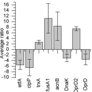
P. aeruginosa, the bacteria were treated with a relatively low concentration (0.12 mM) of ZSB. 2-D DIGE analysis yielded 647 significantly (ANOVA p,0.01, including false discovery rate (FDR) correction q,0.01) differently expressed protein spots, out of which 270 were down-regulated and 377 were up-regulated. From these protein spots, eight of the most significant differentially abundant proteins were identified using mass spectrometry. Protein spots that were identified could be defined as outer membrane proteins or their precursors. Different enzymes responsible for redox potential regulation were identified, as well as a chaperone and an electron transport chain protein, (see figures A to I in File S1). Figure 1 illustrates the mean protein abundance ratios (average ratios) of the ZSB treatedP. aeruginosa
PAO1 in relation to the untreatedP. aeruginosaPAO1.
another related protein (outer membrane protein OprD (Acces-sion: gi|158429225)) was found to be down-regulated.
Microevolution ofP. aeruginosaPAO1 to zinc Schiff-base In order to define the starting point for microevolution experiments, the minimal inhibitory concentration of ZSB was determined forP. aeruginosa PAO1 in LB medium (figure 2). For microevolution a concentration of 0.5 mM ZSB was used as an initial concentration. The concentration of ZSB was then successively increased by 0.2 mM, or 0.4 mM, steps to 1.3 mM ZSB over a period of 45 passages. After each increase of the ZSB concentration, the optical density of the culture sharply decreased and subsequently increased in a gradual manner during the succeeding passages (see File S2).
The minimum inhibitory concentration of ZSB of the ZSB adapted (P45) and non-adapted (P0) P. aeruginosa PAO1 was
determined. The results, depicted in figure 2, indicate that the MIC50value ofP. aeruginosaPAO1 P45increased from 0.86 mM to
1.27 mM and the MIC90 from 1.03 mM to 1.6 mM, which
corresponds to a 1.5 fold increase of MIC50and 1.6 fold of MIC90.
To verify whether the increased tolerance against ZSB was as a result of increased metabolic activity or due to a heritable mutation, the adaptedP. aeruginosaPAO1 P45 was passaged two
times in ZSB free LB medium (P45L). The growth behaviour of
P45L, as well as of the adaptedP. aeruginosaPAO1 P45and
non-adapted P0 strain, were compared in LB medium containing
1.3 mM of ZSB. Growth (OD600.0.5) was found forP. aeruginosa
PAO1 P45after less than 24 h, the P45L showed growth after 28 h.
In the case of the P. aeruginosa PAO1 P0, no growth could be
observed, even after 96 h of incubation.
Proteomic analysis of the adapted and non-adaptedP. aeruginosaPAO1
In order to compare bothP. aeruginosaPAO1 groups (P0and P45)
both strains were grown in the absence of ZSB to mid-log phase to an OD600of 0.65. Then ZSB was added to both strains to a final
concentration of 0 5 mM (addition indicated by arrows in figure 3). Both strains were grown in presence of ZSB for 3 h to 4 h at 37uC. As shown in figure 3 both cultures exhibit a similar growth pattern, with the exception that P45exhibits a prolonged lag phase, most
likely resulting from minor differences in the starting inoculum. Following addition of ZSB, growth of both cultures paused for a short time period, indicated by the plateau in figure 3.
The proteomes of both the adapted and the non-adapted P. aeruginosaPAO1 were subsequently compared for changed protein expression by 2-D DIGE. Only two protein spots were found to be significantly different expressed (p,0.01). The proteins were identified by UHPLC-MS as alkyl hydroperoxide reductase
Figure 1. Average ratios of significantly different expressed proteins in comparative analysis ofP. aeruginosaPAO1 treated with ZSB.2-D DIGE proteome analysis was performed of non-adapted ZSB treated and untreated P0strains. The following proteins exhibiting
significantly different abundance where identified: etfA: electron transfer flavoprotein subunit alpha; clpP: ATP-dependent Clp protease proteolytic subunit; trxA: thioredoxin; fusA1: elongation factor G; acnB: bifunctional aconitate hydratase 2/2-methylisocitrate dehydratase; DnaK: molecular chaperone DnaK; OprD2: OprD2-like porin precursor; OprD: Chain A, crystal structure of the outer membrane protein OprD. Average ratio indicates the standardised volume ratio between two protein spots in two experimental populations [54].
doi:10.1371/journal.pone.0066862.g001
Figure 2. Comparison of growth of the ZSB adapted(P45)and the non-adapted(P0)P. aeruginosaPAO1.LB medium containing 2% (v/v) DMSO was used as control. A non-linear fit algorithm by dose-response function with variable Hill slope was used to fit both curves. doi:10.1371/journal.pone.0066862.g002
Figure 3. Growth curves of adapted P45and non-adapted P0P. aeruginosa PAO1. Black arrows indicate the addition of the ZSB. (n = 4).
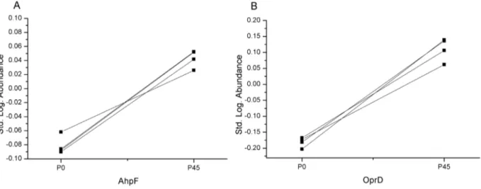
subunit F (AhpF) from P. aeruginosa PAO1 (accession: gi|15595338), which was 1.3 times more abundant in the ZSB adapted strain, and the outer membrane protein OprD fromP. aeruginosa (accession: gi|158429225), which was 2 times more abundant (see figures A to C in File S3). The logarithmic standard abundances of the identified proteins AhpF and OprD are shown in figure 4. As depicted in figure 4, AhpF and OprD are present in significantly lower abundances (q,0.01) in the unpassaged group than in the passaged P45group.
Bacterial susceptibility tests of adaptedP. aeruginosa PAO1
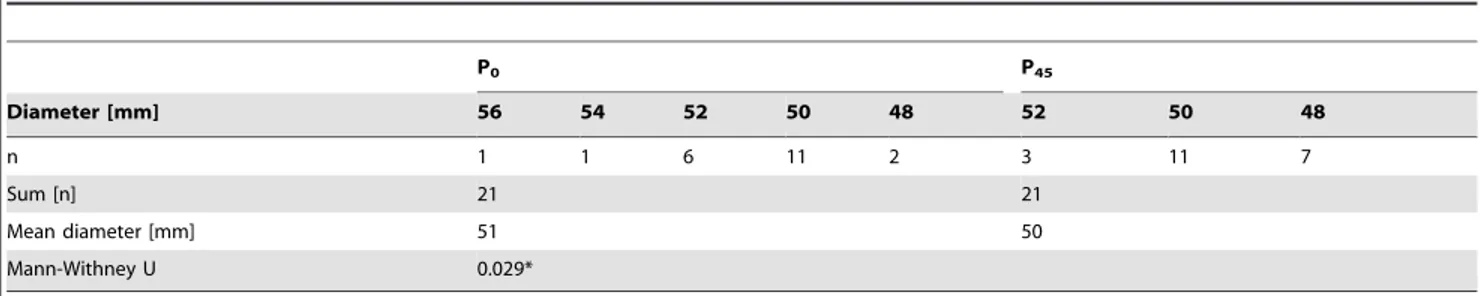
Both 2-D DIGE experiments in combination with mass spectrometry of ZSB adapted (P45) and non-adapted (P0) P. aeruginosaPAO1 gave evidence of oxidative stress response when exposed to ZSB. Results of the comparative proteome analysis were therefore further verified by measuring the tolerance against the oxidative stressor hydrogen peroxide by agar diffusion test in triplicates on seven different days (ntotal = 21). Table 1 shows the
diameters of the individual zone of inhibition and the frequency of their occurrence.
Using the Mann-Whitney-U-Test a p-value ,0.05 was obtained. As indicated in table 1 a small but significant difference was found towards smaller inhibition zones forP. aeruginosaPAO1 P45. Hydrogen peroxide inhibited the growth ofP. aeruginosaPAO1
P45 to a smaller extent than that of P. aeruginosa PAO1 P0.
However, no significant change could be determined in the susceptibility against imipenem. Thus, the observed insensitivity against oxidative stress of P45 does not lead to imipenem
resistance.
Degradation of ZSB by the ZSB adapted and the non-adaptedP. aeruginosa PAO1
An increased abundance of Ahp is known to be involved in facilitating the degradation and tolerance against aromatic compounds [30] [23] [24]. Figure 5 shows the ZSB concentration during incubation with P. aeruginosa PAO1 P0 and P. aeruginosa
PAO1 P45. After five hours no ZSB could be found in case of the
P45cultures, whereas 0.360.1 mM could still be detected in the
case of P0. After 24 h also for P0the ZSB concentration fell below
the analytical limit of detection (data not shown). As shown in figure 5 both strains (P45and P0) were still able to grow in 1.8 mM
ZSB, in a similar manner. Note that the initial cell density was much higher than that used in the experiment shown in figure 2, thus growth of both strains was still possible. No statistically significant difference (p.0.05) in bacterial growth could be determined until the second hour of incubation. In contrast, the actual ZSB concentration differed noticeably (p,0.05) betweenP. aeruginosaP0and P45after 30 minutes of incubation. After 1 h of
incubation the ZSB concentrations differed by a factor of 1.1, and after 2 h by a factor of 1.9 (cP0/cP45). From a statistical point of
view, the bacterial growth behaviour of both groups began to differ from the second hour on. The observed proportion of ZSB degradation at incubation times above 2 hours could have thus been additionally influenced by the slightly increased number of P45 in relation to P0 cells (OD600 of P45/OD600 of P0 after
5 h = 1.1).
Measurement of the zinc uptake by the bacteria from the medium did not show a significant difference for P0and P45(data
not shown).
Discussion
The ability of bacteria to rapidly adapt to environmental conditions is well documented, evidenced by the recent surge of antibiotic resistance exhibited by nosocomial pathogens. Various strategies are under investigation to stem the spread of resistant strains, most especially those antibiotic-resistant bacteria in hospitals. These strategies include implementation of strict hygiene sanctions, and incorporate the prospective use of surface disinfection by employing ‘‘auto-aseptic" surface coatings, such as zinc containing materials [37] [13]. The antimicrobial property of zinc is well known [38], as demonstrated by the zinc-containing material ZSB utilised by Poulter et al. for the deposition of antimicrobially-active coatings [15]. Due to the probable use of ZSB, or related materials, as antimicrobials, the development of resistances against such materials should be considered. For this
Figure 4. Logarithmic standard abundance of identified proteins of adapted and non-adapted P. aeruginosaPAO1. Logarithmic standard abundance is the decadal logarithm of the ratio of an individual protein spot and the internal standard within the same gel [54]. Individual values were in case of AhpF for P020.087709;20.086223;20.06188;20.090214 and P450.051973; 0.05241; 0.025915; 0.041694. For OprD, the
logarithmic abundances were in case of P020.167843;20.172957;20.202919;20.181265 and for P450.105833; 0.0618; 0.139092; 0.136039. Exact
ANOVA q-values (including FDR) for both protein spots were q = 0.008. Excluding FDR the p- value for AhpF was 1.33 1026
and 5.7 1026
for OprD respectively.
reasonP. aeruginosaPAO1 was recurrently incubated (passaged) for approximately 300 generations (45 passages) in ZSB, a material with known antibacterial properties [15]. Passaging yielded a MIC50value increase by factor 1.6, showing adaptation. A study
by Toprak et al. [39], who used a special device (‘‘morbidostat’’) for cultivation along with known biocides such as chloramphen-icol, doxycycline and trimethoprim, demonstrated rising resistance inEscherichia coliwithin 20 days. In comparison to Lenski et al. [4], in our study it is likely that passaging ofP. aeruginosaPAO1 for a higher number of generations could have led to an even greater tolerance against ZSB. Lenski et al. [4] showed, by comparing the fitness of minimal medium adapted E. coli in relation to non-adaptedE. coli, that most of the adaptation occurs during the first 2000 generations. Molecular level mechanism descriptions of e.g. cell size were not mentioned in the latter study [4]. To gain further insight in to the mechanism of adaptation, and in to the mechanism of increased tolerance against ZSB, comparative studies of the proteome and physiology have been performed.
In our experiments the AhpF was found to be up-regulated in the ZSB adapted P. aeruginosa PAO1 P45. AhpF is expressed
together with AhpC as a heterodimer forming the alkyl hydroperoxide reductase [23]. Ahp is known as a scavenger of hydrogen peroxides [40] and one function is the reduction of alkyl hydroperoxides to alcohols [30] [41]. Even AhpF alone can have advantageous effects on the resistance against reactive oxygen species (ROS) as shown by Poole and Ellis [41]. Kang et al. [24] reported that degradation of aromatic compounds such as naphthalene leads to the formation of reactive oxygen species. Moreover, the results of Kang et al [24] and Jacobson et. al [30] showed that over-expression of, amongst others, alkyl hydroper-oxide reductase enhances the degradation of these compounds whilst increasing the tolerance to both naphthalene and ROS. This agrees with our results of the agar diffusion tests against hydrogen peroxide, where a small but significant increase in the tolerance was determined in the case ofP. aeruginosaPAO1 P45.
Moreover, P. aeruginosa PAO1 P45 degraded ZSB significantly
faster than P. aeruginosa PAO1 P0. In accordance with the
literature, our results suggest involvement of Ahp in the defence mechanism against ROS formed during the metabolic degrada-tion of ZSB by the bacteria. The observed up-reguladegrada-tion of AhpF appears to be advantageous for the improved survivability of ZSB adaptedP. aeruginosa PAO1 P45. The literature suggests that
up-regulation of Ahp might also be involved in the observed accelerated degradation of ZSB byP. aeruginosaPAO1 P45.
Proteome analysis of the ZSB adaptedP. aeruginosaPAO1 (P45)
compared to wildtype resulted in an observed increase in the level of the outer membrane protein OprD. Huang and Hancock demonstrated that strong over-expression of OprD leads to elevated susceptibility against carbapenemes [42]. No statistically significant change (p = 0.08) in the susceptibility against the carbapeneme Imipenem could be observed in the case of P45.
The development of resistance against ZSB does not necessarily result in the development of co-resistances. To gain deeper knowledge about the influence of ZSB on the proteome, and to verify the results of the proteome analysis ofP. aeruginosaPAO1 P0
and P45, the non-adaptedP. aeruginosaPAO1 P0was treated with
low concentrations of ZSB for a short period (3.5 h) , and compared to untreated P0. Although working at low
concentra-tions, a dramatic change in the proteome was observed for ZSB treated P. aeruginosa PAO1 P0. Outer membrane proteins and
enzymes connected to oxidative stress were found to be significantly differently expressed. Interestingly, the OprD was found to be down-regulated here. Ochs et al. [43] demonstrated that upon short-time co-incubation with acetylsalicylate, salicylate or benzoate, OprD is repressed. Our results are in good agreement with the latter, as the ZSB ligand SB is a salicylaldehyde [15], and thus possesses a similar structure. In addition, Conejo et al. [44] and Martinez-Martinez et al. [45] showed that upon addition of Table 1.Inhibition zone diameters in relation to their frequency of occurrence in the experiment.
P0 P45
Diameter [mm] 56 54 52 50 48 52 50 48
n 1 1 6 11 2 3 11 7
Sum [n] 21 21
Mean diameter [mm] 51 50
Mann-Withney U 0.029*
*Statistical significance using Student’s T-Test was p = 0.018. doi:10.1371/journal.pone.0066862.t001
Figure 5. Degradation of ZSB by the adaptedP. aeruginosaP45. (a) Degradation of ZSB by P. aeruginosaPAO1 P0andP. aeruginosa
PAO1 P45. (b) Optical density of P. aeruginosa P45 and P0 during
incubation with ZSB. No difference (p.0.05) was found in the growth until the second hour of the experiment. Initial optical density for both experimental groups was 1.7, initial ZSB concentration was 1.8 mM. (n = 4).
zinc (as zinc salt or zinc eluted from catheters) to the culture medium, OprD is also repressed and thus the tolerance for antibiotics of the carbapenem group is increased. This effect was found to be reversible [45].
Our data indicates that short-term exposure to ZSB leads to down-regulation of OprD expression, whereas after adaptation the expression of OprD is less affected by ZSB. It is our view that the changed regulation of OprD is not a result of adaptation to ZSB. It is more likely a result of the adaptation to the pure LB medium. This is supported by the fact that OprD is responsible for uptake of basic amino acids under physiological cell conditions [46] [43] [42]. In our study we observed an increase in the optical densities after six passages ofP. aeruginosaPAO1 P0, after 24 h of growth in
LB. A similar effect was observed in the work of Perron et al. [47] who performed microevolution experiments on an antimicrobial peptide.
The other group of proteins found in the comparative proteome analysis of the short exposure ZSB treatedP. aeruginosaPAO1 P0
could be connected to oxidative stress. The thioredoxin, the bifunctional aconitate hydratase 2/2-methylisocitrate dehydratase, ATP-dependent Clp protease, elongation factor G and DnaK proteins were found to be differentially regulated.
Thioredoxin was found to be up-regulated. Thioredoxins, together with glutaredoxins, are the main groups of proteins responsible for maintaining the reducing potential of a cell [48]. Thioredoxin was found to play a crucial role in the oxidative stress response. Das and Das [32] have shown that thioredoxin on its own is able to scavenge reactive oxygen species, such as hydroxyl radicals. Moreover, mutant and adaptive response analyses have highlighted the importance of increased expression of thioredoxin in response to oxidative stress [49].
Our results have shown that bifunctional aconitate hydratase 2/ 2-methylisocitrate dehydratase (aconitase) was present in higher abundance in the proteome of the ZSB treatedP. aeruginosaPAO1. Experiments on E. coli have shown that aconitase activity is decreased during oxidative stress [50]. In contrast, the abundance of the protein increases with increasing concentration of the oxidative agent [50].
DnaK together with ATP-dependent Clp protease are described as heat shock proteins. In our experiments both proteins were found to be down-regulated. Tamarit et al. [33] showed that DnaK is one of the major targets for oxidative stress by hydrogen peroxide in E. coli,DnaK is post-translational inactivated under oxidative stress conditions in favour of the chaperone Hsp33, which gets activated [36]. Salunkhe et al. [35] suggest as a reason for down-regulation of ATP-dependent Clp proteases to reduce the rate of protein degradation. In the same study it was stated that prolyl-peptidyl isomerases exhibited increased expression to counteract protein dysfunction by misfolding.
Our data revealed a substantial increase in the elongation factor G (EF-G). This protein has an essential function in protein biosynthesis with respect to translocation of the tRNA from the A to the P site of the ribosome [51]. During oxidative stress EF-G is found to be readily inactivated by oxidation in the carboxyl-terminal region [33]. Increased expression of EF-G to compensate for the inactivation during oxidative stress conditions would, therefore, be favourable to maintain translation. Stronger expres-sion of RNAs encoding for ribosomal proteins was also found by Salunkhe et al [35].
One electron transfer flavoprotein was found to be less abundant in the proteome of ZSB treated P. aeruginosa PAO1. The electron transfer flavoproteines can be classified into two groups. In one group it is expressed continuously, as a house-keeping protein; where as in the second group it is only expressed
under defined circumstances [52]. Lower abundance of this protein in presence of ZSB is not surprising, as the protein is involved in energy metabolism and the ZSB treatedP. aeruginosa
PAO1 exhibited delayed growth.
No change in expression of proteins connected to zinc homeostasis was found. In addition, no significant change in the Zn uptake from the medium could be determined.
In summary, proteomic analyses indicate protein expression profiles exhibited by cells undergoing oxidative stress. The stress is caused by the hydrocarbon component of the ZSB. It was demonstrated by an additional experiment on the organic compound of the Schiff-base alone that the bactericidal effect was remarkably reduced by the addition of zinc (see File S4). This effect may be associated with the fact that zinc at certain concentrations is recognized as an antioxidant [53].
According to the literature reactive oxygen species (ROS) occur during degradation of aromatic and aliphatic hydrocarbons [24]: namely the up-regulation of Ahp was reported to be part of the defence mechanism combating ROS [30] and in promoting accelerated degradation of toxic organic compounds [24] [23]. Therefore we assume that the increased ZSB tolerance of the passaged P. aeruginosa PAO1 P45 is apparently related to the
increase of AhpF, or Ahp, respectively.
It is likely that AhpF up-regulation enables P. aeruginosa to reduce hydrocarbon peroxides, formed during degradation of the hydrocarbon part of the ZSB, significantly faster and thus provides an advantage in the growth rate in contrast to the non-adaptedP. aeruginosaPAO1 P0.
Conclusions
We were able to demonstrate adaptation ofP. aeruginosaPAO1 against the ZSB (bis(N-allylsalicylideneiminato)-zinc) within a relatively short time period. Our results indicate thatP. aeruginosa
degrades ZSB, which leads to the formation of ROS. The adapted
P. aeruginosaPAO1 displays a higher tolerance towards hydrogen peroxide and is able to degrade ZSB significantly faster. The slightly higher abundance of AhpF suggests involvement of alkyl hydroperoxide reductase in the defence mechanism against ROS and in the acquired ZSB tolerance. The accelerated degradation of toxic organic compounds has also been associated with up-regulation of Ahp in literature. With reference to the literature this gives some hint that Ahp might also be involved in the observed accelerated ZSB degradation by the ZSB adapted strain. Additional experiments with mutants possessing knocked-out or overexpressed Ahp may elucidate further the role of Ahp, both in the observed accelerated degradation of ZSB and in the defence mechanism against ROS. Further investigations are therefore needed to fully elucidate the underlying mechanism and should thus be part of future work.
In summary, the speed of adaptation of microbes is an additional, and arguably critical, factor that should be considered in the development of antimicrobial substances, as demonstrated in this study.
Supporting Information
File S1 Single channel image of a two-dimensional difference gel
and peptide mass fingerprints. (DOC)
File S3 Single channel image of a two-dimensional difference gel and peptide mass fingerprints.
(DOC)
File S4 Measurement of intracellular hydrogenperoxide forma-tion using 2,7-dichlorodihydro-fluorescein diacetate.
(DOC)
Acknowledgments
The authors would like to thank Dr. G. Mulley, Department of Chemistry and Department of Biology & Biochemistry, University of Bath, UK (now member of the School of Biological Sciences, Faculty of Life Sciences, University of Reading, U.K) for constructive comments about bacterial cultural conditions, Dr. June Mercer-Chalmers, Department of Chemistry University of Bath, Bath, UK, for comments on the manuscript and Dr. A.
T. A. Jenkins and his group, Department of Chemistry, University of Bath, UK, for the supply and the chemical description of the antibacterial compounds as well as of theP. aeruginosaPAO1 (P0) strain, PD Dr. Martin
Hellmich from the Institute of Medical Statistics, Informatics and Epidemiology of the University of Cologne for help with statistical evaluation of the bacterial susceptibility tests. Furthermore we would like to thank J. Thieleke, T. Kieliba and C. Su¨ßenbach, Institute of Legal Medicine, Medical Faculty, University of Cologne, Germany, for their help and assistance with HPLC measurements and B. Cullman for performing the peptide mass fingerprints of the 2-DE spots.
Author Contributions
Conceived and designed the experiments: PC MJ SM KB. Performed the experiments: PC SM. Analyzed the data: PC SM MJ KB. Wrote the paper: PC MJ KB SM.
References
1. Reznick DN, Ricklefs RE (2009) Darwin’s bridge between microevolution and macroevolution. Nature 457: 837–842.
2. Darwin C (1859) On the Origin of Species by Means of Natural Selection, or the Preservation of Favoured Races in the Struggle for Life London: John Murray. 3. Wallace AR (1858) On the tendency of varieties to depart indefinitely from the
original type. J Proc Linnean Soc Zool 3: 53–62.
4. Lenski RE, Travisano M (1994) Dynamics of adaptation and diversification: a 10,000-generation experiment with bacterial populations. Proceedings of the National Academy of Sciences 91: 6808–6814.
5. Poole K (2002) Mechanisms of bacterial biocide and antibiotic resistance. Symp Ser Soc Appl Microbiol 31: 55S–64S.
6. Palumbi SR (2001) Humans as the World’s Greatest Evolutionary Force. Science 293: 1786–1790.
7. ECDC/EMEA (2009) Joint technical report: The bacterial challenge: time to react. Stockholm: European Centre for Disease Prevention and Control. 8. Anderegg TR, Sader HS, Fritsche TR, Ross JE, Jones RN (2005) Trends in
linezolid susceptibility patterns: report from the 2002–2003 worldwide Zyvox Annual Appraisal of Potency and Spectrum (ZAAPS) Program. International Journal of Antimicrobial Agents 26: 13–21.
9. Hancock RE, Woodruff WA (1988) Roles of porin and lactamase in beta-lactam resistance ofPseudomonas aeruginosa. Rev Infect Dis 10: 770–775. 10. Poole K (2001) Multidrug efflux pumps and antimicrobial resistance in
Pseudomonas aeruginosaand related organisms. J Mol Microbiol Biotechnol 3: 255–264.
11. Thomson KS, Smith Moland E (2000) Version 2000: the new beta-lactamases of Gram-negative bacteria at the dawn of the new millennium. Microbes Infect 2: 1225–1235.
12. Vasilev K, Cook J, Griesser HJ (2009) Antibacterial surfaces for biomedical devices. Expert Rev Med Devices 6: 553–567.
13. Dastjerdi R, Montazer M (2010) A review on the application of inorganic nano-structured materials in the modification of textiles: Focus on anti-microbial properties. Colloids and Surfaces B: Biointerfaces 79: 5–18.
14. Karatepe M, Karatas F (2006) Antioxidant, pro-oxidant effect of the thiosemicarbazone derivative Schiff base (4-(1-phenylmethylcyclobutane-3-yl)-2-(2-hydroxybenzylidenehydrazino) thiazole) and its metal complexes on rats. Cell Biochem Funct 24: 547–554.
15. Poulter N, Donaldson M, Mulley G, Duque L, Waterfield N, et al. (2011) Plasma deposited metal Schiff-base compounds as antimicrobials. New Journal of Chemistry 35: 1477–1484.
16. Singh K, Puri P, Kumar Y, Sharma C, Aneja KR (2011) Biological and Spectral Studies of Newly Synthesized Triazole Schiff Bases and Their Si(IV), Sn(IV) Complexes. Bioinorg Chem Appl 2011: 4.
17. Chohan ZH, Sumrra SH (2012) Synthesis, characterization and biological properties of thienyl derived triazole Schiff bases and their oxovanadium(IV) complexes. J Enzyme Inhib Med Chem 27: 187–193.
18. Spiers AJ, Buckling A, Rainey PB (2000) The causes ofPseudomonasdiversity. Microbiology 146 ( Pt 10): 2345–2350.
19. Vincent JL (2000) Microbial resistance: lessons from the EPIC study. European Prevalence of Infection. Intensive Care Med 26 Suppl 1: S3–8.
20. Pournaras S, Iosifidis E, Roilides E (2009) Advances in antibacterial therapy against emerging bacterial pathogens. Semin Hematol 46: 198–211. 21. Doring G, Conway SP, Heijerman HG, Hodson ME, Hoiby N, et al. (2000)
Antibiotic therapy againstPseudomonas aeruginosain cystic fibrosis: a European consensus. European Respiratory Journal 16: 749–767.
22. Branski LK, Al-Mousawi A, Rivero H, Jeschke MG, Sanford AP, et al. (2009) Emerging infections in burns. Surg Infect (Larchmt) 10: 389–397.
23. Fukumori F, Kishii M (2001) Molecular cloning and transcriptional analysis of the alkyl hydroperoxide reductase genes fromPseudomonas putidaKT2442. J Gen Appl Microbiol 47: 269–277.
24. Kang YS, Lee Y, Jung H, Jeon CO, Madsen EL, et al. (2007) Overexpressing antioxidant enzymes enhances naphthalene biodegradation inPseudomonassp. strain As1. Microbiology 153: 3246–3254.
25. Bertani G (2004) Lysogeny at mid-twentieth century: P1, P2, and other experimental systems. J Bacteriol 186: 595–600.
26. Bertani G (1951) Studies on lysogenesis. I. The mode of phage liberation by lysogenicEscherichia coli. J Bacteriol 62: 293–300.
27. Go¨rg A (2007) Two-Dimensional Electrophoresis with Immobilized pH Gradients for Proteome Analysis: Technical University of Munich.
28. (2004) 2-D Electrophoresis Principles and Methods. Buckinghamshire: GE Healthcare Limited.
29. BSAC (2011) BSAC Methods for Antimicrobial Susceptibility Testing. British Society for Antimicrobial Chemotherapy.
30. Jacobson FS, Morgan RW, Christman MF, Ames BN (1989) An alkyl hydroperoxide reductase fromSalmonella typhimuriuminvolved in the defense of DNA against oxidative damage. Purification and properties. Journal of Biological Chemistry 264: 1488–1496.
31. Kumar JK, Tabor S, Richardson CC (2004) Proteomic analysis of thioredoxin-targeted proteins inEscherichia coli. Proceedings of the National Academy of Sciences of the United States of America 101: 3759–3764.
32. Das KC, Das CK (2000) Thioredoxin, a singlet oxygen quencher and hydroxyl radical scavenger: redox independent functions. Biochem Biophys Res Commun 277: 443–447.
33. Tamarit J, Cabiscol E, Ros J (1998) Identification of the major oxidatively damaged proteins inEscherichia colicells exposed to oxidative stress. J Biol Chem 273: 3027–3032.
34. Kojima K, Oshita M, Nanjo Y, Kasai K, Tozawa Y, et al. (2007) Oxidation of elongation factor G inhibits the synthesis of the D1 protein of photosystem II. Mol Microbiol 65: 936–947.
35. Salunkhe P, Topfer T, Buer J, Tummler B (2005) Genome-wide transcriptional profiling of the steady-state response of Pseudomonas aeruginosa to hydrogen peroxide. J Bacteriol 187: 2565–2572.
36. Winter J, Linke K, Jatzek A, Jakob U (2005) Severe Oxidative Stress Causes Inactivation of DnaK and Activation of the Redox-Regulated Chaperone Hsp33. Molecular Cell 17: 381–392.
37. Shim J-W, Kim J-W, Han S-H, Chang I-S, Kim H-K, et al. (2002) Zinc oxide/ polymethylmethacrylate composite microspheres by in situ suspension polymer-ization and their morphological study. Colloids and Surfaces A: Physicochemical and Engineering Aspects 207: 105–111.
38. Soderberg TA, Sunzel B, Holm S, Elmros T, Hallmans G, et al. (1990) Antibacterial effect of zinc oxide in vitro. Scand J Plast Reconstr Surg Hand Surg 24: 193–197.
39. Toprak E, Veres A, Michel JB, Chait R, Hartl DL, et al. (2012) Evolutionary paths to antibiotic resistance under dynamically sustained drug selection. Nat Genet 44: 101–105.
40. Seaver LC, Imlay JA (2001) Alkyl hydroperoxide reductase is the primary scavenger of endogenous hydrogen peroxide inEscherichia coli. J Bacteriol 183: 7173–7181.
41. Poole LB, Ellis HR (1996) Flavin-dependent alkyl hydroperoxide reductase from
Salmonella typhimurium. 1. Purification and enzymatic activities of overexpressed AhpF and AhpC proteins. Biochemistry 35: 56–64.
42. Huang H, Hancock RE (1993) Genetic definition of the substrate selectivity of outer membrane porin protein OprD ofPseudomonas aeruginosa. J Bacteriol 175: 7793–7800.
43. Ochs MM, McCusker MP, Bains M, Hancock REW (1999) Negative Regulation of the Pseudomonas aeruginosa Outer Membrane Porin OprD Selective for Imipenem and Basic Amino Acids. Antimicrob Agents Chemother 43: 1085– 1090.
44. Conejo MC, Garcia I, Martinez-Martinez L, Picabea L, Pascual A (2003) Zinc eluted from siliconized latex urinary catheters decreases OprD expression, causing carbapenem resistance in Pseudomonas aeruginosa. Antimicrob Agents Chemother 47: 2313–2315.
siliconized latex urinary catheters is related to outer membrane protein alterations. Antimicrob Agents Chemother 43: 397–399.
46. Trias J, Nikaido H (1990) Protein D2 channel of thePseudomonas aeruginosaouter membrane has a binding site for basic amino acids and peptides. Journal of Biological Chemistry 265: 15680–15684.
47. Perron GG, Zasloff M, Bell G (2006) Experimental evolution of resistance to an antimicrobial peptide. Proceedings of the Royal Society B: Biological Sciences 273: 251–256.
48. Zeller T, Klug G (2006) Thioredoxins in bacteria: functions in oxidative stress response and regulation of thioredoxin genes. Naturwissenschaften 93: 259–266. 49. Ritz D, Patel H, Doan B, Zheng M, Aslund F, et al. (2000) Thioredoxin 2 is involved in the oxidative stress response inEscherichia coli. J Biol Chem 275: 2505–2512.
50. Tang Y, Guest JR (1999) Direct evidence for mRNA binding and post-transcriptional regulation byEscherichia coliaconitases. Microbiology 145 ( Pt 11): 3069–3079.
51. Spiegel PC, Ermolenko DN, Noller HF (2007) Elongation factor G stabilizes the hybrid-state conformation of the 70S ribosome. RNA 13: 1473–1482. 52. Weidenhaupt M, Rossi P, Beck C, Fischer H-M, Hennecke H (1996)
Bradyrhizobium japonicum possesses two discrete sets of electron transfer flavoprotein genes: fixA, fixB and etfS, etfL. Archives of Microbiology 165: 169–178.