Antibacterial activity of standard and N-doped
titanium dioxide-coated endotracheal tubes: an
in vitro
study
INTRODUCTION
Patients undergoing mechanical ventilation in the intensive care unit (ICU) are at risk of several complications, including ventilator-associated pneumonia (VAP).(1) Despite heterogeneity in its deinition,(2) VAP is a disease that may
afect as many as a quarter of all mechanically ventilated patients(3) and has the
potential to double the risk of mortality and increase the costs of care and the duration of hospitalization and mechanical ventilation.(4) New guidelines and
a bundle of simple interventions are being developed to identify VAP and to reduce its incidence.(5-7)
Valentina Caratto1,2, Lorenzo Ball3, Elisa
Sanguineti1,2,4, Angelo Insorsi3, Iacopo Firpo3,
Stefano Alberti1, Maurizio Ferretti1,2, Paolo Pelosi3
1. Dipartimento di Chimica e Chimica Industriale, Università degli Studi di Genova, Genova, Italy. 2. Istituto CNR SPIN - Genova, Italy. 3. Dipartimento di Scienze Chirurgiche e Diagnostiche Integrate, Università degli Studi di Genova, Genova, Italy.
4. Dipartimento di Scienze della Terra, dell’Ambiente e della Vita, Università degli Studi di Genova, Genova, Italy.
Conflicts of interest: None
Submitted on November 4, 2016 Accepted on January 9, 2017
Corresponding author: Lorenzo Ball
Dipartimento di Scienze Chirurgiche e Diagnostiche Integrate
Largo Rosanna Benzi 8 16100 Genova, Italy
E-mail: [email protected]
Responsible editor: Felipe Dal Pizzol
Atividade antibacteriana de tubos endotraqueais revestidos com
dióxido de titânio padrão e dopados com nitrogênio: um estudo
in vitro
DOI: 10.5935/0103-507X.20170009
Objective: he aim of this study was to assess the antibacterial activity against Staphylococcus aureus and Pseudomonas aeruginosa of two nanoparticle endotracheal tube coatings with visible light-induced photocatalysis.
Methods: Two types of titanium dioxide nanoparticles were tested: standard anatase (TiO2) and N-doped TiO2 (N-TiO2). Nanoparticles were placed on the internal surface of a segment of commercial endotracheal tubes, which were loaded on a cellulose acetate ilter; control endotracheal tubes were left without a nanoparticle coating. A bacterial inoculum of 150 colony forming units was placed in the endotracheal tubes and then exposed to a luorescent light source (3700 lux, 300-700 nm wavelength) for 5, 10, 20, 40, 60 and 80 minutes. Colony forming units were counted after 24 hours of incubation at 37°C. Bacterial inactivation was calculated as the percentage reduction of bacterial growth
compared to endotracheal tubes not exposed to light.
Results: In the absence of light, no relevant antibacterial activity was shown against neither strain. For P. aeruginosa, both coatings had a higher bacterial inactivation than controls at any time point (p < 0.001), and no diference was observed between TiO2 and N-TiO2. For S. aureus, inactivation was higher than for controls starting at 5 minutes for N-TiO2 (p = 0.018) and 10 minutes for TiO2 (p = 0.014); inactivation with N-TiO2 was higher than that with TiO2 at 20 minutes (p < 0.001), 40 minutes (p < 0.001) and 60 minutes (p < 0.001).
Conclusions: Nanosized commercial and N-doped TiO2 inhibit bacterial growth under visible luorescent light. N-TiO2 has higher antibacterial activity against S. aureus compared to TiO2.
ABSTRACT
One of the factors contributing to the pathogenesis of VAP is believed to be the rapid colonization of
bioilm-forming pathogens, such as Pseudomonas
aeruginosa and Staphylococcus aureus, on the surface of
endotracheal tubes (ETT).(8) hese bioilms constitute
a protective environment for bacterial colonies, also possibly contributing to the development of resistance to antibacterial drugs.(9)
Several antibacterial internal coatings have been proposed to reduce ETT bacterial colonization, including silver sulfadiazine and chlorhexidine,(10) antibiotics(11) and
nanoparticle-sized titanium dioxide (TiO2) alone or with silver.(12) Photocatalysis was proposed as an additional
method to further increase the antimicrobial activity of coatings,(12,13) but it has not been employed because
the catalysts investigated in the irst studies required an ultraviolet (UV) light source at the bedside to activate the antibacterial efect. Photocatalysis is more efective on gram-negative than gram-positive bacteria due to diferences in cell wall composition.(14)
Recently, modiied forms of nanoparticle-sized TiO2 are under investigation due to their capability of showing photocatalytic antibacterial activity in the spectrum of visible light,(15-17) thus avoiding the use of potentially
harmful UV light and obtaining a bactericidal efect
with conventional luorescent light illumination,(18)
widely available in hospital environments. TiO2 exhibits its catalytic activity when irradiated with ultraviolet and visible light, while the action of other catalysts, such as Cu(19,20) or Ag(21) nanoparticles, is mainly explained by their
chemical composition due to the release of metal ions in the suspension medium. In fact, for metal nanoparticles, it is crucial to control size, shape, composition, chemical and physical properties during the synthesis process; therefore, TiO2 was used for its cost-efectiveness(22,23) and its easy
synthetic process, which appears to be much easier than the techniques used to synthesize metal nanoparticles, such as sacriicial anode electrolysis or thermal plasma techniques.
he aim of this study was to assess in vitro the eicacy of two forms of TiO2 as internal coating agents of ETTs segments by examining their ability to inhibit the growth of two bacteria commonly involved in VAP, S. aureus and
P. aeruginosa, through photocatalysis under visible light.
We hypothesized that N-doping of TiO2 could potentiate the antibacterial efect, also allowing photocatalysis under visible light, especially in gram-positive bacteria, where photocatalysis is less eicient.
METHODS
In this study, two diferent forms of TiO2 nanoparticles were used: commercially available anatase (Sigma Aldrich,
USA) and self-produced N-doped TiO2 (N-TiO2)
crystallized in the anatase structure. N-TiO2 nanoparticles were synthesized using the sol-gel method, as described in a previous study.(24) Briely, 37.5mL titanium isopropoxide
Aldrich) with 70mL of 2-propanol
(Sigma-Aldrich) and 9mL aqueous solution of NH3 (15% V/V)
were stirred at room temperature for 4 hours, then washed with deionized water and dried at 105°C for 12 hours. he xerogel was inally crushed into a ine powder and calcined at 350°C for 1 hour to complete the crystallization process.
Difuse relectance spectrophotometry (JASCO V-570 UV-VIS-NIR, Jasco Int. Co. Ltd., Japan), Brunauer-Emmett-Teller (BET, ASAP 2000, Micromeritics, US), X-ray powder difraction (XRPD, Philips PW 1830 generator, 40 kV, 30 mA) and transmission electron microscopy (TEM, JEOL JEM 2010, lanthanum boride crystal operated at 200 kV) were used to characterize the nanoparticles. Scanning electron microscopy (SEM) with energy-dispersive X-ray spectroscopy (EDS) (StereoScan 360 microscope, Leica Cambridge Instruments, United Kingdom) was used to study the nanoparticles and the samples. EDS analysis was performed on diferent areas of a representative sample to estimate the chemical composition corresponding to each observed morphological pattern.
Preparation of endotracheal tube segments and bacterial inoculation
Titanium dioxide was placed on the internal surface of a 5cm segment of an 8.0mm polyvinyl chloride commercial ETT (Rüsch, Kernen, Germany) loaded on a cellulose acetate ilter with a 0.45µm pore size (Sartorius Stedim Biotech, Gottingen, Germany) at a concentration of 0.052mg/cm2 as previously described.(17,25)
initiate bioilm formation in vitro.(26) he ilter was later
inserted in the ETT segment. A small bacterial inoculum was chosen to simulate the initial colonization of the tube, subsequently leading to the formation of bioilm. Sample ETTs were internally coated with TiO2 or N-TiO2 on cellulose acetate, and controls were only coated with cellulose acetate.
Photocatalytic activity
Sample and control ETTs were exposed to luorescent light generated by a commercial luorescent light source, a neon tube with 300 - 700nm wavelength emission spectrum and 3700 lux illuminance (Neon L36W/640, Osram, Münich, Germany) placed 15cm from the ETT outlet for diferent exposure times: 5, 10, 20, 40, 60 and 80 minutes. Figure 1 illustrates the experimental setup and the nanoparticle deposition on acetate ilter.
At the end of each exposure time, acetate ilters with bacterial colonies were removed from the ETT and placed at 37°C on appropriate selective agar media: MacConkey agar for P. aeruginosa and mannitol-salt agar for S. aureus. After 24 hours of incubation at 37°C, CFUs were counted.
Blank ETT segments were coated with cellulose acetate without TiO2, placed in a dark room after bacterial inoculation, and analyzed at the same time points as the sample and control tubes: they were used as references to calculate contingent bacterial inactivation not attributable to the photocatalysis processes.
he percentage of inactivation (I) due to the photocatalytic activity was calculated, as previously described,(17) as the percent reduction of CFUs in the
samples and control tubes compared to the blank ETTs, with the formula I(%) = (NB-N)/NB· 100, where NB is the number of CFUs on the ilter without TiO2 and without light exposure (blank), and N is the number of CFUs on examined nanoparticle-treated (sample) or untreated (control) ETTs.
Statistical analysis
he kinetics of bacterial inactivation were calculated from the data of three independent repeated measurements. Data were compared by two-way analysis of variance (ANOVA) with Tukey’s post hoc test, and statistical signiicance was considered at p < 0.05. Data were reported as mean ± standard deviation or as the percentage of inactivation if not otherwise stated. Experimental data were analyzed using Statistical Package for the Social Science (SPSS) version 21 (IBM Corp, USA).
RESULTS
TiO2 nanoparticle characterization
Difuse relectance analysis showed wavelength
absorption values of 384nm and 413nm for TiO2
and N-TiO2, respectively, thus representing a shift of
the absorption edge towards the visible region for the
N-TiO2. BET analysis revealed speciic surface area
values of 120m2g-1 and 96.8m2g-1 for TiO
2 and N-TiO2
nanoparticles, respectively. XRPD and TEM analyses
revealed that both the commercial TiO2 and N-TiO2
crystal structure contained anatase, with a particle size of 19 ± 2nm and 17 ± 2nm, respectively.
Bacterial inactivation kinetics
In the absence of light, no relevant antibacterial activity was shown against both strains, with bacterial inactivation
after 80 minutes remaining below 3.5% for both TiO2
and N-TiO2.
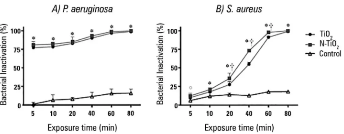
he inactivation kinetics of the two bacterial strains under luorescent light irradiation are illustrated in igure 2. In EETs exposed to irradiation without TiO2 (controls), bacterial inhibition ranging from 1% to 18% was found due to the dehydration induced by irradiation.
For P. aeruginosa, all TiO2-coated ETTs showed higher
bacterial inactivation when compared to controls (p < 0.001 at all time points); no signiicant diference was found between commercial TiO2 and N-TiO2 (p > 0.20 at all time points).
For S. aureus, inactivation was higher than for controls
starting at 5 minutes for N-TiO2 (p = 0.018) and 10
minutes for TiO2 (p = 0.014). For exposure times longer than 10 minutes, both coatings were more eicient than controls (p < 0.001 at each time point). N-TiO2 showed an inactivation higher than that for TiO2 at 20 minutes (p < 0.001), 40 minutes (p < 0.001) and 60 minutes (p < 0.001) of light exposure.
Figure 3 shows representative SEM images of P.
aeruginosa deposed on the internal surface of an ETT
coated with commercial TiO2 before (a) and after (b) 10 minutes of luorescent light irradiation. Figure 4 shows a representative sample of N-TiO2-loaded ETT inoculated with P. aeruginosa after 10 minutes of light exposure with semi-quantitative EDS spectral analysis.
DISCUSSION
The main findings of this study are the following: 1) in the absence of light, neither N-TiO2 nor TiO2 could inhibit bacterial growth within 80 minutes; 2) photocatalysis under fluorescent visible light inhibited bacterial growth in ETTs; 3) no difference in bacterial
inactivation efficacy was observed between N-TiO2
and TiO2 for P. aeruginosa; and 4) bacterial inactivation
was higher in N-TiO2-coated tubes compared to TiO2
for S. aureus.
his is the irst study to investigate a potential clinical application of N-TiO2. Diferently from previous studies, a small CFU count was deposed in the inoculum, mimicking the initial tube contamination that might
occur in vivo. Absorbance measurements conirmed a
shift of the absorption edge towards the visible region in N-TiO2 compared to commercial TiO2 due to a decrease of the energy gap of the lattice, which explains the better eiciency of N-TiO2 found in the present study.
Bacterial inactivation mechanism
Both TiO2 and N-TiO2 were anatase, as previously reported elsewhere,(17,24) and in presence of light uncoated
Figure 2 - Inactivation kinetics of Pseudomonas aeruginosa (A) and Staphylococcus aureus (B) under fluorescent light. * The inactivation of TiO2 and N-TiO2 was significantly higher than that in controls (p < 0.05). ° Only N-TiO2 was more active than controls
Figure 3 - Scanning electron microscopy images (magnification: x 4000; extra high tension: 20 kV) of untreated Pseudomonas aeruginosa cells (A) and treated Pseudomonas aeruginosa cells (B) upon fluorescent light illumination in the presence of commercial TiO2 for 10 minutes.
Figure 4 - Scanning electron microscopy images of a representative sample of endotracheal tubes inoculated with Pseudomonas aeruginosa after 10 minutes of fluorescent light irradiation (left panel). P indicates an area where only the bacterial layer is visible, T represents an area where only TiO2 is visible, and I represents an intermediate region. The right panel shows element weight fractions (%) according to the energy-dispersive X-ray spectroscopy spectra analysis of a representative sample of Pseudomonas aeruginosa
after 10 minutes of treatment with TiO2 under fluorescent light irradiation.
control showed a minimal inactivation activity, due to dehydration.(25) As illustrated in igure 3, before
irradiation (left panel), the TiO2 was not visible and was entirely covered by a bacterial mat. After exposure to visible light (right panel), the white areas corresponding
to the underlying TiO2 were visible and surrounded
by bacterial debris as well as intact cells. Upon EDS analysis, the presence of Na and Cl was mainly due to the solution in which bacteria were suspended (0.9% NaCl) and appeared homogeneously distributed in the three regions, whereas C and N were attributable to bacterial
hese indings suggest that the direct contact between bacteria and TiO2 on the ilter surface increased the extent of oxidative damage, enhancing killing of bacteria in a short time according to mechanism proposed by Foster et al.(27) he authors suggested a mechanism involving
initial damage in the contact areas between the cells and
TiO2, afecting membrane permeability, followed by
increased damage to all cell wall layers, allowing leakage of small molecules such as ions. Further membrane damage allows the leakage of higher molecular weight components, such as proteins. his may be followed by protrusion of the cytoplasmic membrane into the surrounding medium through degraded areas of the peptidoglycan and, eventually, lysis of the cell. Degradation of the internal components of the cell then occurs, followed by complete mineralization.
he advantages of N-TiO2 compared to TiO2 in terms of bacterial inactivation were not explained by diferent crystalline forms, as both nanoparticles were made of anatase with comparable sizes. he slight diference between the BET values indicated that the diferent performances of the two nanoparticles might then be explained by the lower value of the energy gap, which allows major eiciency in the visible photon absorption and in the photocatalytic antimicrobial activity. It must also be noticed that the presence of further defects, such as doping atoms, in the TiO2 lattice enhances electron trapping, which is one of the main mechanisms involved in photocatalytically activated processes.
Bacterial inactivation kinetics
In the absence of luorescent light, bacteria impregnated on the internal surface of endotracheal tubes were insensitive to the two nanoparticle coatings in the examined time window.
In the present study, both TiO2 and N-TiO2
demonstrated the inactivation of P. aeruginosa growth higher than 77% after 5 minutes of exposure (Figure 2). he bactericidal activity began within 5 or 10 minutes depending on the strain and coating and increased with a longer irradiation time. he photocatalytic inactivation appears to be similar to that obtained in a previous study
on Escherichia coli.(17)
In ETTs inoculated with S. aureus, bacterial
inactivation rates comparable to those obtained for P.
aeruginosa were achieved after 40 minutes and 60 minutes
in samples treated with N-TiO2 and TiO2, respectively. As
shown in igure 2, after 5 minutes of light exposure, there were no signiicant diferences between TiO2 and control ETTs. After 20, 40 and 60 min, N-TiO2 was more active than TiO2, and for longer exposure times (80 min), the photocatalytic inactivation reached a plateau at nearly 100% (99 ± 1)%. Previous studies evaluated commercial TiO2 without light exposure supplementation and found a lack of activity against S. aureus.(12)
he kinetics of the photocatalytic inactivation of P.
aeruginosa were equal for TiO2 and N-TiO2 and faster
than those of S. aureus, in accordance with data found by other authors who observed that gram-positive bacteria are more resistant to photocatalysis than gram-negative bacteria.(12,27,28)
Clinical implications
According to our indings, luorescent light irradiation may be suicient to trigger the photocatalytic activity of TiO2 and N-TiO2 without the need for an ultraviolet light source. N-TiO2 shows a higher and faster bacterial inactivation than TiO2 when tested against S. aureus. Our results suggest that photocatalysis under luorescent visible light, especially with N-TiO2, could have clinical applications.
hese results showed that nanosized N-TiO2 can
efectively prevent the growth of a small bacterial inoculum when exposed to conventional luorescent light: this inding could have several practical applications. Nanoparticles-coated materials could be used to prevent or reduce bacterial colonization of medical devices as well as furniture and working surfaces, especially in environments like the ICU, where the high prevalence of multi-resistant strains of bacteria requires non-pharmaceutical measures to control the spread of infection.
It is therefore important to optimize the chemical, physical and morphological properties of this material to further improve disinfection under visible light and study the activity of other microorganisms. In this bench-top study, nanoparticles were loaded in the ETT deposed on an acetate ilter. For clinical applications, the development of a method to depose the nanoparticles on the polymeric matrix of the tube is necessary.
Limitations of the study
photocatalytic process. Since ETTs are placed deeply inside the trachea, further studies should investigate whether ambient light transmission through the tube wall is suicient to activate photocatalysis or whether an especially designed light source should be placed nearby to allow the antimicrobial activity. In the latter case, the fact that N-TiO2 is more efectively activated by luorescent light without the need for a UV source would increase the clinical feasibility of such an approach. he eicacy of the coating was not tested against an active comparator, such as an antibiotic-loaded coating. However, the emergence of multi-drug resistant strains of bacteria in the ICU encourages research in infection control strategies not implying the use of antibiotics. Moreover, further studies are warranted to investigate the antibacterial activity of N-TiO2 against other bacterial strains and its applicability
in vivo.
CONCLUSIONS
he photocatalytic inactivation of P. aeruginosa and
S. aureus by commercial TiO2 and synthesized N-TiO2
was analyzed. Nanosized commercial TiO2 and N-TiO2
inhibit bacterial growth under visible luorescent light. N-TiO2 has higher antibacterial activity against S. aureus
compared to TiO2. he results of this study show that
photocatalysis with the N-TiO2 method was an efective tool for bacteria inactivation. According to our indings, natural light irradiation or room light could be suicient for N-TiO2 activation, so this disinfection method could be constantly operating.
ACKNOWLEDGMENTS
he authors would like to express their gratitude to Cristina Bernini and Laura Negretti for their assistance in SEM characterization. he work was partially supported by the Italian MIUR through the FIRB Project RBAP115AYN “Oxides at the nanoscale: multifunctionality and applications.”
Authors’ contributions
V Caratto and L Ball equally contributed to this
manuscript. V Caratto, L Ball and E Sanguineti
designed the study, collected the data, and wrote and revised the manuscript. M Ferretti and P Pelosi designed the study, interpreted the results and wrote and revised the manuscript. I Firpo and S Alberti collected the data and revised the manuscript. L Ball and A Insorsi performed data analysis and revised the manuscript. All the authors revised the final version of the manuscript.
Objetivo: Avaliar a atividade antibacteriana contra Staphylo-coccus aureus e Pseudomonas aeruginosa de dois revestimentos endotraqueais com nanopartículas e fotocatálise sob luz visível.
Métodos: Testaram-se dois tipos de nanopartículas de titânio: anatase padrão (TiO2) e TiO2 nano-dopada (N-TiO2). As nanopar-tículas foram colocadas em superfície interna de segmentos de tubos endotraqueais comerciais, aplicadas sobre um iltro de acetato de ce-lulose; os tubos endotraqueais controle foram deixados sem revesti-mento de nanopartículas. Em cada tubo endotraqueal foi inoculado um total de 150 unidades formadoras de colônia e, a seguir, estes foram expostos a uma fonte de luz luorescente (3700 lux, compri-mento de onda de 300 - 700nm) por 5, 10, 20, 40, 60 e 80 minu-tos. Contaram-se as Unidades Formadoras de Colônia após 24 horas de incubação a 37ºC. A inativação bacteriana foi calculada como a redução porcentual do crescimento bacteriano em comparação a tubos não expostos à luz.
Resultados: Na ausência de luz, não se observou qualquer atividade antibacteriana relevante contra qualquer das cepas
RESUMO
Descritores: Pneumonia associada à ventilação mecânica; Intubação intratraqueal/instrumentação; Materiais revestidos biocompatíveis; Antibacterianos
estudadas. Para P. aeruginosa, ambos os revestimentos tiveram inativação bacteriana mais elevada do que o controle em qual-quer dos momentos de avaliação (p < 0,001), sendo que não se observaram diferenças entre o revestimento padrão e nano--dopado. Para S. aureus, a inativação foi maior que os controles, começando a partir de 5 minutos para nano-dopado (p = 0,018) e 10 minutos para o revestimento padrão (p = 0,014); a inati-vação com a forma nano-dopada foi maior do que com a forma padrão aos 20 minutos (p < 0,001), 40 minutos (p < 0,001) e 60 minutos (p < 0,001).
REFERENCES
1. Klompas M, Anderson D, Trick W, Babcock H, Kerlin MP, Li L, Sinkowitz-Cochran R, Ely EW, Jernigan J, Magill S, Lyles R, O’Neil C, Kitch BT, Arrington E, Balas MC, Kleinman K, Bruce C, Lankiewicz J, Murphy MV, E Cox C, Lautenbach E, Sexton D, Fraser V, Weinstein RA, Platt R; CDC Prevention Epicenters. The preventability of ventilator-associated events. The CDC Prevention Epicenters Wake Up and Breathe Collaborative. Am J Respir Crit Care Med. 2015;191(3):292-301.
2. Dalmora CH, Deutschendorf C, Nagel F, dos Santos RP, Lisboa T. Defining ventilator-associated pneumonia: a (de)construction concept. Rev Bras Ter Intensiva. 2013;25(2):81-6.
3. Rello J, Ollendorf DA, Oster G, Vera-Llonch M, Bellm L, Redman R, Kollef MH; VAP Outcomes Scientific Advisory Group. Epidemiology and outcomes of ventilator-associated pneumonia in a large US database. Chest. 2002;122(6):2115-21.
4. Safdar N, Dezfulian C, Collard HR, Saint S. Clinical and economic consequences of ventilator-associated pneumonia: a systematic review. Crit Care Med. 2005;33(10):2184-93.
5. American Thoracic Society; Infectious Diseases Society of America. Guidelines for the management of adults with hospital-acquired, ventilator-associated, and healthcare-associated pneumonia. Am J Respir Crit Care Med. 2005;171(4):388-416.
6. Eom JS, Lee MS, Chun HK, Choi HJ, Jung SY, Kim YS, et al. The impact of a ventilator bundle on preventing ventilator-associated pneumonia: a multicenter study. Am J Infect Control. 2014;42(1):34-7.
7. Ataee RA. To: The use of 2% chlorhexidine gel and toothbrushing for oral hygiene of patients receiving mechanical ventilation: effects on ventilator-associated pneumonia. Rev Bras Ter Intensiva. 2014;26(4):438-9. 8. Adair CG, Gorman SP, Feron BM, Byers LM, Jones DS, Goldsmith CE,
et al. Implications of endotracheal tube biofilm for ventilator-associated pneumonia. Intensive Care Med. 1999;25(10):1072-6.
9. Loo CY, Lee WH, Young PM, Cavaliere R, Whitchurch CB, Rohanizadeh R. Implications and emerging control strategies for ventilator-associated infections. Expert Rev Anti Infect Ther. 2015;13(3):379-93.
10. Berra L, Kolobow T, Laquerriere P, Pitts B, Bramati S, Pohlmann J, et al. Internally coated endotracheal tubes with silver sulfadiazine in polyurethane to prevent bacterial colonization: a clinical trial. Intensive Care Med. 2008;34(6):1030-7.
11. Tarquinio K, Confreda K, Shurko J, LaPlante K. Activities of tobramycin and polymyxin E against Pseudomonas aeruginosa biofilm-coated medical grade endotracheal tubes. Antimicrob Agents Chemother. 2014;58(3):1723-9.
12. Tarquinio KM, Kothurkar NK, Goswami DY, Sanders RC Jr, Zaritsky AL, LeVine AM. Bactericidal effects of silver plus titanium dioxide-coated endotracheal tubes on Pseudomonas aeruginosa and Staphylococcus aureus. Int J Nanomedicine. 2010;5:177-83.
13. Berra L, Curto F, Li Bassi G, Laquerriere P, Pitts B, Baccarelli A, et al. Antimicrobial-coated endotracheal tubes: an experimental study. Intensive Care Med. 2008;34(6):1020-9.
14. Machado MC, Tarquinio KM, Webster TJ. Decreased Staphylococcus aureus biofilm formation on nanomodified endotracheal tubes: a dynamic airway model. Int J Nanomedicine. 2012;7:3741-50.
15. Wolfrum EJ, Huang J, Blake DM, Maness PC, Huang Z, Fiest J, et al. Photocatalytic oxidation of bacteria, bacterial and fungal spores, and model biofilm components to carbon dioxide on titanium dioxide-coated surfaces. Environ Sci Technol. 2002;36(15):3412-9.
16. Li Q, Mahendra S, Lyon DY, Brunet L, Liga MV, Li D, et al. Antimicrobial nanomaterials for water disinfection and microbial control: potential applications and implications. Water Res. 2008;42(18):4591-602. 17. Caratto V, Aliakbarian B, Casazza AA, Setti L, Bernini C, Perego P, et al.
Inactivation of Escherichia coli on anatase and rutile nanoparticles using UV and fluorescent light. Mater Res Bull. 2013;48(6):2095-101. 18. Liou JW, Chang HH. Bactericidal effects and mechanisms of visible
light-responsive titanium dioxide photocatalysts on pathogenic bacteria. Arch Immunol Ther Exp (Warsz). 2012;60(4):267-75.
19. Ren G, Hu D, Cheng EW, Vargas-Reus MA, Reip P, Allaker RP. Characterisation of copper oxide nanoparticles for antimicrobial applications. Int J Antimicrob Agents. 2009;33(6):587-90.
20. Cioffi N, Torsi L, Ditaranto N, Tantillo G, Ghibelli L, Sabbatini L, et al. Copper Nanoparticle/polymer composites with antifungal and bacteriostatic properties. Chem Mater. 2005;17(21):5255-62.
21. Sondi I, Salopek-Sondi B. Silver nanoparticles as antimicrobial agent: a case study on E. coli as a model for Gram-negative bacteria. J Colloid Interface Sci. 2004;275(1):177-82.
22. Mathur A, Parashar A, Chandrasekaran N, Mukherjee A. Nano-TiO2 enhances biofilm formation in a bacterial isolate from activated sludge of a waste water treatment plant. Int Biodeterior Biodegrad. 2017;116:17-25. 23. Qiang L, Shi X, Pan X, Zhu L, Chen M, Han Y. Facilitated bioaccumulation
of perfluorooctanesulfonate in zebrafish by nano-TiO2 in two crystalline phases. Environ Pollut. 2015;206:644-51.
24. Caratto V, Setti L, Campodonico S, Carnasciali MM, Botter R, Ferretti M. Synthesis and characterization of nitrogen-doped TiO2 nanoparticles prepared by sol-gel method. J Sol-Gel Sci Technol. 2012;63(1):16-22. 25. Caballero L, Whitehead KA, Allen NS, Verran J. Inactivation of Escherichia
coli on immobilized TiO2 using fluorescent light. J Photochem Photobiol Chem. 2009;202(2-3):92-8.
26. Barros J, Grenho L, Manuel CM, Ferreira C, Melo L, Nunes OC, et al. Influence of nanohydroxyapatite surface properties on Staphylococcus epidermidis biofilm formation. J Biomater Appl. 2014;28(9):1325-35. 27. Foster HA, Ditta IB, Varghese S, Steele A. Photocatalytic disinfection using
titanium dioxide: spectrum and mechanism of antimicrobial activity. Appl Microbiol Biotechnol. 2011;90(6):1847-68.