Printed version ISSN 0001-3765 / Online version ISSN 1678-2690 www.scielo.br/aabc
http://dx.doi.org/10.1590/0001-3765201520140122
Altered hyaluronic acid content in tear luid
of patients with adenoviral conjunctivitis
JULIANA L. DREYFUSS1,2*, CAIO V. REGATIERI1,3,5*, BRUNO COELHO1, JOSÉ B. BARBOSA3, DENISE DE FREITAS3, HELENA B. NADER1 and JOÃO R. MARTINS1,4
1Departamento de Bioquímica, Disciplina de Biologia Molecular, Universidade Federal de São Paulo, Rua Três de Maio, 100, 4º andar, 04044-020 São Paulo, SP, Brasil
2
Departamento de Informática em Saúde, Grupo Interdisciplinar de Ciências Exatas em Saúde, Universidade Federal de São Paulo, Rua Botucatu, 862, 04023-062 São Paulo, SP, Brasil
3
Departamento de Oftalmologia, Universidade Federal de São Paulo, Rua Botucatu, 821, 04023-062 São Paulo, SP, Brasil
4Departamento de Medicina, Disciplina de Endocrinologia, Universidade Federal de São Paulo, Rua Borges Lagoa, 800, 04038-001 São Paulo, SP, Brasil
5
Department of Ophthalmology, New England Eye Center, Tufts Medical Center, 800 Washington Street, 02111 Boston, MA, USA
Manuscript received on March 13, 2014; accepted for publication on October 24, 2014
ABSTRACT
The adenoviral conjunctivitis is one of the biggest causes of conjunctival infection in the world. Conjunctivitis causes relatively nonspecific symptoms, as hyperaemia and chemosis. Even after biomicroscopy, complex laboratory tests, such as viral culture, are necessary to identify the pathogen or its etiology. To contribute to the better understanding of the pathobiology of the adenoviral conjunctivitis, the tear fluids of patients with unilateral acute adenovirus conjunctivitis (UAAC), normal donors (control) and patients with allergic conjunctivitis were analyzed. Tear samples were collected with Schirmer strips from control, allergic conjunctivitis and UAAC patients, diagnosed by clinical signs. UAAC tears were tested positive in viral cultures. After the elution, HA was quantified using an ELISA-like luorometric assay and the protein proile was determined by SDS-PAGE. A profound increase in the HA tear content in UAAC patients was found when compared to control and ALC. This HA increase in UAAC tears remarkably was not observed in tears from contralateral eyes without clinical signs, nor in allergic conjunctivitis. In addition a distinct profile of UAAC tear proteins was observed in patients with UAAC. The quantification of HA in the tear fluid is a rapid, sensitive and specific test. This molecule might be a biomarker candidate for acute conjunctivitis.
Key words: conjunctivitis, glycosaminoglycans, hyaluronic acid, tear ilm.
Correspondence to: Juliana L. Dreyfuss E-mail: jdreyfuss@unifesp.br
*Both authors contributed equally to this work INTRODUCTION
The adenovirus conjunctivitis is one of the most common conjunctival infection. Normally it does not
A healthy ocular surface requires a functional
tear ilm. The normal function of tear luid is due
to its complex biochemical composition consisting of buffered electrolytes and a diversity of proteins and glycoconjugates (Van Haeringen 1981, Baker et al. 2006).
Hyaluronic acid (HA), an important com-ponent of the extracellular matrix, is a large glycosaminoglycan composed of repeating units of
β-D-glucuronic acid and N–acetyl-D-glucosamine. HA plays an important role in tissue development,
cell migration, cell proliferation and inlammation
(Inoue and Katakami 1993, Gomes et al. 2004).
HA is increased in the tear luid when corneal
epithelium erosion is present, and may play an important role in corneal epithelium wound healing
(Oya et al. 1995, Miyauchi et al. 1996). Tear luid
consistency (gel-like) can be attributed to HA (Itano et al. 1999). Another important characteristic of this GAG is its chemical structure that can attract ions and water due to a negative charge density (Frescura et al. 1994, Yoshida et al. 1996).
Endogenous hyaluronan is present in virtually all corneal disorders (Fitzsimmons et al. 1994). HA is normally detected only in the corneal endothelium, and the presence of any amount of this compound in the epithelium or stroma is likely to indicate altered tissue (Inoue and Katakami 1993). Vitreous humor contains HA and its receptor (CD44), and these molecules are also present at the apical surface of corneal endothelium (Tengblad 1979).
Tear proteins also play an important role in maintaining eye surface integrity and in patients with external eye diseases (Avisar et al. 1981, Van Haeringen 1981, Ballow et al. 1987). The protein fraction of normal tears contains antimicrobial factors which are important for protecting the external eye from infection. These substances are produced by the main and accessory lacrimal glands. Important components of the hosts defense system for the external eye include complement proteins, immunoglobulins, especially secretory IgA (sIgA), lysozyme, lipocalin (TSPA)
and lactoferrin (Friedman 1990, Kuizenga et al. 1991, Tragoulias et al. 2005). Lactoferrin, an iron complexing protein in normal tears, has bacteriostatic, bactericidal and complement (C) inhibitory activity properties which make this tear protein an important component
of the nonspeciic host defense system of the external
eye (Ballow et al. 1987, Flanagan and Willcox 2009). Human tear prealbumin, now called tear lipocalin, was
described as a major protein in tear luid, and is the
main lipid binding protein in tears. It exerts important
functions in eyelid lubriication, and acting as a
general protection factor for cornea and conjunctiva
epithelia (Redl 2000). IgA is the only signiicant immunoglobulin found in tear ilm. The antibodies are
postulated to play a role in preventing adherence of microorganisms to the ocular surface (Alizadeh et al. 2001, Knop and Knop 2005). Another component of
tears is lysozyme, a bacteriolytic protein irst described
by Fleming (Fleming 1922, McClellan 1997, Caffery et al. 2008). An allergen exposure can cause a marked increase in lysozyme secretion (Proud et al. 1998).
The aim of this study was to quantify HA
content and verify the protein proile of tear luids
from patients with adenoviral conjunctivitis and compare them with normal donors.
MATERIALS AND METHODS
SUBJECTS
presented all of the following signs and symptoms: conjunctival hyperemia, ocular secretion, no corneal epithelium erosion, conjunctival follicular reaction, no use of any ocular medication and symptoms that had started for no more than 3 days prior. In the contralateral eye group, we included contralateral eyes from patients with UAAC that didn’t have any clinical signs of acute conjunctivitis. Diagnosis of allergic conjunctivitis was based on clinical history and evaluation of signs and symptoms. Patients
included in the study were in an active inlammatory phase of the disease with active limbal iniltrates
and were free of topical antihistamines and mast cell stabilizers for at least 3 days, topical corticosteroids for at least 7 days, and systemic antiallergic treatment
for at least 2 week at the time of irst presentations.
The control group constituted of patients without any ocular or systemic diseases.
The ethics Committee of the Federal University of São Paulo approved this protocol and a written informed consent was obtained from each subject. The study protocol adhered to the tenets of The Declaration of Helsinki.
SAMPLE COLLECTION
For collecting the tears,Schirmer strips were placed in the temporal side of each eyeunder the eyelid, during 5 minutes, without any use of topicalanesthetics. The strips were dried at room temperatureand stored at –20°C until analysis. The same procedure was performed for the three groups.
ADENOVIRAL CULTURE
At the same time, conjunctival swabs were collected in the affected eye group in medium appropriate for adenoviral culture, using Hep-2 cells as described by de Paiva et al. 1992.
TEAR SAMPLE PREPARATION
Tear compounds were eluted from the Schirmer strips using 100 µL of distilled water, and HA and protein content analyses were performed.
HAMEASUREMENT
HA content in tear luids was assayed by a non-competitive and non-isotopic luoroassay (Martins et al. 2003). Eluted tear luids and standard
concentrations of HA (Sigma, St. Louis, MO) were added to 96 multiwell plates (FluoroNUNC Maxisorp-microtiterplates, Roskilde, Denmark) previously coated with HA-binding protein. The plates were then sequencially incubated with biotinylated HA-binding protein and europium-labeled streptavidin (Amershan, Piscataway, NJ). Afterwards, the europium remaining in the solid phase was released by an enhancement solution and
the luorescence was measured using a time-resolved luorometer (Perkin–Elmer Life Sciences-Wallac Oy,
Turku, Finland). The data (counts/s) were processed automatically using the MultiCalc software program (Perkin-Elmer Life Sciences-Wallac Oy) and values are expressed as ng/mg protein.
PROTEIN ANALYSIS
Total tear protein concentration was determined using a colorimetric assay kit according to the manufacturer’s instructions (Protein Assay Kit
from Bio-Rad, Hercules, CA). The protein proile
was analyzed through sodium dodecylsulfate polyacrylamide gel electrophoresis (SDS-PAGE)
as previously described (Laemmli 1970). Briely,
10 µg of protein from the tear samples were applied to a 3-20% linear gradient polyacrylamide gel under reducing conditions. After electrophoresis, Patients
UAAC
Patients ALC
Donors (both eyes)
No. 15 15 23
Age (mean ± SD) 32.6 ± 14.7 11.8 ± 3.72 25.2 ± 3.02 Sex
F 9 4 15
M 6 11 8
TABLE I
Patients and donors characteristics.
the gels were stained by comassie blue (Bio-Rad,
Hercules, CA). Each protein band was quantiied
by densitometry using the software ImageJ Version 10.2 for Mac (U.S. National Institutes of Health, Bethesda, Maryland, USA). The results are expressed by arbitrary densitometric units (ADU).
STATISTICAL ANALYSIS
Data are expressed as mean ± standard error of the mean. Statistical analyses were performed using One-way ANOVA with Bonferroni´s Multiple Comparison test, using Graph Pad Prism 5.0
software for Mac. A 95% conidence interval and a 5% level of signiicance were adopted; therefore,
results with P-value less than or equal to 0.05 were
considered signiicant.
RESULTS
COMPARISON OF HYALURONIC ACID CONTENT IN TEAR FLUID FROM PATIENTS AFFECTED BY CONJUNCTIVITIS AND NORMAL DONORS
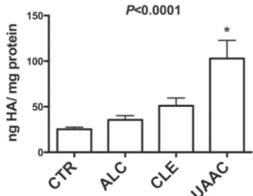
The HA content in tears from eyes with UAAC (n=15), from contralateral eyes (n=15), from eyes ofnormal donors (n=46) and from allergic conjunctivitis (n=26) was: 102.9 ± 19.9 ng/ mg protein; 51.02 ± 8.5 ng/mg protein, 25.2 ± 2.3 ng/mg protein and 35.5 ± 4.5 ng/mg protein, respectively. The comparison of HA content among
the three groups showed a signiicant difference
(P< 0.0001) (Fig. 1). UAAC tears presented higher HA concentrations when compared to either normal tears (P< 0.0001), tears from contralateral eyes or tears from allergic conjunctivitis. HA content in tears from contralateral eyes did not differ (P> 0.05) from tears from normal donors or from tears from patients affected by allergic conjunctivitis.
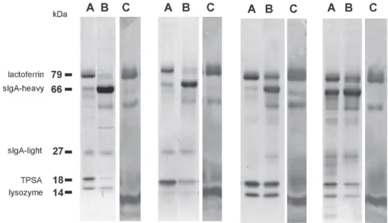
PROTEIN CONTENT AND QUALITATIVE ANALYSIS OF TEARS The protein analysis showed distinct protein
proiles when comparing tears from UAAC eyes
to tears from contralateral eyes. Polypeptide
bands were identiied by matching their migrations
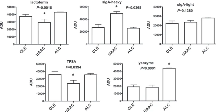
to those of molecular weight standards. Tear protein patterns from normal donors have been exhaustively reported by previous investigators (Ballow et al. 1987, Lopez-Cisternas et al. 2006, Mann and Tighe 2007) and are represented by lactoferrin (79 kDa), sIgA-heavy chain (66 kDa), sIgA-light chain (27 kDa), lipocalin (TSPA, 18 kDa), and lysozyme (14 kDa) (Fig. 2). Figure 3 shows that eyes affected by UAAC presented higher amounts of sIgA-heavy chain when compared to the contralateral eyes (CLE) or allergic conjunctivitis (ALC) (P=0.0368). On the other hand, UAAC presented lower quantities of lipocalin or TSPA (P=0.0394) and lactoferrin (P=0.0018) when compared to CLE. No differences were found in the protein content of sIgA-light chain among UAAC, CLE and ALC. Finally, lysozyme is increased in tears from ALC compared to UAAC or CLE (P<0.0001).
DISCUSSION
We show that UAAC leads to a signiicant increase
in tear HA content. The difference found between Figure 1 - Hyaluronic acid content in tear luid of UAAC patients, normal donors and allergic conjunctivitis. HA was
measured in tears by a non-competitive luorometric
HA tear content in UAAC was 2 times greater than that from contralateral eyes without clinical signs and 4 times higher than in the tears from normal donors indicating that HA could be a marker for subclinical
inlammation. HA levels in tears of patients affected
by allergic conjunctivitis showed no differences compared to tears from normal subjects.
Our results show that the levels of HA content
in the tear luid are related to the viral injury and conjuctival inlammation, suggesting that higher
levels lead to a situation in which the viscoelastic properties of HA may protect corneal epithelium and help promote wound healing (Laurent and Fraser 1992, Miyauchi et al. 1996).
We may also consider that HA may be playing
an important role in promoting inlammatory cell
migration to the conjunctiva, possibly modulating
inlammatory cytokine release (Mummert 2005). UAAC, CLE and ALC tear protein proiles
showed 79, 66, 27, 24, 18, and 14 kDa bands, which were a constant feature with different amounts
of each protein (Fig. 2). The protein proile of
tears is well established in normal eye (Kuizenga et al. 1991). In accordance to the data reported by other authors (Janssen and Van Bijsterveld 1981, Kuizenga et al. 1991), we propose that the proteins observed in the present study correspond to lactoferrin (79 kDa), sIgA-heavy chain (66 kDa), sIgA-light chain (27 kDa), lipocalin (TSPA, 18 kDa), and lysozyme (14 kDa), respectively.
A higher content of sIgA was found in UAAC, when compared to CLE. Secretory IgA-enriched
luid presumably augments the effectiveness of
the external barrier to microbial adherence and
increases the eficiency by which pathogens are processed by the immune and inlammatory
systems (Sack et al. 1992). No differences were found in sIgA light chain content.
The decrease in lactoferrin and lipocalin levels
in UAAC tears may occur evoked by major relex tearing caused by inlammation (de Paiva et al.
1992). Furthermore, lactoferrin has been shown to
Figure 2 - Protein proile in tear of allergic and adenoviral conjunctivitis.Electrophoretic proiles of
human tear luid on SDS-PAGE revealed by Coomassie blue staining. In A (contralateral eye), B (acute
adenoviral conjunctivitis eye) and C (allergic conjunctivitis), the corresponding proiles of 4 different
subjects are shown. Protein bands were identiied by matching their migrations to those of molecular
possess antiviral activity against adenovirus. This possible down regulation may be an evolutionary mechanism used by adenovirus to survive in ocular tissue. There was no difference between UAAC and CLE regarding lysozyme secretion, but it was increased in ALC tears, since an allergen exposure can cause a marked increase in lysozyme secretion (Proud et al. 1998).
CONCLUSIONS
In this study we were able to identify an important increase in HA tear content in patients affected by UAAC, compared with normal donors or allergic conjunctivitis, using a rapid and non invasive method. Regarding protein analysis we could verify
a distinct protein proile in tear luid of UAAC
patients. Taken together these results indicate that the increase in HA content seems to play a role in
the tear luid of UAAC patients. The quantiication of HA in the tear luid is a rapid, sensitive and
speciic test. This molecule might be a biomarker
candidate for acute conjunctivitis.
ACKNOWLEDGMENTS
This work was supported by grants from Brazilian funding agencies Fundação de Amparo à Pesquisa do Estado de São Paulo (FAPESP), Conselho Nacional
de Desenvolvimento Cientíico e Tecnológico
(CNPq) and Coordenação de Aperfeiçoamento de Pessoal de Nível Superior (CAPES). We are indebted to Aline Mendes, M.Sc. for technical assistance, and to Vivien Jane Coulson-Thomas, Ph.D., for the English revision. Prof. Martins had full access to all the data in the study and takes responsibility for the integrity of the data and the accuracy of the data analysis.
ABBREVIATIONS:
UAAC, unilateral acute adenovirus conjunctivitis; CLE, contralateral non-affected eye; ALC, allergic Figure 3 - Protein quantiication in tearof allergic and adenoviralconjunctivitis. Protein bands of human tear luid on SDS-PAGE
were quantiied by densitometry: lactoferrin (79 kDa), serum albumin and sIgA-heavy chain (66 kDa), sIgA-light chain (27 kDa),
conjunctivitis; HA, hyaluronic acid; GAG, glyo-saminoglycan; SDS-PAGE, sodium dodecylsulfate polyacrylamide gel electrophoresis.
RESUMO
A conjuntivite causada por adenovírus é uma das maiores causas de infecção da conjuntiva no mundo. A conjuntivite provoca sintomas relativamente inespecíicos, como hiperemia e quemose. Mesmo depois de biomicroscopia, testes laboratoriais complexos, como cultura viral, são necessários para identiicar o patógeno ou sua etiologia. Para contribuir para o melhor entendimento da isiopatologia da conjuntivite causada por adenovírus, lágrimas de pacientes com conjuntivite aguda unilateral causada por adenovírus (UAAC), de doadores normais (controle) e de pacientes com conjuntivite alérgica foram analisadas. As amostras foram coletadas com tiras de Schirmer de doadores normais, pacientes com conjuntivite alérgica e pacientes com UAAC diagnosticados por sinais clínicos e testes positivos em culturas virais. Após a eluição das lágrimas, o HA foi quantiicado utilizando um ensaio luorométrico semelhante ao ELISA e o peril da proteína foi determinado por SDS-PAGE. Um aumento profundo no conteúdo de HA em lágrima de pacientes com UAAC foi encontrado quando comparado com o controle ou a conjuntivite alérgica. Este aumento de HA em lágrimas de pacientes com UAAC não foi observado em lágrimas do olho contralateral sem sinais clínicos ou de pacientes com conjuntivite alérgica. Além disso observou-se um peril distinto de proteínas nas lágrimas de pacientes com UAAC. A quantiicação de HA no luido lacrimal é um ensaio rápido, sensível e especíico. Esta molécula pode ser um bom candidato a biomarcador para conjuntivite aguda.
Palavras-chave: conjuntivite, glicosaminoglicanos, ácido hialurônico, lágrima.
REFERENCES
ALIZADEH H, APTE S, EL-AGHA MS, LI L, HURT M, HOWARD K, CAVANAGH HD, MCCULLEY JP AND NIEDERKORN JY. 2001. Tear IgA and serum IgG antibodies against Acanthamoeba in patients with Acanthamoeba keratitis. Cornea 20: 622-627.
AVISAR R, MENACHE R, SHAKED P AND SAVIR H. 1981. Lysozyme content of tears in some external eye infections. Am J Ophthalmol 92: 555-558.
BAKER GR, MORTON M, RAJAPASKA RS, BULLOCK M, GULLU S, MAZZI B AND LUDGATE M. 2006. Altered tear composition in smokers and patients with graves ophthalmopathy. Arch Ophthalmol 124: 1451-1456.
BALLOW M, DONSHIK PC, RAPACZ P AND SAMARTINO L. 1987. Tear lactoferrin levels in patients with external
inlammatory ocular disease. Invest Ophthalmol Vis Sci
28: 543-545.
BUTT AL AND CHODOSH J. 2006. Adenoviral keratocon-junctivitis in a tertiary care eye clinic. Cornea 25: 199-202. CAFFERY B, JOYCE E, BOONE A, SLOMOVIC A, SIMPSON T,
JONES L AND SENCHYNA M. 2008. Tear lipocalin and lysozyme in Sjogren and non-Sjogren dry eye. Optom Vis Sci 85: 661-667.
DE PAIVA TM, TAKIMOTO S, ISHIDA MA, DE SOUZA MC, ISHIMARU T, NEUMANN J AND KALIL J. 1992. Comparative study of adenoviruses with monoclonal antibodies. Rev Inst Med Trop Sao Paulo 34: 19-26.
FITZSIMMONS TD, MOLANDER N, STENEVI U, FAGERHOLM P, SCHENHOLM M AND VON MALMBORG A. 1994. Endogenous hyaluronan in corneal disease. Invest Ophthalmol Vis Sci 35: 2774-2782.
FLANAGAN JL and Willcox MD. 2009. Role of lactoferrin in
the tear ilm. Biochimie 91: 35-43.
FLEMING A. 1922. On a Remarkable Bacteriolytic Element Found in Tissues and Secretions. Proceedings of the Royal Society of London. Series B, Containing Papers of a Biological Character 93: 306-317.
FRESCURA M,BERRY M, Corield A, Carrington S and Easty DL. 1994. Evidence of hyaluronan in human tears and secretions of conjunctival cultures. Biochem Soc Trans 22: 228S.
FRIEDMAN MG. 1990. Antibodies in human tears during and after infection. Surv Ophthalmol 35: 151-157.
GOMES JA, AMANKWAH R, POWELL-RICHARDS A AND DUA HS. 2004. Sodium hyaluronate (hyaluronic acid) promotes migration of human corneal epithelial cells in vitro. Br J Ophthalmol 88: 821-825.
INOUE M AND KATAKAMI C. 1993. The effect of hyaluronic acid on corneal epithelial cell proliferation. Invest Ophthalmol Vis Sci 34: 2313-2315.
ITANO N ET AL. 1999. Three isoforms of mammalian hyaluronan synthases have distinct enzymatic properties. J Biol Chem 274: 25085-25092.
JANSSEN PT AND VAN BIJSTERVELD OP. 1981. Comparison of electrophoretic techniques for the analysis of human tear
luid proteins. Clin Chim Acta 114: 207-218.
KNOP E AND KNOP N. 2005. The role of eye-associated lymphoid tissue in corneal immune protection. J Anat 206: 271-285.
LAEMMLI UK. 1970. Cleavage of structural proteins during the assembly of the head of bacteriophage T4. Nature 227: 680-685.
LAURENT TC AND FRASER JR. 1992. Hyaluronan. FASEB J 6: 2397-2404.
LOPEZ-CISTERNAS J, CASTILLO-DIAZ J, TRAIPE-CASTRO L AND LOPEZ-SOLIS RO. 2006. Use of polyurethane minisponges
to collect human tear luid. Cornea 25: 312-318.
MANN AM AND TIGHE BJ. 2007. Tear analysis and lens-tear
interactions. Part I. Protein ingerprinting with microluidic
technology. Cont Lens Anterior Eye 30: 163-173. MARTINS JR, PASSEROTTI CC, MACIEL RM, SAMPAIO LO,
DIETRICH CP AND NADER HB. 2003. Practical determination
of hyaluronan by a new noncompetitive
luorescence-based assay on serum of normal and cirrhotic patients. Anal Biochem 319: 65-72.
MCCLELLAN KA. 1997. Mucosal defense of the outer eye. Surv Ophthalmol 42: 233-246.
MIYAUCHI S, MORITA M, KURAMOTO K AND HORIE K. 1996. Hyaluronan and chondroitin sulfate in rabbit tears. Curr Eye Res 15: 131-135.
MUMMERT ME. 2005. Immunologic roles of hyaluronan. Immunol Res 31: 189-206.
NATIVIDAD A, COOKE G, HOLLAND MJ, BURTON MJ, JOOF HM, ROCKETT K, KWIATKOWSKI DP, MABEY DC AND BAILEY RL. 2006. A coding polymorphism in matrix metalloproteinase 9 reduces risk of scarring sequelae of ocular Chlamydia trachomatis infection. BMC Med Genet 7: 40.
OKADA AA AND FORRESTER JV. 2000. Ocular inlammatory disease in the new millennium. Arch Ophthalmol 118: 116-119.
OYA T, OBATA H, MIYATA K, TSURU T AND MIYAUCHI S. 1995. Quantitative analyses of glycosaminoglycans in tear luids
in normal human eyes and eyes with corneal epithelial disorders. Nippon Ganka Gakkai Zasshi 99: 302-307. PROUD D, REYNOLDS CJ, LICHTENSTEIN LM, KAGEY
-SOBOTKA A AND TOGIAS A. 1998. Intranasal salmeterol inhibits allergen-induced vascular permeability but not
mast cell activation or cellular iniltration. Clin Exp
Allergy 28: 868-875.
REDL B. 2000. Human tear lipocalin. Biochim Biophys Acta 1482: 241-248.
SACK RA, TAN KO AND TAN A. 1992. Diurnal tear cycle:
evidence for a nocturnal inlammatory constitutive tear luid. Invest Ophthalmol Vis Sci 33: 626-640.
TENGBLAD A. 1979. Afinity chromatography on immobilized hyaluronate and its application to the isolation of hyaluronate binding properties from cartilage. Biochim Biophys Acta 578: 281-289.
TRAGOULIAS ST, ANDERTON PJ, DENNIS GR, MIANO F AND MILLAR TJ. 2005. Surface pressure measurements of
human tears and individual tear ilm components indicate
that proteins are major contributors to the surface pressure. Cornea 24: 189-200.
VAN HAERINGEN NJ. 1981. Clinical biochemistry of tears. Surv Ophthalmol 26: 84-96.