Prevalência de conjuntivite adenoviral em
clínica oftalmológica no município de Viçosa/MG
Prevalence of adenoviral conjunctivitis at the
ophthalmologic clinic on municipality of Viçosa/MG
Euldes Nei Rosado-Filho
1, Silvia Almeida Cardoso
2, Lorena Nacif Marçal
3, Eliziária Cardoso dos Santos
3, Eduardo de
Almeida Marques da Silva
4, Sérgio Oliveira de Paula
4, Leandro Licursi de Oliveira
4,1, 4 Department of General Biology, Universidade Federal de Viçosa (UFV) – Viçosa (MG), Brazil;
2, 3 Associate Professor, Department of General Biology, Universidade Federal de Viçosa (UFV) – Viçosa (MG), Brazil.
Received for publication 29/10/2012 - Accepted for publication 28/01/2013
The authors declare no conflists of interest
A
BSTRACTObjective: The aim of this study was to evaluate the prevalence of Adenovirus as a etiologic agent of conjunctivitis on a ophthalmic clinic in Viçosa, Minas Gerais, Brazil. Methods: Samples of conjunctival secretion from 91 patients clinically diagnosed with conjunctivitis were subjected to polymerase chain reaction (PCR) using degenerate primers targeted to the gene encoding the structural protein II. Positive samples were subsequently subjected to sequencing and genotyping. Results: PCR results showed 36.3% prevalence of Adenovirus. No differences between the sexes and was found to be higher in the age group 26-65 years with 60.60% of the positive cases. Sequencing of positive cases showed the presence of Adenovirus serotypes 3, 4, 7, 8, and 34 circulating in the region. Conclusion: In Viçosa two in five cases of conjunctivitis has Adenovirus as etiologic agent.
Keywords: Virology; Adenovirus/epidemiology; Conjunctivitis viral/etiology; Conjunctivitis viral/diagnosis; Polimerase chain reaction
R
ESUMOObjetivo: O objetivo deste estudo foi avaliar a prevalência de Adenovirus como agente etiológico da conjuntivite, em clínica médica oftalmológica especializada, em Viçosa, Minas Gerais, Brasil. Métodos: Amostras da secreção conjuntival de 91 pacientes clinicamente diagnosticados com conjuntivite foram submetidas à reação em cadeia da polimerase (PCR) utilizando primers degenerados para a região codificadora do gene da proteína estrutural II. Posteriormente as amostras positivas foram submetidas a sequenciamento e genotipagem. Resultados: A análise dos resultados de PCR revelou prevalência de 36,3% de Adenovirus. Não havendo distinção entre os sexos e com maior prevalência na faixa etária de 26 a 65 anos com 60,60% dos casos positivos. O sequenciamento dos casos positivos por Adenovirus revelaram a presença dos sorotipos 3, 4, 7, 8 e 34 circulante na região. Conclusão: No município de Viçosa, dois em cada cinco casos de conjuntivite são de etiologia adenoviral.
Descritores: Virologia; Adenovirus/epidemiologia; Conjuntivite viral/etiologia; Conjuntivite viral/diagnóstico;Reção em cadeia da polimerase
I
NTRODUCTIONC
onjunctivitis is considered one of the most common disorders of ophthalmic emergency worldwide (1). It hasviruses and bacteria as its main etiologic agents, is highly contagious and can occur in outbreaks(2,3). The viral conjunctivitis
has in most cases the adenovirus (AdVs) as the causative agent, which are viruses with double-stranded DNA, non-enveloped, lytic and with icosahedral morphology.
There are more than 55 AdVs serotypes already identified. They are classified into seven distinct groups (AdV A-F), based on their biological phytochemical, and genetic characteristics(4).
Adenoviral conjunctivitis cases are caused most often by AdV-3 (AdV-B), AdV -4 (AdV-E), AdV -8, -19a, -AdV-37, -5AdV-3, and -54 (AdV-D)(4, 5).
Conjunctivitis can also be caused by other viruses such as Herpes Virus, Coxsackievirus, Rhinovirus, Echovirus, Enterovirus, Molluscum Contagiosum Virus, among others. The Enterovirus 70 and Coxsackievirus A4 are both related to the hemorrhagic form of viral conjunctivitis(6).
Most eye disorders secondary to the infection with Adenovirus are presented in the form of simple follicular conjunctivitis, faringoconjuntival fever and epidemic keratoconjunctivitis. Simple follicular conjunctivitis is usually self-limited, transitory and non-associated to systemic dysfunctions(7).
The serotypes 1 to 11 and 19 are the primary cause of non-specific follicular conjunctivitis(8-10). Faringoconjuntival fever, most
commonly caused by serotypes 3, 4, 5 and 7 of the Adenovirus, is characterized by fever, headache, pharyngitis, follicular conjunctivitis, and preauricular adenopathy. In some isolated cases, the systemic signs can mimic an infection caused by the influenza virus(11,12). The epidemic keratoconjunctivitis is often
caused by serotypes 8, 19 and 37 of group AdV-D, with significant involvement of the cornea. In most patients it is presented in the bilateral form, preceded by upper respiratory tract infection(13).
The assessment of systemic signs associated with precise molecular biology techniques is key for the clinical diagnosis and prevention of epidemics during the acute phase of adenoviral conjunctivitis(3). Some of the most important
conventional techniques for the clinical diagnosis of viral conjunctivitis include, among others, cytological investigation of the conjunctiva, assessment of the cytopathic effect in susceptible cell lines, detection of antigens in the cells of conjunctiva by direct fluorescence or detection of antibodies. Despite the sensitivity of detection of the same, these techniques are time consuming and costly from an economic point of view, what becomes a challenge for detection and a rapid diagnosis of AdV as caused by conjunctivitis. Other important, relatively simple, fast, highly sensitive and low cost diagnostic method which has been used a lot in the last decade is the polymerase chain reaction (PCR)(13-16).
Considering the importance of accuracy of early diagnosis and specific treatment of adenoviral conjunctivitis, the objective of the present study is to evaluate the frequency of Adenovirus in the conjunctival secretion of the patients assisted in eye clinic located in the municipality of Viçosa, Minas Gerais, Brazil, in the period from May 2009 to January 2011, using PCR as the technique.
M
ETHODSThis is a cross-sectional study on patients assisted in a specialized eye clinic located in the municipality of Viçosa-MG, in the period from May 2009 to January 2011. A total of 91 patients of both genders aged between 1 and 74 years with symptoms indicative of viral conjunctivitis (hyperemic eyes, watery discharge and discomfort) underwent ocular inspection followed by thorough ophthalmologic assessment. After assessment, patients with diffuse conjunctival hyperemia presenting ocular discomfort, palpebral edema and tearing with watery secretion, follicular reaction in the tarsal conjunctiva, satellite lymphadenopathy and keratitis with multiple sub-epithelial infiltrates were considered as possible cases of conjunctivitis of adenoviral etiology.
After the diagnosis confirmation, the patients were informed about the possibility of Adenovirus prevalence as the causative agent of the disease, and were invited to participate in a clinical research for the detection of the same. The 91 patients who volunteered signed a written consent to participate in the study after being aware of the clinical implications of the same. The principles of Bioethics were guaranteed to all participants, according to regulation 196/96 of the National Health Council determining the ethical aspects necessary for conducting research in humans (CEPH-167741).
Subsequently, a conjunctival secretion was collected from the anterior eyeball of each patient with aid of a sterile rayon swab soaked in solution of 0.9% sodium chloride, using slit lamp (Topcon 3E). The material obtained was placed in sterile 1.5 ml polypropylene tubes previously identified and containing phosphate buffered saline solution (PBS), and freezed for subsequent viral DNA extraction, PCR and gene sequence analysis for the identification of the serotype responsible for the infection.
The viral DNA of all samples of conjunctival secretion supposedly infected by Adenovirus have been extracted using a specific kit for viral RNA and DNA extraction (QIAamp UltraSens Virus-Quiagen). The extraction was carried out according to the manufacturer’s instructions. Then, 500mL of sample were treated with 500mL of buffer AR and 20mL of proteinase K, and incubated at 40°C for 10 min. Later 300mL of buffer AB were added and the material was applied to a QIAamp affinity column, washed and eluted with 30mL of buffer AVE. The eluted material was used for the PCR reactions; samples of patients with a healthy conjunctiva and with no sign of infection were used as negative control.
The total DNA extracted was then subjected to PCR using degenerate primers hex1deg (5’-GCC (C,G)CA (AG)TG G(G,T)C (A,T)TA CAT GCA CAT C-3’) with 25 nucleotides and hex2deg (5’-CAG CAC (C,G)CC (A,T,C,G)GG (A,G)AT GTC AAA-3’) with 21 nucleotides(17). The primers generate an
Ladder as a standard molecular marker (Invitrogen, LT) with 60 mV for approximately 1.5 hours as sieving parameters. After the electrophoresis sieving the gel was visualized under a UV irradiation chamber and with digital photo documentation.
The bands of the positive samples confirmed by electrophoresis were cut out of the gels and subjected to sequencing for confirmation of identity of the viral isolate. Sequencing was performed on ABI Prism 377 DNA Sequencer device according to the Protocol described in the kits used (Templiphi DNA amplification, Amersham Biosciences; Big DyeTM Terminator Cycle Sequencing Ready Reaction, PE Applied
Biosystems).
The nucleotide sequences obtained were aligned with the aid of the program CAP 3 to obtain the final consensus sequence of viral DNA. The identity was analyzed using the Blast program of the ‘National Center for Biotechnology Information’ (NCBI) (http://www.ncbi.nlm.nih.gov). The alignment of the consensus sequences was performed by the program Multalin (INRA) (http://prodes.toulouse.inra.fr/multalin/multalin.html).
R
ESULTSOf the 91 patients clinically diagnosed with conjunctivitis and who had the conjunctival secretion collected for analysis, 36.3% (33) of the cases were confirmed as having conjunctivitis from adenoviral source. The distribution of positive cases in the PCR analysis during the study was more prevalent between May and June 2009, a period in which there was an outbreak of the disease in the city. The presence of Adenovirus in the secretions collected from patients was also found considerable in the months of February, November, December 2010, and in a lesser proportion in January 2011 (Figure 1).
The prevalence of adenoviral conjunctivitis, when compared between genders in the period of the study, was a little different, and the percentage of women and men with the disease confirmed represented 55.17% and 45.83% respectively (Figure 2A). Regarding the distribution of conjunctivitis cases by age group, a higher prevalence among individuals in the economically active age group (26 to 65 years) was observed, accounting for
60.60% of cases confirmed (Figure 2B). There was a high incidence of confirmed cases by Adenovirus in children under two years of age (75%).
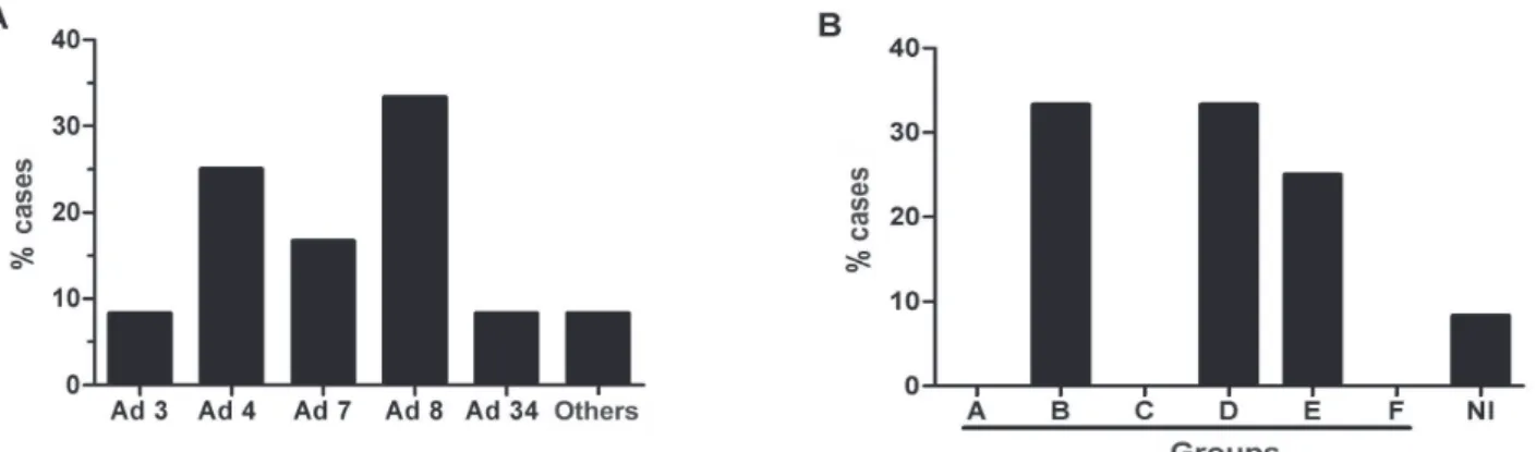
Regaring the different serotypes of Adenovirus as causative of conjunctivitis in the municipality, serotypes 3, 4, 7, 8 and 34 were identified in the samples from confirmed cases (Figure 3A). Of these, serotype 8 proved to be more prevalent with approximately 33.33% of the cases evaluated, followed by serotype 4 with 25%, serotype 7 presented 17% and serotypes 3 and 34 presented less than 10% of cases. Other unidentified serotypes in the sequencing (8.33%) were also detected in the period of the study (Figure 3A). Regarding the group of Adenovirus as causative of conjunctivitis, groups B and D presented 33.33% of cases each, followed by group E with 25% of cases; groups A, C and F were not found in the present study, and 8.33% of non-identified Adenovirus were group NI (Figure 3B).
The signs and symptoms presented by patients during the detailed ophthalmic assessment before the collection and analysis of conjunctival secretion were used to supplement the laboratory detection diagnosis of Adenovirus in the secretions analyzed (table 1). All patients assessed (91 cases) had ocular hyperemia and tearing, common signs of conjunctivitis. When the signs and symptoms strongly related to adenoviral conjunctivitis were analysed, it was found that patients with satellite lymphadenopathy 66.67% (22 cases), conjunctival chemosis 81.82% (27 cases) and nummular keratitis 75.76% (25 cases) were more expressive in the cases confirmed of adenoviral conjunctivitis.
Figure 1: Distribution of conjunctivitis cases from May 2009 to January 2011. The samples were tested by PCR for positive or negative reaction to the detection of Adenovirus in conjunctival swabs.
Figure 3: Identification of the etiological agent causative of adenoviral conjunctivitis. Serotypes identified by sequencing (A). Prevalence of conjunctivitis causative groups (B).
Non-adenoviral conjunctivitis Adenoviral conjunctivitis
Signs and symptoms Nº cases % Nº cases % (n=58) (n=33)
Ocular hyperemia 58 100.00 33 ‘100.00 Tearing 58 100.00 33 100.00
Serous secretion 51 87.93 32 96.97
Purulent secretion 3 5.17 1 3.03
Satellite lymphadenopathy 12 20.69 22 66.67
Conjunctival chemosis 21 36.21 27 81.82
Conjunctival membrane 3 5.17 7 21.21
Subconjunctival haemorrhage 3 5.17 7 21.21
Discomfort 35 60.34 15 45.45
Moderate pain 18 31.03 11 33.33
Severe pain 0 0.00 7 21.21
Corneal wound 1 1.72 4 12.12
Nummular keratitis 7 12.07 25 75.76
Fever 7 12.07 6 18.18
General malaise 22 37.93 16 48.48
Table 1
Evaluation of frequency of conjunctivitis symptoms
D
ISCUSSIONIt is established in the scientific literature that Adenovirus is responsible for epidemic outbreaks of conjunctivitis worldwide, affecting all age groups in varied chronology and in different orders of gravity (18-20). The early diagnosis and efficacy in
controlling the infection is a key process to minimize the incidence of the disease(17,20).
One of the methods considered as gold standard to detect Adenovirus is the qPCR and the real-time PCR due to the fast analysis, high sensitivity and precision on molecular identification of the different serotypes involved in ocular disease (20-22).
According to Allard et al. degenerate primers for the coding region of the structural protein gene II of Adenovirus has been shown to be effective in highlighting different serotypes of AdVs in epidemiological studies(17). Considering these findings, the PCR
reaction using these primers was the method used to analyze the
frequency and detect different serotypes causative of adenoviral conjunctivitis in the municipality of Viçosa/MG in the period from May 2009 to January 2011 without the need of culture of viral isolation.
The prevalence of positive samples analyzed for adenoviral conjunctivitis was 36.3% in the present study. Previous studies have demonstrated prevalence ranging from 15%(23), 70%(24),
and 82%(20). Different environmental factors may be related to
these discrepancies.
Scientific literature has reported differences in the seasonality for the emergence of Adenovirus as a causative agent of conjunctivitis worldwide. According to Matsui et al. in a study conducted in Japan, Adenovirus keeps a low frequency throughout the year; however an outbreak was reported in September(20). Another study by Maranhão et al. in Brazil which
outbreak in April 2004, with no constancy over the years of study(3). The present study presented the differences in relation
to this seasonal distribution, and AdV was more frequent in the months of May and June, a period of disease outbreak in the city. A possible explanation for said differences may be related to weather conditions, because a higher prevalence of cases has occurred in early winter.
Allard et al. analyzed secretion samples from 40 patients with clinical symptoms of conjunctivitis caused by AdV considering the age of the patients, and they found a very heterogeneous distribution, with a frequency ranging from 9 months to 74 years of age(17). This finding is in agreement with
our approach, despite the higher frequency of cases in this study has occurred in the age group between 26 and 65 years.
In order to determine the serotypes present in the samples confirmed of adenoviral conjunctivitis, the PCR products were characterized by sequencing(17). There was a predominance of
serotype 8 (33.33%), followed by 4 (25%) associated to keratitis, as well as in the study of Jin et al. in Hanoi, Vietnam(25). The
presence of serotype 7 (17%) brought particular concern, since this serotype has been associated to more severe cases of infection by Adenovirus, such as pneumonia and myocarditis, with a possible adverse development to respiratory and cardiovascular failure(5).
The main clinical symptoms presented by the patients with conjunctivitis were ocular hyperemia, serous secretion and tearing. Patients with adenoviral conjunctivitis showed a greater frequency of satellite lymphadenopathy, conjunctival chemosis and nummular keratitis. Keratitis usually appear on the 5th day after the onset of conjunctivitis, followed by the rise of corneal nummular opacity on day 7, as it is reported by various authors(26,27).
C
ONCLUSIONThe present study revealed that in Viçosa two out of five cases of conjunctivitis are of adenoviral etiology. Considering the possibility of adenoviral conjunctivitis outbreaks throughout the year, and the different serotypes that can lead to eye disease, the study highlights the importance of conducting rapid diagnostic tests such as PCR for the recognition of the serotype and specific treatment as a potential measure to reduce the virus and consequently the potential for transmission.
A
CKNOWLEDGEMENTSThe funding agencies: the State of Minas Gerais Research Support Foundation (FAPEMIG) and the National Council of Scientific and Technological Development (CNPq).
R
EFERENCES1. Adlhoch C, Schöneberg I, Fell G, Brandau D, Benzler J. Increas-ing case numbers of adenovirus conjunctivitis in Germany, 2010. Euro Surveill. 2010;15(45):pii: 19707.
2. González-Sotero J, Rojas-Álvarez E, Correa-Rojas O, Iviricu-Tielves R. Resistencia antimicrobiana en oftalmología. Rev Mex Oftalmol. 2011;85(3):148-55.
3. Maranhão AG, Soares CC, Albuquerque MC, Santos N. Molecular epidemiology of adenovirus conjunctivitis in Rio de Janeiro, Brazil, between 2004 and 2007. Rev Inst Med Trop Sao Paulo. 2009;51(4):227-9.
4. Aoki K, Kaneko H, Kitaichi N, Ohguchi T, Tagawa Y, Ohno S. Clinical features of adenoviral conjunctivitis at the early stage of infection. Jpn J Ophthalmol. 2011;55(1):11-5.
5. Robinson CM, Shariati F, Zaitshik J, Gillaspy AF, Dyer DW, Chodosh J. Human adenovirus type 19: genomic and bioinformatics analysis of a keratoconjunctivitis isolate. Virus Res. 2009;139(1):122-6.
6. Flint SJ, Enquist LW, Racaniello VR, Skalka AM. Principles of virology: molecular biology, pathogenesis, and control of animal viruses. 2nd ed. Washington: ASM Press; 2003.
7. Weber CM, Eichenbaum JW. Acute red eye. Differentiating viral conjunctivitis from other, less common causes. Postgrad Med. 1997;101(5):185-6, 189-92, 195-6. Comment in Postgrad Med. 1997;102(3):63-4.
8. Takeuchi S, Itoh N, Uchio E, Aoki K, Ohno S. Serotyping of adenoviruses on conjunctival scrapings by PCR and sequence analysis. J Clin Microbiol. 1999;37(6):1839-45.
9. Weiss A, Brinser JH, Nazar-Stewart V. Acute conjunctivitis in childhood. J Pediatr. 1993;122(1):10-4.
10. Wood SR, Sharp IR, Caul EO, Paul I, Bailey AS, Hawkins M, et al. Rapid detection and serotyping of adenovirus by direct im-munofluorescence. J Med Virol. 1997;51(3):198-201.
11. Bell JA, Rowe WP, Engler JI, Parrott RH, Huebner RJ. Pharyngoconjunctival fever; epidemiological studies of a re-cently recognized disease entity. J Am Med Assoc. 1955;157(13):1083-92.
12. Weiss A. Acute conjunctivitis in childhood. Curr Probl Pediatr. 1994;24(1):4-11. Review.
13. Elnifro EM, Cooper RJ, Klapper PE, Bailey AS, Tullo AB. Diag-nosis of viral and chlamydial keratoconjunctivitis: which labora-tory test? Br J Ophthalmol. 1999;83(5):622-7. Review. 14. Allard A, Girones R, Juto P, Wadell G. Polymerase chain
reac-tion for detecreac-tion of adenoviruses in stool samples. J Clin Microbiol. 1990;28(12):2659-67. Erratum in J Clin Microbiol. 1991;29(11):2683.
15. Pring-Akerblom P, Adrian T. Type- and group-specific polymerase chain reaction for adenovirus detection. Res Virol. 1994;145(1):25-35.
16. Van Rij G, Klepper L, Peperkamp E, Schaap GJ. Immune elec-tron microscopy and a cultural test in the diagnosis of adenovi-rus ocular infection. Br J Ophthalmol. 1982;66(5):317-9. 17. Allard A, Albinsson B, Wadell G. Rapid typing of human
adenoviruses by a general PCR combined with restriction en-donuclease analysis. J Clin Microbiol. 2001;39(2):498-505. 18. Cheung D, Bremner J,Chan JT. Epidemic keratoconjunctivitis—do
outbreaks have to be epidemic? Eye (Lond). 2003;17(3):356-63. 19. Matsui K, Saha S, Saitoh M, Mizuki N, Itoh N, Okada E, et al. Isolation and identification of adenovirus from conjunctival scrapings over a two-year period (between 2001 and 2003) in Yokohama, Japan. J Med Virol. 2007;79(2):200-5.
20. Matsui K, Shimizu H, Yoshida A, Nagaoka E, Nishio O, Okuda K. Monitoring of adenovirus from conjunctival scrapings in Ja-pan during 2005—2006. J Med Virol. 2008;80(6):997-1003. 21. Cooper RJ, Yeo AC, Bailey AS, Tullo AB. Adenovirus polymerase
22. Heim A, Ebnet C, Harste G, Pring-Akerblom P. Rapid and quantita-tive detection of human adenovirus DNA by real-time PCR. J Med Virol. 2003;70(2):228-39. Erratum in J Med Virol. 2003;71(2):320. 23. Aoki K, Tagawa Y. A twenty-one year surveillance of adenoviral
conjunctivitis in Sapporo, Japan. Int Ophthalmol Clin. 2002;42(1):49-54.
24. Mejía-López H, Matías-Florentino M, Vélez-Montoya R. [Iden-tification of adenovirus associated with conjunctivitis by mo-lecular methodology]. Arch Soc Esp Oftalmol. 2006;81(7):375-82. Spanish.
25. Jin XH, Ishiko H, Nguyen TH, Ohguchi T, Akanuma M, Aoki K, et al. Molecular epidemiology of adenoviral conjunctivitis in Hanoi, Vietnam. Am J Ophthalmol. 2006;142(6):1064-6.
Corresponding author
Leandro Licursi de Oliveira
Av. PH Rolfs, s/n Campus Universitário ZIP Code: 36570-000-Viçosa (MG), Brazil. E-mail: [email protected]
26. Durand M, Weber DJ, Rutala WA. Nosocomial ocular infections. In: Mayhall CG,editor. Hospital epidemiology and infection control. 3rd ed. Baltimore: Lippincott, Williams & Wilkins; 2004.