Journal of Evolution of Medical and Dental Sciences/ eISSN- 2278-4802, pISSN- 2278-4748/ Vol. 4/ Issue 97/ Dec. 03, 2015 Page 16215
EFFECT OF SYSTEMIC ADMINISTRATION OF HMG Co A REDUCTASE INHIBITOR ATORVASTATIN ON
CENTRAL RETINAL ARTERY FLOW DYNAMICS, INTRA OCULAR PRESSURE AND VISUAL FIELD
CHANGES IN PRIMARY OPEN ANGLE GLAUCOMA AND NORMAL TENSION GLAUCOMA PATIENTS
Deepak Kumar Sharma1, K. P. Chaudhary2, P. C. Negi3, Rajeev Tuli4
1Eye Surgeon, Civil Hospital, Palampur, H. P.
2Professor & HOD, Department of Ophthalmology, IGMC, Shimla, H. P. 3Professor & HOD, Department of Cardiology, IGMC, Shimla, H. P.
4Professor & HOD, Department of Ophthalmology, Dr. RPGMC Kangra at Tanda, H. P.
ABSTRACT PURPOSE
To study the effect of systemic administration of HMG CoA reductase inhibitor Atorvastatin on Central Retinal Artery (CRA) flow dynamics, Intraocular Pressure (IOP) and Visual Field (VF) changes in Primary Open Angle Glaucoma (POAG) and Normal Tension Glaucoma (NTG) patients.
MATERIALS AND METHODS
Prospective randomized placebo controlled double blind parallel group study consisting of 80 eyes of 40 patients suffering from POAG and NTG was conducted. After baseline clinical evaluation and Color Doppler Imaging (CDI) of CRA subjects were randomized to receive 40mg/day of Atorvastatin (Tonact) or matching Placebo for 3 months and followed at two weeks, one month and three months. Main outcome measures were Peak Systolic Velocity (PSV), End Diastolic Velocity (EDV), Resistive Index (RI), IOP and Mean Deviation (MD) in VF.
RESULTS
Atorvastatin group showed decrease in IOP (P=0.0009 in right eye and P=0.0049 in left eye) and in RI (P=0.0005 in right eye and P=0.0008 in left eye), while there was increase in RI in the placebo group (P=0.0006 in right eye and P=0.0007 in left eye) after 3 months. No significant change in MD of VF was noticed in both groups.
CONCLUSION
Atorvastatin has favorable effect in POAG and NTG patients causing decrease in IOP and resistance of CRA with increase in CRA flow dynamics.
KEYWORDS
Atorvastatin, Glaucoma, Intraocular Pressure, Resistive Index, Visual Field.
HOW TO CITE THIS ARTICLE: Deepak Kumar Sharma, K. P. Chaudhary, P. C. Negi, Rajeev Tuli. Effect of Systemic Administration of HMG Co A Reductase Inhibitor Atorvastatin on Central Retinal Artery Flow Dynamics, Intra ocular Pressure and Visual Field Changes in Primary Open Angle Glaucoma and Normal Tension Glaucoma Patients. Journal of Evolution of Medical and Dental Sciences 2015; Vol. 4, Issue 97, December 03; Page: 16215-16220, DOI: 10.14260/jemds/2015/2386
INTRODUCTION
Disturbance in the blood flow velocity in Ophthalmic Artery (OA) and Central Retinal Artery (CRA) have been documented in Primary Open Angle Glaucoma (POAG) and in Normal Tension Glaucoma (NTG) using Color Doppler Imaging (CDI) and pulsatile ocular blood flow measurements.[1],[2],[3],[4],[5],[6], [7],[8]
Statin drugs are 3-Hydroxy-3-Methylglutaryl Coenzyme A (HMG-CoA) reductase inhibitors have got pleiotropic effect by up regulating Endothelial Nitric Oxide Synthase (eNOS) in cultured endothelium and modulate Nitric Oxide (NO) production from the vascular endothelium.
They reduce the levels of various intermediary products of cholesterol biosynthesis, i.e. Isoprenoids which regulate various cellular proteins playing a significant role in regulating cellular contraction,
Financial or Other, Competing Interest: None. Submission 15-11-2015, Peer Review 16-11-2015, Acceptance 25-11-2015, Published 01-12-2015. Corresponding Author:
Dr. Deepak Kumar Sharma, Eye Surgeon,
Civil Hospital, Palampur Dist., Kangra, H. P.
E-mail: [email protected] DOI:10.14260/jemds/2015/2386
actin cytoskeletal organization and cell and cell-to-extracellular interactions. This enzymatically regulated post-translational modification is essential for the activities and functionality of the Rho-GTPase in vital cellular interactions.
All the observed cellular changes induced by statins were associated with increased aqueous outflow facility in organ cultured porcine eye anterior segment models, increase in retinal blood flow and plasma nitrite/nitrate levels and decrease in IOP.[9],[10],[11] The present study was
planned to study the variation of blood flow velocity in CRA using CDI, Intraocular Pressure (IOP) and VF of glaucomatous patients after administration of Atorvastatin and compare it with matched control subjects. Peak Systolic Velocity (PSV), End Diastolic Velocity (EDV) and Resistive Index (RI) in CRA, IOP and Mean Deviation (MD) of VF were main outcome measures.
MATERIALS AND METHODS
A prospective randomized placebo controlled parallel group double blind study was conducted in the Department of Ophthalmology and Department of Cardiology at our institute over a period extending from March 2007 to August 2008.
Journal of Evolution of Medical and Dental Sciences/ eISSN- 2278-4802, pISSN- 2278-4748/ Vol. 4/ Issue 97/ Dec. 03, 2015 Page 16216
study drug and study was approved by the Institutional Review Committee.
They were divided into two groups:
1. Group I patients (20 in number) constituted Atorvastatin Group.
2. Group II patients (20 in number) constituted Placebo Group.
Each patient of Atorvastatin group was matched for sex, hypertensive status and type of antiglaucoma medication already taking with Placebo group. Type of antiglaucoma medication patients already taking was continued as such.
Patients with glaucomatous Optic Nerve Head (ONH) damage in at least one eye (which includes localized notch, diffuse erosion of Neuro Retinal Rim (NRR), shallow saucerized cupping and enlargement of cup or retinal nerve fiber layer defect), bilateral open anterior chamber angle and reproducible Humphrey VF defect with 24-2 full threshold strategy or 30-2 Swedish Interactive Threshold Algorithm strategy were included in this study.
Patients with any ophthalmological or general condition, in which IOP could not be recorded using Goldmann Applanation tonometer with significant lenticular and vitreous opacities obstructing optic disc examination and VF analysis, active ocular infections or inflammations with any contraindication for treatment with Atorvastatin (Myopathy and liver disease) and with pregnancy or breastfeeding were not considered for the study.
Before subjecting the patients to study detailed medical history, general physical examination, baseline blood biochemistry (FBS, SGOT, SGPT, Alkaline Phosphatase and Serum Lipid Profile), and Pulse Doppler Examination of CRA and VF analysis was done. Visual acuity was measured using
Snellen’s chart placed at a distance of six meters and best corrected visual acuity was documented.
IOP was measured using Goldmann Applanation Tonometer mounted on Haag Streit–900 slit lamp as per criteria laid down procedure in the manual for recording of IOP using Goldmann Applanation Tonometer. IOP measurement was done at 11:00 A.M. by same observer and same instrument. IOP was measured three times five minutes apart and average of the three readings was noted. VF analysis was done with the help of automated perimeter
(Humphrey Perimeter Carl Zeiss Meditec HFA ІІ 720-6294 3.5/3.5); 30-2 Swedish Interactive Threshold Algorithm Strategy was used for VF analysis pre and post treatment in all subjects.
Posterior segment evaluation was done after dilating pupils with Tropicamide 0.8% w/v and Phenylephrine 5% w/v (Tropac–P Optho Remedies) with direct and indirect ophthalmoscope. CDI studies were performed by 7.5MHz vector array transducer of HDI-3000 machine (Advanced Technologies Limited, Seattle, U.S.A.). The transducer power output and the maximum spatial peak time average intensity was kept to a minimum within FDA safety guidelines. The transducer is applied with contact jelly to the closed eyelid patient in supine position and a B-scan image of the eye and the orbit is obtained on a VDU screen. By using the color flow imaging as a map, the CRA is identified and interrogated in the center of the ONH shadow as close to the retinal surface as possible.
The blood velocity is calculated from Doppler frequency shifts, which are displayed as spectral waveforms. From the spectral display, PSV and EDV can be measured. The RI can be calculated according to the method of Pourcelot viz., PSV minus EDV divided by PSV.[2] The RI is a ratio and is therefore
angle independent. It represents a quantification of end organ resistance and formula is suited to low resistance beds typical
of cerebrovascular region.[12] The observers doing CDI, IOP
and VF analysis were blinded for the type of treatment subjects were receiving. After detailed baseline evaluation, subjects were randomized to receive 40mg/day of Atorvastatin (Tonact) or matched placebo (Provided by Lupin Pinnacle Pharma Pvt. Ltd.) for three months.
All subjects were reviewed after two weeks and four weeks period for assessment of any adverse effects and Liver Function Tests (LFT) were done at the end of two weeks. Creatine Phosphokinase (CPK) was estimated if patients complained of muscle ache or weakness. If LFT were observed, deranged (elevated three times the upper limit of normal range) dose of Atorvastatin was to be reduced to 20mg/day and SGOT/SGPT enzyme levels were to be repeated after one week. If repeat SGOT/SGPT levels remained elevated three times the upper limit, the study drug was to be stopped.
If SGOT/SGPT levels were within normal limits 20mg/day dose was to be continued for three months. But not even one case with adverse drug reaction or deranged LFT more than three times the upper limit was noticed. So all the patients were continued on 40 mg/day of Atorvastatin for 3 months. At the end of 3 months clinical evaluation, IOP measurement, VF analysis and Doppler examination of CRA was repeated.
Data collected was managed on Microsoft excel worksheet. Discrete variables were expressed as percent and continuous variables were expressed as mean±SD. The analysis was performed with the help of Statistical Package for Social Sciences (SPSS) software 10.00 for windows package software. Paired t–test was used to assess the significance of difference in means of the continuous variables. Chi square was used to assess the significance of difference in means of the discrete variables. Mean±SD in the beginning of the study and at the end of the study along with p value was expressed. A p value less than 0.05 was considered statistically significant.
RESULTS
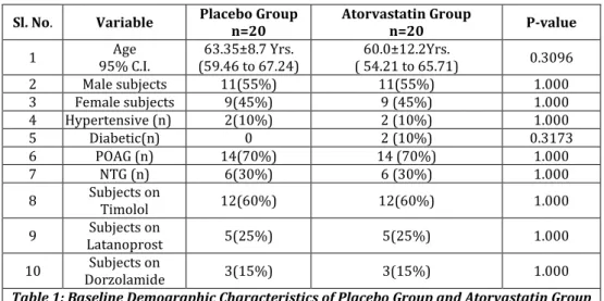
The baseline values of all variables of placebo group and Atorvastatin group are depicted in Table 1 and Table 2. Mean age for placebo group was 63.35±8.7 yrs. (95% C I; 59.46 to 67.24 yrs.) and for Atorvastatin group was 60.0±12.2 yrs. (95% C I; 54.21 to 65.71 yrs.) (p=0.306). There were 11(55%) males and 9(45%) females in both groups; 14(70%) patients were of POAG and 6(30%) were of NTG, 12(60%) patients were on Timolol, 5(25%) were on Latanoprost and 3(15%) patients were on Dorzolamide in both groups. Mean duration for which subjects were on antiglaucoma medication at baseline was 40.95±40.00 months and for placebo group was 59.15±65.04 months. Three patients in Atorvastatin group and five patients in placebo group were on antiglaucoma medication for a period less than a month.
Three months post intervention data of placebo group are depicted in Table 3 and that of Atorvastatin group in Table 4.
Effect on IOP
Decrease in IOP was noticed from 16.2±5.9mmHg (95% C I; 13.4 to 19.04mmHg) to 11.7±1.3mmHg (95% C I; 11.05 to 12.34mmHg) (p=0.0009) in right eye and from 18.9±13.2 mmHg (95% C I; 12.71 to 25.08mmHg) to 14.3±8.4 mmHg (95% C I; 10.32 to 18.30mmHg) (p=0.0049) in left eye in Atorvastatin group. No significant difference was noticed in IOP in placebo group after three months of study.
Effect on CRA Flow Dynamics
Journal of Evolution of Medical and Dental Sciences/ eISSN- 2278-4802, pISSN- 2278-4748/ Vol. 4/ Issue 97/ Dec. 03, 2015 Page 16217
cm\s (95% C I; 8.66 to 11.22cm\s) to 10.51±2.51cm\s (95% C I; 9.34 to 11.69cm\s) (p=0.3747) in right eye and from 10.15±2.63cm\s (95% C I; 8.92 to 11.38cm\s) to 10.97± 2.26cm\s (95% C I; 9.92 to 12.04cm\s ) (p=0.1056) in left eye. In Atorvastatin group change in PSV was from 10.6±2.4 cm\s (95% C I; 9.46 to 11.76cm\s) to 11.1±3.1 cm\s (95% C I; 9.66 to 12.64cm\s) (p = 0.4152) in right eye and from 10.2±3.1 cm\s (95% C I; 8.79 to 11.74cm\s) to 10.4±2.1cm\s (95% C I; 9.45 to 11.47cm\s) (p=0.7992) in left eye. There was increase in EDV in both eyes [Right eye from 3.6 ± 1.0cm\s (95% C I; 3.2 to 4.1cm\s) to 4.4±1.2cm\s (95% C I; 3.9 to 5.0cm\s) (p=0.0085) and in left eye from 3.23±0.94cm\s (95% C I; 2.79 to 3.67cm\s) to 4.0±1.2cm\s (95% C I; 3.44 to 4.59cm\s) (p=0.0095)] in patients taking Atorvastatin. No significant difference was noticed in EDV in placebo group.
RI increased in both eyes in placebo group. In right eye from 0.70±0.075 (95% C I; 0.67 to 0.74) to 0.74±0.067 (95% C I; 0.72 to 0.77) (p=0.0006) and in left eye from 0.69±0.074 (95% C I; 0.66 to 0.72) to 0.74±0.061 (95% C I; 0.72 to 0.77) (p = 0.0007). RI decreased in Atorvastatin group from 0.65 ± 0.06 (95% C I; 0.63 to 0.69 ) to 0.59±0.08 (95% C I; 0.55 to 0.63 ) (p=0.0005) in right eye and from 0.67±0.06 (95% C I; 0.64 to 0.70) to 0.61±0.10 (95% C I; 0.55 to 0.66) (p=0.0008) in left eye.
Effect on VF
No significant difference in MD was observed in both groups after three months of postinterventional trial. In Placebo group MD depressed from 14.68±7.7dB (95% C I; 18.42 to -10.94dB) to -14.81±7.0dB (95% C I; -18.36 to -11.24dB) (p=0.8260) in right eye and from 13.36±8.4 dB (95% C I; -17.88 to -8.84 dB) to -13.42 ± 7.0 dB (95% C I; -17.56 to -9.26 dB) (p=0.8991) in left eye. In Atorvastatin group change in MD was from-11.32 ± 9.0dB (95% C I; -16.09 to -6.54dB) to 11.68 ± 9.0dB (95% C I; -16.28 to -7.00 dB) (p = 0.4765) in right eye and from -17.07 ± 11.0dB (95% C I; -23.02 to -12.37 dB) to -18.43 ± 11.0 dB (95% C I; -23.40 to -12.40dB) (p=0.4754 ) in left eye.
Effect on Cardiovascular Risk Variables
In placebo group Systolic Blood Pressure (SBP) and diastolic blood pressure (DBP) remained unchanged after a period of three months. Our study reported decrease in SBP from 135.50±12.0mmHg (95% C I; 129.90 to 141.19mmHg) to 120.60±8.1mmHg (95% C I; 116.77 to 124.43mmHg) (p=0.0001) and DBP from 84.30±7.8mmHg (95% C I; 80.64 to 87.96mmHg) to 78.20±4.3mmHg (95% C I; 76.14 to 80.25mmHg ) (p=0.0008) three months post administration of Atorvastatin. There was decrease in S.Cholesterol (p=0.0021), TG-C (p=0.0001), LDL-C (p=0.0924), VLDL-C (p= 0.0033) and increase in HDL-C (p=0.0011) three months post administration of Atorvastatin. Cholesterol HDL ratio also decreased (p= 0.0014).
DISCUSSION
HMG-CoA reductase inhibitors are extensively used in patients of atherosclerotic vascular diseases and have been found to be safer and effective in reducing morbidity and mortality in patients of coronary artery diseases. [13],[14]
The present study was undertaken with the hypothesis that study drug Atorvastatin at a dose of 40mg once a day used for 3 months would improve the CRA flow dynamics and may have impact in at least checking the progression of underlying disease process.
The findings of the present study suggest that Atorvastatin is effective in improving the flow of CRA. This improves the perfusion of ONH and arrests the progression of glaucomatous damage through favorable modification of
cardiovascular risk factors and decrease in IOP partly and partly may be mediated through its pleiotropic effect.
The reduction in Blood Pressure (BP), IOP and CRA resistance could be mediated through improvement in the endothelial function by Atorvastatin. Atorvastatin induces the expression of eNOS that in turn induces NO synthesis, which is vasodilator, antithrombotic and anti-inflammatory.[15] After
administration of Mevastatin for a period of 28 days Hanjani SA, et al.[15] found significant increase in the level of eNOS
mRNA and decrease in the infarct size and neurological deficit in a dose and time dependent manner.
In the previous studies reproducibility, accuracy, evaluation of severity of disease, its progression, efficacy of treatment in glaucoma patients and suitability of CDI in imaging retrobulbar vasculature has been studied.[16],[17] It
was found that Retinal Blood Flow (RBF) parameter measurement may not be feasible in all the retinal vessels and significant changes in various haemodynamic parameters were found in CRA.[5],[17] In our study, we scanned CRA to
measure retinal artery RI and flow. In total group of 40 patients, there were 28 patients of POAG and 12 patients of NTG. Rankin SJA.[6] did not find any difference in the
haemodynamic parameters of NTG and POAG in our study also we did not notice any significant difference.
In this study various parameters which we studied were IOP, PSV, EDV, RI, MD of VF and BP. Rankin SJA.[1] studied CDI
of CRA and found PSV 11.4±2.6cm/s, EDV 2.9±1.1cm/s and RI 0.75 in right eye. In left eye PSV was 10.6±2.9cm/s, EDV was 2.5±1.3cm/s and RI was 0.76±0.10.
Nicolela M.T. et al.[7] found lower blood flow velocities
in CRA in glaucoma patients compared to normal subjects. In this study PSV was 10.40±2.78cm/s, EDV was 2.75±0.85cm/s and RI was 0.74±0.05. In normal subjects PSV was 15.35±4.44cm/s, EDV was 4.00±1.17cm/s and RI was 0.74±0.05. Both of these investigators studied only baseline parameters in glaucoma patients without doing any intervention. In our study, we studied baseline as well post intervention parameters. In right eye PSV was 10.6±2.4cm/s, EDV was 3.6 ±1.0cm/s and RI was 0.65±0.06. In left eye PSV was 10.2±3.1cm/s, EDV was 3.23±0.94cm/s and RI was 0.67±0.06. These baseline parameters were comparable to the parameters previously documented.
Nagaoka T. et al.[11] found no significant effect of
administration of Simvastatin 20mg/day on BP. In our study we reported decrease in SBP from 135.50±12.0mmHg (95% C I; 129.90 to 141.19mmHg) to 120.60± 8.1mmHg (95% C I; 116.77 to 124.43mmHg) (p=0.0001) and DBP from 84.30± 7.8mmHg (95% C I; 80.64 to 87.96 mmHg) to 78.20± 4.3mmHg (95% C I; 76.14 to 80.25mmHg) (p=0.0008). This effect may be related to the release of NO having pleiotropic effect leading to decrease in BP as described previously also. They also found significant decrease in the levels of S. Cholesterol, LDL, TG and VLDL. They found no significant change in the levels of HDL-C. In our study, we noticed decrease in the levels of S. Cholesterol (p=0.0021), LDL-C (p=0.0924), TG (p=0.0001), VLDL-C (p=0.0034) and cholesterol and HDL ratio (p=0.0014). HDL-C increased (p=0.0011) in Atorvastatin group compared to Placebo group. Senn B. et al.[18] administered Simvastatin for a period of
three months in hypercholesterolemic subjects and found no significant change in the RBF. McGwin et al.[19] reported that
statin use may be associated with reduced risk of glaucoma, but author did not provide any data on the effect of statins on IOP. Nagaoka T. et al.[11] found significant decrease in IOP
Journal of Evolution of Medical and Dental Sciences/ eISSN- 2278-4802, pISSN- 2278-4748/ Vol. 4/ Issue 97/ Dec. 03, 2015 Page 16218
It was also found that many statins inhibit the activity of Rho-Kinase, which leads to an enhanced aqueous outflow and thereby lowering IOP.[9] These previous findings may support
the Atorvastatin mediated IOP lowering effect that we observed in our study. Alternatively, statins may also decrease the IOP via an increase in the plasma NO levels.[11]
Since we could not measure the levels of NO, so in our study we are not able to correlate IOP reduction with NO release.
In our study we found increase in EDV in both eyes and decrease in RI in both eyes in comparison with our Placebo group, in which there was decrease in EDV and increase in RI. RI is an adequate measure of distal vascular resistance in vivo and in vitro.[17]
The vascular bed of the ONH consists almost entirely of capillaries. Capillaries pose highest vascular resistance within the circulation due to vasospasm in OAG and NTG.
RI is correlated well with perfusion pressure. Increase in RI decreases the perfusion pressure and vice versa. Decrease in RI may be associated with increase in perfusion pressure of the ONH.[6]
Because LDL-C can impair NO release by endothelial cells through down regulation of eNOS mRNA and protein.,[20]
the increased RBF may be a consequence of improving NO release by the endothelial cells. We noticed significant decrease in the levels of LDL-C, which may be associated with enhanced NO effect. Therefore it is possible that not only the increase in NO levels, but also decrease in the levels of cholesterol induced by Atorvastatin are associated with the increase in flow velocities and decrease in RI of retinal vessels that we observed. In previous clinical trials, positive effects of statins in the improvement of endothelial function were observed within 3 days.[21] and within 7 days.[11] of
treatment Robinson F. et al.[22] noticed increase in retinal
vascular resistance with the increase in blood pressure suggesting effect of change in blood pressure on retinal blood flow. Thus difference noticed in RBF parameters may also be blood pressure mediated.
In our study, we noticed that except for significant difference in IOP in right eye all other 95% CIs overlap each other. Peter C. Austin, et al.[23] has demonstrated that 95% CI
for two means can overlap and yet the two means can be statistically significantly different from one another at the p=0.05. Mark E. Payton, et al.[24] in their study concluded that
caution should be exercised when the results of an experiment are displayed with confidence intervals, whether or not these intervals overlap does not imply the statistical significance of the parameters of the interest. Rory Wolfe, et al.[25] also concluded that it is misconception if two
independent 95% CIs overlap each other, the findings are not statistically significant at the 5% level. For two independent samples even if CI overlap, the findings may be significantly different. So it will not be inappropriate to state that these differences are significant and are not merely due to small sample size.
In view of above observations, it is evident that after 3 months post intervention trial with Atorvastatin 40mg/day there is increase in the flow velocities and decrease in RI in CRA in POAG and NTG patients. IOP decreased in Atorvastatin group as compared to Placebo group. This decrease in IOP may be attributed to decreased outflow obstruction due to its pleiotropic effect.[15] on endothelial lining of juxtacanalicular
part of Trabecular Meshwork (TM) and decreased cell to cell interaction due to inhibition of Rho-Kinase.[9]
No significant change in the MD of VF was recorded in both groups. Since gap period between two fields charting was only 3 months, hence probable deterioration or improvement in MD of VF could not be commented.
CONCLUSION
Atorvastatin causes decrease in IOP and decrease in RI of CRA with increase in CRA flow dynamics. It appears to have an ocular hypotensive effect and increases ONH blood flow when given in combination with other antiglaucoma medication with no side effects in POAG and NTG patients.
REFERENCES
1. Rankin SJA, Drance SM, Buckley AR, Walman BE. Visual Field Correlation with Color Doppler Studies in Open Angle Glaucoma. Journal of Glaucoma
1996;vol.5.no.1:15-21.
2. Kaiser HJ, Schoetzau A, Stumpfig D, Flammer J. Blood flow velocities of the extraocular vessels in patients with High-tension and Normal-tension Primary Open Angle Glaucoma. American Journal of Ophthalmology 1997;123:320-327.
3. Charles D, James J. Migraine and low tension glaucoma. Invest Ophthalmol Vis Sci 1985;26:1105-1108.
4. Riva CE, Grunwald JE, Petrig BL. Auto regulation of human retinal blood flow. An investigation with Laser Doppler Velocimetry. Invest Ophthalmol Vis Sci 1986;27:1706-1712.
5. Grunwald JE, Riva CE, Brucker AJ. Total retinal volumetric blood flow rate in diabetic patients with poor glycemic control. Invest Ophthalmol Vis Sci 1992;33:356-363.
6. Rankin SJA, Walman BE, Bruckley AR, Drance SM. Color Doppler Imaging and Spectral Analysis of Optic Nerve Vasculature in Glaucoma. American Journal of Ophthalmology 1995;119:685-693.
7. Nicolela MT, Walman BE, Buckley AR, Drance SM. Ocular hypertension and primary open angle glaucoma: A comparative study of their retrobulbar blood flow velocity. Journal of Glaucoma 1996;vol.5 no.5:308-310. 8. Kerr J, Nelson P, O’Brien C. A comparison of ocular
blood flow in untreated Primary Open Angle Glaucoma and Ocular Hypertension. American Journal of Ophthalmology 1998;126:42-51.
9. Rao PV, Deng PF, Kumar J, Epstein DL. Modulation of aqueous humor outflow facility by the Rho Kinase specific inhibitor Y-27632. Investigative Ophthalmology and Visual Sciences 2001;42:1029-1037.
10. Miyahara S, Kiryu J, Yamashiro K, Miyamoto K, Hirose F, Tamura H, et al. Simvastatin inhibits leukocyte accumulation and vascular permeability in the retinas of rats with Streptozotocin induced diabetes. American Journal of Pathology 2004;164:1697-1706.
11. Nagaoka T, Takashashi A, Sato E, Izumi N, Hein TW, Kuo L, et al. Effect of systemic administration of Simvastatin on retinal circulation. Archives of Ophthalmology 2006;124:665-670.
12. Rankin SJA. Color Doppler Imaging of the retrobulbar circulation in Glaucoma. Survey of Ophthalmology 1999;43, Supplement1:S176-S182.
13. Henry NG, Ira JG. Disorders of lipid metabolism. In: Kasper, Braunwald, Fauci, Hauer, Longo, Jameson,
editors. Harrison’s Principles of Internal Medicine th
ed. International Edition McGraw- Hill Companies, Inc.1998;Vol.2:2146-2147.
14. Law M, Rudnicka AR. Statin Safety: A Systemic Review. Am.J.Cardiology 2006;97:S52-S60.
Journal of Evolution of Medical and Dental Sciences/ eISSN- 2278-4802, pISSN- 2278-4748/ Vol. 4/ Issue 97/ Dec. 03, 2015 Page 16219
16. Baxter GM, Williamson TH, McKillop G, Dutton GN. Color Doppler Ultrasound of orbital and optic nerve blood flow. Effects of Posture and Timolol 0.5%. Invest Ophthalmol Vis Sci 1992;33:604-610.
17. Harris A, Sergot RC, Spaeth GL, Katz JL, Schoemaker JA, Martin BJ. Color Doppler analysis of ocular vessel blood velocity in Normal Tension Glaucoma. American Journal of Ophthalmology 1994;118:642-649.
18. Senn B, Orgul S, Keller U. Retrobulbar and peripheral capillary blood flow in hypercholesterolemic subjects. Am J Ophthalmol 1999;128:310-316.
19. McGwin G, McNeal S, Owsley C, Girkin C, Epstein D, Lee PP. Statins and other cholesterol lowering medications and the presence of Glaucoma. Archives of Ophthalmology 2004;122(6):822-826.
20. Vidal F, Colome C, Gonzalez JM, Badimon L. Atherogenic concentration of native low density lipoprotein down regulates nitric oxide synthase m-RNA and protein levels in endothelial cells. European Journal of Biochemistry 1998; vol.252, issue3: 378-384.
21. Tsunekawa T, Hayashi T, Kano H, Sumi D. Cerivastatin, a HMG-CoA reductase inhibitor improves endothelial function in elderly diabetic patients within three days. Circulation 2001;104:376-379.
22. Robinson F, Riva CE, Grunwald JE, Petrig BL, Sinclair SH. Retinal blood flow autoregulation in response to an acute increase in blood pressure. Invest Ophthalmol Vis Sci 1986;27:722-726.
23. Peter C Austin, Janet E Hux. A brief note on overlapping confidence intervals. J Vasc Surgery 2002;36:194-5. 24. Mark E Payton, Matthew H Greenstone, Nathaniel
Schenker. Overlapping confidence intervals or standard error intervals: what do they mean in terms of statistical significance. J Insect Sci 2003;3:34.
25. Rory Wolfe, James Hanley. PRACTICE If we’re so different, why do we keep overlapping? When one plus
one doesn’t make two. Canadian Medical Association Journal January 8, 2002;166(1).
Sl. No. Variable Placebo Group
n=20
Atorvastatin Group
n=20 P-value
1 Age
95% C.I.
63.35±8.7 Yrs. (59.46 to 67.24)
60.0±12.2Yrs.
( 54.21 to 65.71) 0.3096 2 Male subjects 11(55%) 11(55%) 1.000 3 Female subjects 9(45%) 9 (45%) 1.000 4 Hypertensive (n) 2(10%) 2 (10%) 1.000
5 Diabetic(n) 0 2 (10%) 0.3173
6 POAG (n) 14(70%) 14 (70%) 1.000
7 NTG (n) 6(30%) 6 (30%) 1.000
8 Subjects on
Timolol 12(60%) 12(60%) 1.000
9 Subjects on
Latanoprost 5(25%) 5(25%) 1.000
10 Subjects on
Dorzolamide 3(15%) 3(15%) 1.000 Table 1: Baseline Demographic Characteristics of Placebo Group and Atorvastatin Group
N- Number of subjects, POAG – Primary Open Angle Glaucoma, NTG – Normal Tension Glaucoma, C.I. – Confidence Interval.
Sl.
No. Variable Placebo Group Atorvastatin Group P-value
1 IOP Rt. Eye 95% C.I.
16.95±8.24 (13.09 to 20.18 )
16.2±5.9
(13.45 to 19.04) 0.7750
2 IOP Lt. Eye 95% C.I.
17.40±11.27 (12.12 to 22.68)
18.9±13.2
(12.71 to 25.08) 0.6835
3 PSV Rt. Eye 95% C.I.
9.94±2.74 cm\s (8.66 to 11.22 )
10.6±2.4 cm\s
(9.46 to11.76) 0.4936
4 PSV Lt Eye 95% C.I.
10.15±2.63 cm\s (8.92 to 11.38)
10.2±3.1 cm\s
(8.79 to 11.74) 0.8886
5 EDV Rt Eye 95% C.I.
2.88±0.99 cm\s ( 2.42 to 3.35)
3.6±1.0 cm\s
(3.2 to 4.1 ) 0.0761
6 EDV Lt. Eye 95% C.I.
3.03±0.81 cm\s (2.65 to 3.41)
3.23±0.94 cm\s
(2.79 to 3.67) 0.4433
7 RI Rt. Eye 95% C.I.
0.70±0.075 (0.67 to 0.74)
0.65±0.06
(0.62 to 0.68) 0.0515
8 RI Lt Eye 95% C.I.
0.69±0.074 (0.66 to 0.72)
0.67±0.06
(0.64 to 0.70) 0.4257
9 MD Rt. Eye 95% C.I.
-14.68±7.7 dB (-18.42 to -10.94 )
-11.32±9.6 dB
(- 16.09 to -6.54) 0.3067
10 MD Lt Eye 95% C.I.
-13.36±8.4 dB (-17.68 to -8.84)
-17.70±11.0 dB
(-23.02 to -12.37) 0.3886 Table 2: Baseline Clinical Characteristics of Placebo
Group and Atorvastatin Group
Journal of Evolution of Medical and Dental Sciences/ eISSN- 2278-4802, pISSN- 2278-4748/ Vol. 4/ Issue 97/ Dec. 03, 2015 Page 16220
Sl.
No. Variable Baseline Three Months P-value
1 IOP Rt. Eye 95% C.I.
16.95±8.24 (13.09 to 20.81)
15.55±3.18
(14.06 to 17.04) 0.4456
2 IOP Lt. Eye 95% C.I.
17.40±11.27 (12.12 to 22.68)
16.50±3.88
(14.68 to 18.32) 0.6493
3 PSV Rt. Eye 95% C.I.
9.94±2.74 cm\s (8.66 to 11.22)
10.51±2.51 cm\s
(9.34 to 11.69) 0.3747
4 PSV Lt Eye 95% C.I.
10.15±2.63 cm\s (8.92 to 11.38 )
10.97±2.26 cm\s
(9.92 to 12.04) 0.1056
5 EDV Rt Eye 95% C.I.
2.88±0.99 cm\s (2.42 to 3.35)
2.55±0.68 cm\s
(2.24 to 2.87) 0.1234
6 EDV Lt. Eye 95% C.I.
3.03±0.81 cm\s (2.65 to 3.41)
2.73±0.52 cm\s
(2.49 to 2.98) 0.1929
7 RI Rt. Eye 95% C.I.
0.70±0.075 (0.67 to 0.74)
0.74±0.067
(0.72 to 0.77) 0.0006*
8 RI Lt Eye 95% C.I.
0.69±0.074 (0.66 to 0.72)
0.74±0.061
(0.72 to 0.77) 0.0007*
9 MD Rt. Eye 95% C.I.
-14.68±7.7 dB (-18.42 to -10.94)
-14.81±7.0 dB (-18.36 to -11.24)
0.8260
10 MD Lt Eye 95% C.I.
-13.36±8.4 dB (-17.88 to -8.84)
-13.42±7.0 dB
(-17.56 to -9.26) 0.8991 Table 3: Comparison of Ocular Dynamics Three Months
Post Intervention in Placebo Group
IOP – Intraocular Pressure in mm of Hg, PSV- Peak Systolic Velocity, EDV – End Diastolic Velocity, RI - Resistive Index, MD – Mean Deviation of Visual Field, dB – deciBel, Rt. – Right, Lt. – Left. Parameter with * show significant difference.
Sl.
No. Variable Baseline Three Months P-value
1 IOP Rt. Eye 95% C.I.
16.2±5.9 (13.4 to 19.04)
11.7±1.3
(11.05 to 12.34) 0.0009*
2 IOP Lt. Eye 95% C.I.
18.9±13.2 (12.71 to 25.08)
14.3±8.4
(10.32 to 18.30) 0.0049*
3 PSV Rt. Eye 95% C.I.
10.6±2.4 cm\s (9.46 to 11.76)
11.1±3.1 cm\s
(9.66 to 12.64) 0.4152
4 PSV Lt Eye 95% C.I.
10.2±3.1 cm\s (8.79 to 11.74)
10.4±2.1 cm\s
(9.45 to 11.47) 0.7992
5 EDV Rt Eye 95% C.I.
3.6±1.0 cm\s (3.2 to 4.1)
4.4±1.2 cm\s
(3.9 to 5.0) 0.0085*
6 EDV Lt. Eye 95% C.I.
3.23±0.94 cm\s (2.79 to 3.67)
4.0±1.2 cm\s
(3.44 to 4.59) 0.0095*
7 RI Rt. Eye 95% C.I.
0.65±0.06 (0.63 to 0.69)
0.59±0.08
(0.55 to 0.63) 0.0005*
8 RI Lt Eye 95% C.I.
0.67±0.06 (0.64 to 0.70)
0.61±0.10
(0.55 to 0.66) 0.0008*
9 MD Rt. Eye 95% C.I.
-11.32±9.6 dB (-16.09 to – 6.54)
-11.65±9.0 dB
(- 16.28 to – 7.00) 0.4765 10 MD Lt Eye
95% C.I.
-17.70±11.0 dB (-23.02 to – 12.37)
-18.4±11.0 dB
(- 23.40 to -12.40) 0.4754 Table 4: Comparison of Ocular Dynamics Three Months
Post Intervention in Atorvastatin Group