CASE REPORT
Abstract:
Dyskeratosis Congenita (DC) is a rare inherited disorder of ectodermal dysplasia. It consists of a classical mucocutaneous triad of abnormal skin pigmentation, nail dystrophy and leukoplakia. Pulmonary disease is seen in 10-15%. It is characterized by Idiopathic Pulmonary Fibrosis (IPF), or Idiopathic Familial Pulmonary Fibrosis (IFPF). Non-specific Interstitial Pneumonia (NSIP) has been reported rarely in children with DC and in an isolated adult patient. Our patient had classical clinical presentation of DC with pancytopenia and portal hypertension and clinic-radiological features of NSIP which is a rare association.
Keywords: Dyskeratosis Congenita, Idiopathic Pulmonary Fibrosis, Familial Pulmonary Fibrosis
Introduction:
Dyskeratosis Congenita (DC) is an inherited disorder of ectodermal dysplasia with a classical mucocutaneous triad of abnormal skin pigmentation, nail dystrophy and leukoplakia [1]. Lung involvement in the form of Interstitial Lung Disease (ILD) may be seen in 10-15% [2]. ILD subtypes like Idiopathic Pulmonary Fibrosis (IPF) and Familial Pulmonary Fibrosis (FPF) have been commonly reported [3]. Non-specific Interstitial Pneumonia (NSIP) is reported rarely with DC [4]. We report a rare association of DC and NSIP in our patient.
Case History
A thirty-eight year-old man was referred to our
Dyskeratosis Congenita Associated Non-Specific Interstitial Pneumonia
1 1*
Unnati D. Desai , Jyotsna M. Joshi
1
Department of Pulmonary Medicine, TNMC & BYL Nair Hospital, AL Nair Road, Mumbai Central, Mumbai-400008 (Maharashtra) India
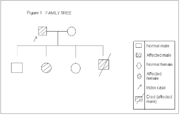
cough, chest X-ray opacities. He was symptomatic with exertional dyspnea, dry cough since 5-6 months. He was a known case of Dyskeratosis Congenital (DC) since eight years diagnosed in view of classical mucocutaneous triad of skin pigmentation, nail dystrophy, leukoplakia. Patient had preexisting pancytopenia, early liver cirrhosis with portal hypertension and right limb non healing ulcer. He was treated for pulmonary tuberculosis 15 years ago. Patient had two children affected with dyskeratosis congenita but two of his progeny were normal. The affected and normal children consisted of one daughter and one son each respectively suggesting a probable autosomal dominant mode of inheritance. The family tree is illustrated in Fig. 1. This was taken into consideration and genetic testing was already offered to the patient and his wife by the geneticist during the fifth pregnancy which was of five months gestation when the patient presented to us for his respiratory illness. There was no significant personal or occupational history. General examination revealed pallor, grade 1 clubbing, post exercise desaturation, reticulate skin pigmentation, nail dystrophy and leukoplakia of tongue due to dyskeratosis congenita (Fig. 2a, b, c). Respiratory system examination had bibasilar fine crepitations on auscultation. Investigations revealed hemoglobin of 6.2gm/dl, total count of
3
function tests, arterial blood gas were normal and was non reactive to HIV antibodies. Chest X- ray showed bibasilar reticular opacities with fibrocalcific lesion in right midzone (Fig. 3). High Resolution Computed Tomography (HRCT) of thorax showed ill defined areas of intralobular, interstitial septal thickening involving lung parenchyma bilaterally showing a peripheral pattern of distribution with no significant zonal predominance suggesting mostly NSIP pattern of ILD (Fig. 4). Focal calcification with adjacent focal fibrosis and patchy air trapping in superior segment of right lower lobe, sequelae to past infection, were additional findings. Spirometry demonstrated a restrictive abnormality with forced vital capacity of 54% of predicted and diffusion capacity for carbon monoxide of 57% predicted. The clinical history with examination findings of clubbing, post exercise desaturation and bibasilar fine crepitations and spirometry suggest ILD.
Classical apico-basal gradient (basal subpleural predominance), honeycombing, traction bronchiectasis characterize definite Usual Interstitial Pneumonitis (UIP) pattern. Exclusion of the definite UIP pattern in presence of homogenous, uniform peripheral distribution with no zonal predominance pattern of intralobular interstitial and septal thickening pointed towards NSIP pattern of ILD on HRCT though immediate subpleural sparing which is very specific for NSIP was not observed. Lung biopsy was not performed due to thrombocytopenia and as DC is known cause for ILD. Our patient was diagnosed as a case of Dyskeratosis Congenita associated NSIP pattern of ILD based on clinic-radiological correlation. In view of clinical worsening and declining lung functions on follow-up patient was initiated on therapy with Prednisolone and N-acetyl cysteine.
Unnati D. Desai & Jyotsna M. Joshi JKIMSU, Vol. 6, No. 1, January-March 2017
Fig. 2a: showing Typical Reticulate Skin Pigmentation
Fig. 2b: showing Leukoplakia of Tongue
Fig. 3: Chest X-ray showing Bilateral Reticulonodular Opacities
Fig. 4: HRCT Thorax showing Ill Defined Areas of Intralobular, Interstitial Septal Thickening Involving Lung Parenchyma Bilaterally showing A Peripheral Pattern of
Discussion:
DC is a rare inherited disorder of ectodermal dysplasia. Also known as Zinsser-Engman-Cole syndrome it consists of a classical mucocutaneous triad of abnormal skin pigmentation, nail dystrophy and leukoplakia [1]. Bone marrow failure, liver cirrhosis and malignancy are observed commonly. Skin changes are characterized by reticulate skin pigmentation, which may be telangiectatic, affecting neck, upper chest, and upper arms with surrounding patches of pale, atrophic skin. Hematological abnormalities range from anemia, thrombocytopenia progressing to pancytopenia [1]. Pulmonary fibrosis has been reported in 10-15% of patients [2, 3]. Though the exact pathology of the disease is not yet fully understood, most evidence points to it being primarily a disorder of poor telomere maintenance [4]. To date, DKC1, Telomerase RNA Component (TERC), Telomerase Reverse Transcriptase (TERT), Nucleolar Protein 10 (NOP10), and NHP2, TINF2, TCAB1 and Regulation of Telomere Elongation Helicase1 (RTEL1) are the eight genes in which mutations are known to cause DC [5]. Three modes of inheritance have been recognized: X-linked, autosomal dominant, autosomal recessive. If the disease-causing mutation in the family has been identified,
prenatal testing for pregnancies at increased risk is possible [4]. IPF has been the common type of ILD reported with DC [2, 6]. Armanios et al. (2005) have also reported familial IPF in DC [7]. It is postulated that loss of telomere length may lead to early loss of epithelial alveolar cells causing subsequent pulmonary fibrosis. NSIP has been reported rarely in children with DC and in one adult patient [8, 9]. IIP and IPF have been generally associated with genetic disorders of telomere length [10]. Treatment guidelines are unclear and literature review suggests therapy according to the severity of pulmonary functions. Steroids and lung transplantation are available treatment modalities for DC associated ILD [2, 11]. Our patient had classical clinical presentation of DC with pancytopenia and portal hypertension and clinic-radiological features of NSIP which is a rare association. It is also worthy to note that presence of pulmonary symptoms with CXR opacities in presence of genetically linked skin/other diseases should raise suspicion of ILD. As a lack of awareness of the same can be misdiagnosed as due to infectious causes such as tuberculosis especially in high burden countries.
References 1. Davidson HR, Connor JM. Dyskeratosis congenita. J
Med Genet 1988; 25: 843-46
2. Kim HJ, Kim KJ, Lee KH, Kyeong-Cheol Shin, Chung JH, Hyun MS et al. Interstitial Lung Disease in a Patient with Dyskeratosis Congenita. Tuberc Respir Dis 2013; 74:70-73
3. Imokawa S, Sato A, Toyoshima M, Yoshitomi A, Tamura R, Suda T et al. Dyskeratosis congenita showing usual interstitial pneumonia. Internal Medicine 1994; 33:226-30
4. Savage SA. Dyskeratosis Congenita. 2009 [Updated 2013 Jan 3]. In: Pagon RA, Adam MP, Bird TD, et al. editors. GeneReviews™ [Internet]. Seattle (WA): University of Washington, Seattle; 1993-2014. Available from: http://www.ncbi.nlm.nih.gov/books/NBK22301/ 5. Dokal I. Dyskeratosis congenita. Hematology Am Soc
Hematol Educ Program 2011; 2011:480-86
6. A Fukuhara, Y Tanino, T Ishii, Y Inokoshi, K saito, N Fukuhara et al. Pulmonary fibrosis in dyskeratosis congenita with TINF2 gene mutation. Eur Respir J
2013; 42: 1757-59.
* nd
Author for Correspondence: Dr. Jyotsna M. Joshi Department of Pulmonary Medicine, 2 floor, OPD bldg,
TNMC & BYL Nair Hospital, AL Nair Road, Mumbai Central, Mumbai- 400008 Email- [email protected] Phone:02223003095
7. Armanios M, Chen JL, Chang YP, et al. Haploin sufficiency of telomerase reverse transcriptase leads to anticipation in autosomal dominant dyskeratosis congenita. Proc Natl Acad Sci USA 2005; 102: 15960–64
8. AS Brody. Imaging Considerations: Interstitial Lung Disease in Children. Radiol Clin N Am 2005;43: 391–403
9. S Hisata, M Ebina. A novel missense mutation in dyskeratosis congenita with pulmonary disease.
AJRCCM 2012; 185: A5419.
10. Alder JK, Chen JJ, Lancaster L, Danoff S, Su SS, Cogan JD et al. Short telomeres are a risk factor for idiopathic pulmonary fibrosis. Proc Natl Acad sci USA
2008; 105: 13051-56.