Case Report
Prosthetic Management of a Child with Hypohidrotic
Ectodermal Dysplasia: 6-Year Follow-Up
Antonione Santos Bezerra Pinto,
1Moara e Silva Conceição Pinto,
2Cinthya Melo do Val,
3Leonam Costa Oliveira,
4Cristhyane Costa de Aquino,
5and Daniel Fernando Pereira Vasconcelos
21Department of Morphology, Faculty of Medicine, Federal University of Cear´a, Fortaleza, CE, Brazil
2Department of Histology and Embryology, Faculty of Biomedicine, Federal University of Piau´ı, Parna´ıba, PI, Brazil 3Department of Genetic and Applied Toxicology, Lutheran University of Brazil, Canoas, RS, Brazil
4Department of Medical Skills, Faculty of Medicine, Federal University of Piau´ı, Parna´ıba, PI, Brazil
5Laboratory of the Biology of Tissue Healing, Ontogeny and Nutrition, Department of Morphology and Institute of Biomedicine,
School of Medicine, Federal University of Cear´a, Fortaleza, CE, Brazil
Correspondence should be addressed to Antonione Santos Bezerra Pinto; [email protected]
Received 3 June 2016; Accepted 8 September 2016
Academic Editor: Asja Celebi´c
Copyright © 2016 Antonione Santos Bezerra Pinto et al. his is an open access article distributed under the Creative Commons Attribution License, which permits unrestricted use, distribution, and reproduction in any medium, provided the original work is properly cited.
Ectodermal dysplasia (ED) is a genetically heterogeneous condition resulting from clinical anomalies of structures derived from the ectoderm, such as the hair, nails, sweat glands, and teeth. his clinical report presents the case of a child diagnosed with hypohidrotic ED at 2 years of age; clinical and imaging evaluation was performed with 6-year follow-up, and we present details of the prosthetic dental care, with a 12-month follow-up. he patient’s masticatory capacity had improved, leading to the child gaining 4 kg. In conclusion, prosthetic management was noninvasive and appeared to lead to developmental beneits for the patient.
1. Introduction
Ectodermal dysplasia (ED) comprises a large group of clin-ical and genetclin-ically heterogeneous diseases afecting the ectoderm-derived structures, such as the hair, nails, sweat glands, and teeth [1]. he prevalence is estimated between 1,6 and 22 per 100,000 persons [2].
he most common forms of ED are the X-linked hypo-hidrotic (Christ-Siemens-Touraine syndrome) and hypo-hidrotic (Clouston syndrome) types. he former presents with hypodontia or anodontia, hypotrichosis, and hypohidrosis or anhidrosis, while the latter is more severe, involving nail dystrophy, hypotrichosis, and palmoplantar keratoderma [3, 4].
Prosthetic dental treatment for ED is necessary to improve chewing, facial aesthetics, speech, nutrition, and social integration, the last of which can inluence the patient’s emotional health [5]. For these reasons, the technique is
widely discussed in the literature. he therapy must be adapted to each case and varies from simple restorations to prostheses and implants [6–9].
Herein, we report the case of a child with hypohidrotic ED (HED) who underwent imaging and prosthodontic treatment and was followed up for 6 years; we also examine the inluence of the procedure on the child’s development, consider his ability to contribute to clinical management at an early age, and discuss tailoring oral rehabilitation to the idiosyncrasies of each patient’s facial bone with similar facial anomalies.
2. Clinical Report
he patient began visiting the dentist at 2 years of age. At this time, his mother complained that several teeth had not erupted.
he mother described several episodes of hyperthermia, lack of sweating, crying with few tears, constant water intake,
Hindawi Publishing Corporation Case Reports in Dentistry
(a)
(b) (c)
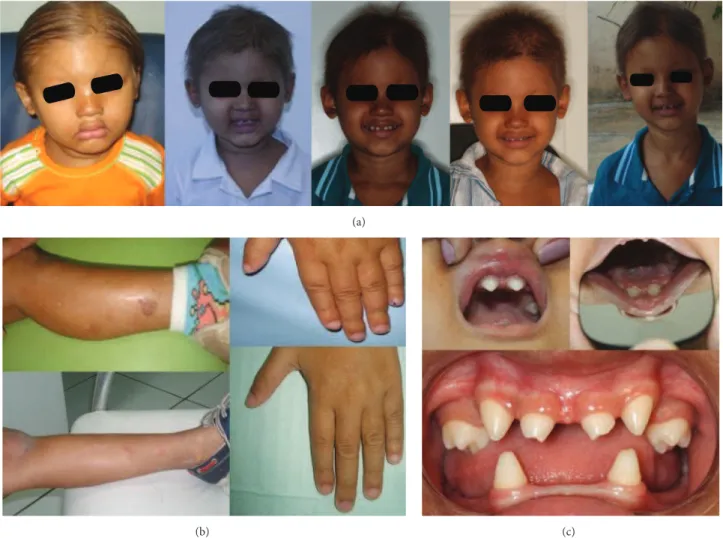
Figure 1: (a) Patient’s visage at 2, 3, 4, 5, and 6 years of age with sparse hair, frontal bossing, saddle nose, and everted prominent lips. (b) Bright, dry skin on the leg; normal nails. (c) Intraoral examination showing variation in the timing and shape of the teeth, hypodontia, and ogival palate.
and frequent bathing. During viral infections, the mother had observed gelatinous, yellow-brown secretions from the respiratory tract. No anomalies were reported concerning the child’s neurological development. He had been attending pediatric appointments, but the condition had never been suspected.
Ater anamnesis, wherein the boy’s medical history was recalled by his mother, both extra- and intraoral examina-tions were performed (Figure 1); on this basis, the child was diagnosed with HED.
Initial dental treatment was based on maintaining oral health, which involved instructing both the boy and his mother in oral hygiene in order to preserve the teeth against oral diseases such as caries and periodontal disease. he mother was further instructed to seek the advice of a medical geneticist who would conirm the diagnosis of HED, request additional tests, and contribute to the patient’s treatment.
From the age of 3 to 6 years, the child periodically attended clinical and imaging exams (Figures 1 and 2) to assess bone development and identify when to begin oral rehabilitation.
When the patient was 6 years old, a cephalometric analysis was carried out (Figure 2(b)) revealing that he had a convex proile, as well as a Class I skeletal malocclusion. Using the Eklof and Ringertz index and a carpal radiograph (Figure 2(d)), we determined that the patient’s bone age matched his chronological age. What is more, cone-beam computed tomography (CBCT) revealed both hypertrophy in the tonsils and mandibular micrognathia (Figures 2(e) and 2(f)).
he patient was of school age and was willing to cooperate with treatment, especially since the characteristic phenotype of the syndrome, and in particular the dental condition, was afecting him emotionally—he felt diferent from his classmates.
Informed consent was given by the boy’s mother, and the treatment consisted of restorations of the upper and lower teeth and the installation of a lower removable partial denture (Figure 4) when the boy was 7 years old.
An impression was taken using alginate (Figure 3(a))
(Jel-tratePlus; Dentsply) to obtain study cast models (Durone
Case Reports in Dentistry 3
(a)
(b) (c)
(d) (e)
(f) (g)
(a)
(b) (c)
Figure 3: Stages of prosthetic rehabilitation. (a) Upper and lower impression. (b) Template for maxillary tooth restorations. (c) Mandibular partial dentures.
(a) (b)
(c)
Case Reports in Dentistry 5
the upper anterior teeth was created to facilitate planning of the lower prosthesis.
A further impression of the diagnostic wax-up was made, using addition silicon (Adsil; Vigodent) to construct a template that would guide restorations of the deciduous teeth (Figure 3(b)). he maxillary conoid teeth were reconstructed by increasing their dimensions, but maintaining the spaces characteristic of the deciduous dentition. he restorations were made using composite resin AO.5 (Opallis; FGM) so as to be white and opaque.
he lower partial denture (Figure 3(c)) was supported by the mandibular primary canines, which received increments
of resin A3 (IPS EmpressDirect; Ivoclar Vivadent), so as
to resemble the premolars. he appropriate tooth color was then selected (tooth color 60; SPG pop) and the gum was characterized (number 15; Tomaz Gomes System, VIPI).
At the installation of the prosthesis, both the patient and his mother were instructed regarding prosthesis placement, removal, and hygiene.
Adjustments were made 48 hours ater installation. he prosthesis showed good retention, and the patient adapted well to it. hen, the next visit was scheduled at 6 months for follow-up and the patient’s mother was advised to arrange additional visits in the case of complications.
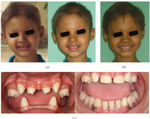
Six months ater installation of the prosthesis, the peri-odontal condition of the teeth, occlusion, functional adap-tation, and cephalometrics were evaluated (Figure 2(c)). Clinical evaluation was repeated over six months when the patient was 8 years old (Figure 4(b)) and new adjustments in prosthesis were performed.
According to his mother, the child’s chewing ability had improved and he was able to enjoy foods that he could not chew before, such as meat, cheese, and ibrous vegetables. his resulted in a gain of 4 kg—from 16 kg to 20 kg—during the irst 6 months of adaptation.
Both the family and the teacher noticed the behav-ioral diferences, and the classmates approved the child’s new appearance, which suggested impact on his socializa-tion.
It must be noted that the patient’s mother, as his legal guardian, read and signed an informed consent form autho-rizing the use of data, images, and all information relating to the dental patient follow-up for use in publications and/or scientiic events.
3. Discussion
Important dental defects are associated with the syndrome. During childhood particularly, this condition is a major cause of frustration that impacts the intellectual and psychological maturity of the patient [5, 6].
Many children require extensive treatment; however, treatment cost should be evaluated. All changes in the dental arches, including alveolar bone growth in response to tooth eruption, should be monitored to ensure appropriate adjust-ments are made. Long-term treatment is an active process that must be constantly adapted to the child’s development and growth [10].
In the present case, ater the prosthetic rehabilitation, the child experienced a wider variety of foods. he relationship between the upper and lower jaws, esthetics, and self-esteem were all improved. Children with these prostheses should perform standard oral hygiene and take care of the devices by themselves.
ED treatment using removable partial dentures is indi-cated for children and adults [6, 7]. Nonetheless, the use of partial dentures has been described in a 2-year-old HED patient; such early rehabilitation prevents growth abnormal-ities and improves socialization [7].
he installation of implants in adult ED patients has been widely reported as a treatment option with high success rate and the associated beneits of rehabilitation [8]. Nonetheless, the high cost, possible failure of osseointegration, and need for complete bone growth are disadvantages, particularly if dental implants are placed before dental and skeletal maturation [9].
In this case, dental implants were not indicated, because CBCT (Figures 2(e) and 2(f)) revealed both maxillary and mandibular micrognathism caused by the absence of teeth. he cephalometric analysis (Figures 2(b) and 2(c)) indicated that the patient should be followed up to assess bone growth ater placement of the prosthesis.
Ater prosthetic rehabilitation, masticatory function improves, as in the present case, where the patient was able to eat meat, ish, ibrous vegetables, and cheese. In general, dentures are well accepted by parents and patients, and the psychological beneits ater oral rehabilitation are many [5–7]. he authors are unanimous in recommending early prosthetic treatment in patients with this syndrome, especially when the child already has a social life, as is the case with children of school age.
4. Conclusion
A removable partial denture and restoration were a noninva-sive, reversible, cost-efective, viable, and eicient treatment for a child with hypodontia and HED. Moreover, the treat-ment appeared to lead to marked developtreat-mental beneits for the patient.
Competing Interests
he authors have no potential competing interests to disclose.
References
[1] M. L. Mikkola, “Molecular aspects of hypohidrotic ectodermal
dysplasia,”American Journal of Medical Genetics, Part A, vol.
149, no. 9, pp. 2031–2036, 2009.
[2] M. Nguyen-Nielsen, S. Skovbo, D. Svaneby, L. Pedersen, and J. Fryzek, “he prevalence of X-linked hypohidrotic ectodermal
dysplasia (XLHED) in Denmark, 1995–2010,”European Journal
of Medical Genetics, vol. 56, no. 5, pp. 236–242, 2013.
[3] S. N. de Aquino, L. M. R. Parana´ıba, M. S. O. Swerts, D. R. B. Martelli, L. M. de Barros, and H. M. J´unior, “Orofacial features
of hypohidrotic ectodermal dysplasia,”Head and Neck
vol. 56, no. 3, pp. 329–337, 2015.
[5] N. A. Alencar, K. R. Reis, A. G. Antonio, and L. C. Maia, “Inlu-ence of oral rehabilitation on the oral health-related quality of
life of a child with ectodermal dysplasia,”Journal of Dentistry for
Children, vol. 82, no. 1, pp. 36–40, 2015.
[6] M. V. Carvalho, J. R. S. de Sousa, F. P. C. de Melo et al., “Hypo-hidrotic and “Hypo-hidrotic ectodermal dysplasia: a report of two
cases,”Dermatology Online Journal, vol. 19, no. 7, article 18985,
2013.
[7] M. A. Derbanne, M. C. Sitbon, M. M. Landru, and A. Naveau, “Case report: early prosthetic treatment in children with
ecto-dermal dysplasia,”European Archives of Paediatric Dentistry,
vol. 11, no. 6, pp. 301–305, 2010.
[8] G. Kearns, A. Sharma, D. Perrott, B. Schmidt, L. Kaban, and K. Vargervik, “Placement of endosseous implants in children and
adolescents with hereditary ectodermal dysplasia,”Oral Surgery,
Oral Medicine, Oral Pathology, Oral Radiology and Endodontics, vol. 88, no. 1, pp. 5–10, 1999.
[9] I. P. Sweeney, J. W. Ferguson, A. A. Heggie, and J. O. Lucas, “Treatment outcomes for adolescent ectodermal dysplasia
patients treated with dental implants,”International Journal of
Paediatric Dentistry, vol. 15, no. 4, pp. 241–248, 2005.
[10] C. Dellavia, F. Catti, C. Sforza, D. G. Tommasi, and V. F. Ferrario, “Craniofacial growth in ectodermal dysplasia. An 8 year
longi-tudinal evaluation of Italian subjects,”The Angle Orthodontist,
Submit your manuscripts at
http://www.hindawi.com
Hindawi Publishing Corporation
http://www.hindawi.com Volume 2014
Oral Oncology
Journal ofDentistry
International Journal ofHindawi Publishing Corporation
http://www.hindawi.com Volume 2014
Hindawi Publishing Corporation
http://www.hindawi.com Volume 2014
International Journal of
Biomaterials
Hindawi Publishing Corporation
http://www.hindawi.com Volume 2014
BioMed
Research International
Hindawi Publishing Corporation
http://www.hindawi.com Volume 2014 Case Reports in Dentistry
Hindawi Publishing Corporation
http://www.hindawi.com Volume 2014
Oral Implants
Journal ofHindawi Publishing Corporation
http://www.hindawi.com Volume 2014
Anesthesiology Research and Practice
Hindawi Publishing Corporation
http://www.hindawi.com Volume 2014
Radiology
Research and Practice Environmental and
Public Health Journal of
Hindawi Publishing Corporation
http://www.hindawi.com Volume 2014
The Scientiic
World Journal
Hindawi Publishing Corporation
http://www.hindawi.com Volume 2014
Hindawi Publishing Corporation
http://www.hindawi.com Volume 2014
Dental Surgery
Journal ofDrug Delivery
Journal ofHindawi Publishing Corporation
http://www.hindawi.com Volume 2014
Hindawi Publishing Corporation
http://www.hindawi.com Volume 2014
Oral Diseases
Journal ofHindawi Publishing Corporation
http://www.hindawi.com Volume 2014
Computational and Mathematical Methods in Medicine
Scientifica
Hindawi Publishing Corporationhttp://www.hindawi.com Volume 2014
Pain
Research and Treatment
Hindawi Publishing Corporation
http://www.hindawi.com Volume 2014
Preventive MedicineAdvances in
Hindawi Publishing Corporation
http://www.hindawi.com Volume 2014
Endocrinology
International Journal ofHindawi Publishing Corporation
http://www.hindawi.com Volume 2014
Hindawi Publishing Corporation