MICHELE CORRÊA BERTOLDI
ANTIOXIDANT CAPACITY, ANTICANCER EFFECTS AND ABSORPTION OF MANGO (Mangifera Indica L.) POLYPHENOLS IN VITRO
Tese apresentada à Universidade Federal de Viçosa, como parte das exigências do Programa de Pós-Graduação em Ciência e Tecnologia de Alimentos, para obtenção do título de
Doctor Scientiae.
VIÇOSA
MICHELE CORRÊA BERTOLDI
ANTIOXIDANT CAPACITY, ANTICANCER EFFECTS AND ABSORPTION OF MANGO (Mangifera Indica L) POLYPHENOLS IN VITRO
Tese apresentada à Universidade Federal de Viçosa, como parte das exigências do Programa de Pós-Graduação em Ciência e Tecnologia de Alimentos, para obtenção do título de
Doctor Scientiae.
APROVADA: 9 de dezembro de 2009.
_________________________ _____________________________ Profa. Tânia Toledo de Oliveira Profa.Susanne U Mertens-Talcott
(Coorientadora) (Coorientadora)
_________________________ ______________________________ Prof. José Carlos Gomes Profa. Nilda de Fátima Ferreira Soares
_____________________________ Prof. Paulo Cesar Stringheta
To my son Vitor, for his unconditional love. To my husband Hernani, for his love, care and friendship.
ACKNOWLEDGMENTS
I would like to thank GOD, for everything I have, light and protection.
I would like to express my gratitude to my chair Paulo Cesar Stringheta for his friendship, scientific support, opportunities and assistance during my academic trajectory.
I would like to thank my co-chairs Susanne U Mertens-Talcott and Stephen T. Talcott for their scientific support, friendship and the generous assistance with the Doctoral training in USA. They really help my family and I to overcome the challenge of living a new culture.
I would like to thank the Universidade Federal de Viçosa and the Department of Food Science and Technology for the opportunity and also to CAPES Foundation (Ministry of Education, Brazil) for providing the fellowship to conduct the research experiment in the USA (Grant n. BEX 130-08-7).
I very appreciate the National Mango Board support for providing plant material used in this study as well as for the financial support.
I would like to thank the members of my doctoral committee and other professors for their scientific support. José Carlos Gomes, Nilda de Fátima Ferreira Soares, Tânia Toledo de Oliveira, Valéria Paula Rodrigues Minim, Afonso Mota Ramos, Frederico José Vieira Passos, Júlio Maria de Andrade Araújo,Mônica Ribeiro Pirozzi, José Benício Paes Chaves, among others, have been taught value scientific background as well as academic skills. I would especially like to thank the professors Nilda de Fatima Ferreira Soares, Paulo Cesar Stringheta, José Benício Paes Chaves, among others, for providing the necessary support and viabilizing the participation of Dr. Susanne Talcott in my thesis defense.
Washington, Mirian, Roney, Aurelia, Taila, Danilo, Luciana, Flavia, Joesse, Roberta, Alice, Leonardo, Meliza, Laura, Roberta, Rita, Fabiana, Jonson, Milton, Márcia, Marília, Solange, Fernanda, Bruna, Mauricio, Igor, Alexandre, for their friendly presence at the Department of Food Technology, UFV.
I really appreciate the receptivity, generous help in the lab and friendly presence of my co-works Gabriela, Armando, Emily, Lisbeth Pacheco, Chris Duncan, Jorge Cardona, Kim, Salvador, Michelle, Patricia, Luis Fernando, Warda, Keily at the Department of Food Science and Nutrition and at Centeq Research Parkway, Texas A&M University, USA. I would like to express my gratitude to Giuliana Norato
and Kimmy for the friendship and generous technical assistance with the experiment analysis.
I am very grateful to the friends Paulo, Carmem, Frederico, Adolfo, Cristiane and Helena for their friendship and help in USA.
I very much appreciate the technical support with the flow cytometry analysis of Dr. Roger Smith, Texas A&M University, College Station, USA.
I highly value the collaborators of the Department of Food Science and Technology, UFV, for their continuous support. Among names and nicknames: Adão, Tineca, Juarez, Geralda, Vaninha, Sueli, Bilico, Lelé, Perereca, Luiz, Zé Geraldo, Maria Rita, Divino, Piu, Pi. I really appreciate Gilcemir, Salvadora, Sueli and Geralda for their help with CAPES process. They also did not measure efforts to viabilize Dr. Susanne trip to Brazil. I also want to thank Alice for my English classes.
I want to express my gratitude to my family Liliane, Edson, Regina, Vicência, José Antônio, Nayarah, Túlio, among others, for their support and care.
Last, but not least, I am very grateful to my lovely parents, Helena and Dirceu, my husband Hernani Santana, and my son Vítor, for their active participation and constant support. Their love, patience, dedication, care and friendship are the main reason of my constant motivation.
BIOGRAPHICAL SKETCH
TABLE OF CONTENTS
LIST OF TABLES ... ix
LIST OF FIGURES ... xi
RESUMO ... xiv
ABCTRACT ...xvii
CHAPTER 1. INTRODUCTION ... 1
Carcinogenesis ... 1
Polyphenols in cancer prevention ... 2
Bioactive effects of mango phytochemicals ... 3
Oxidative stress and cancer... 6
Bioavailability of polyphenols ... 7
Apoptosis ... 11
Extrinsic death receptor pathway... 11
Mitochondrial pathway ... 14
Cell cycle regulation ... 15
Anticarcinogenic effects of polyphenols in normal cells as compared to cancer cells... 18
Objectives ... 19
2. ANTICARCINOGENIC EFFECTS OF POLYPHENOLS FROM DIFFERENT
MANGO (Mangifera indica L.) VARIETIES ... 37
Abstract ... 37
Introduction... 38
Material and Methods ... 39
Chemicals... 39
Plant material ... 39
Extraction of polyphenols ... 40
Antioxidant capacity (ORAC assay)... 41
HPLC-DAD and HPLC-ESI/MSn Analysis ... 42
Cell culture... 43
Cell proliferation ... 43
Cell cycle kinetics ... 44
Quantitative RT-PCR... 44
Reactive oxygen species (ROS)... 45
Statistical analysis ... 45
Results and Discussion ... 46
Total polyphenols and antioxidant activity of mango varieties ... 46
HPLC-DAD and HPLC-ESI/MSn analysis of Haden and Ataulfo polyphenols ... 47
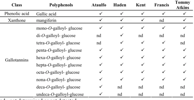
Cell-growth supressive activity of Haden and Ataulfo mango polyphenols on different cancer cell lines ... 49
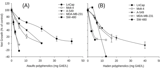
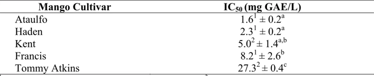
Cell-growth supressive activity of mango polyphenols from different mango varieties on colon cancer cells ... 51
Cell-growth supressive activity of ataulfo polyphenols on cancer cells as compared to normal cells ... 54
Cell cycle regulation ... 55
Gene transcriptional regulation... 57
Protective effects against reactive oxygen species (ROS) ... 60
Conclusion ... 62
3. ABSORPTION AND BIOLOGICAL ACTIVITIES OF POLYPHENOLS FROM DIFFERENT MANGO (Mangifera indica L.) VARIETIES as affected by β
-GLUCOSIDASE hydrolysis ... 70
Abstract ... 70
Introduction... 72
Material and Methods ... 73
Plant material ... 73
Extraction of polyphenols ... 74
Enzymatic hydrolysis... 75
Fractionation of mango phenolic extracts... 76
HPLC-DAD and HPLC-ESI/MSn Analysis ... 77
Antioxidant capacity (ORAC assay)... 78
Cell culture... 79
Cell proliferation ... 79
Transepithelial transport model ... 80
Statistical analysis ... 81
Results and Discussion ... 81
Mango pulp phenolic content and antioxidant activity ... 81
Enzymatic hydrolysis of mango pulp polyphenols ... 82
Phenolic content and antioxidant capacity ... 82
HPLC-DAD and HPLC-ESI/MSn Analysis ... 84
Transepithelial transport model ... 89
Cell-growth supressive activity of mango pulp polyphenols... 93
Cell-growth supressive activity of low and high molecular weight polyphenols-rich fraction ... 99
Conclusion ... 105
References... 107
4. SUMMARY AND GENERAL CONCLUSIONS... 120
LIST OF TABLES
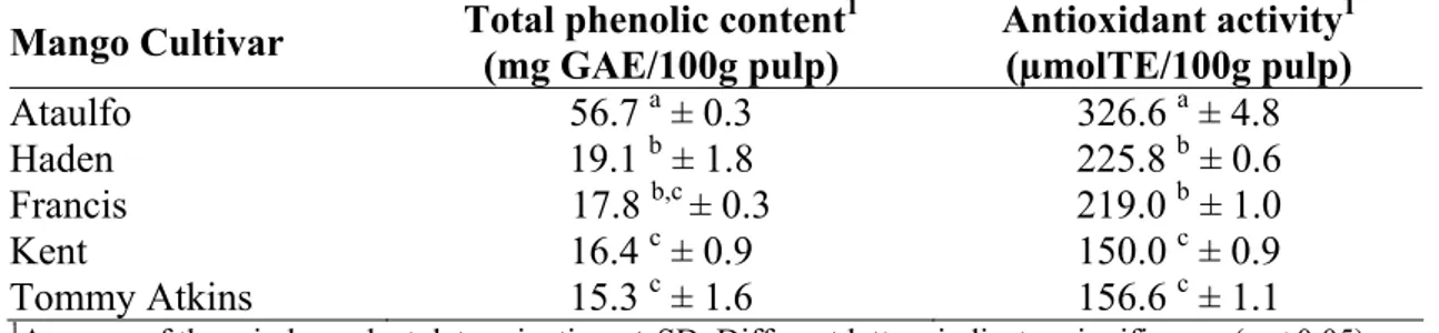
Table 1: Total polyphenols and antioxidant activity (ORAC) of different mango varieties ... 46
Table 2: Polyphenols profile from the mango varieties Ataulfo, Haden, Kent, Francis, and Tommy Atkins determined by DAD and HPLC-ESI-MSn analysis ... 49
Table 3: IC50 values of polyphenols extracted from Ataulfo and Haden mango
varieties for growth suppression of different human cancer cell lines . 50
Table 4: IC50 values of polyphenols extracted from mango varieties for growth
suppression of human SW-480 colon cancer cells... 53
Table 5: Total polyphenols and antioxidant activity (ORAC) of different mango varieties ... 82
Table 6: Total phenolic content and antioxidant activity of hydrolyzed and control mango phenolic extracts ... 83
Table 7: HPLC-DAD and HPLC-ESI-MSn of mango phenolic extracts (Control) from different mango varieties... 86
Table 9: Absorption (%) of phenolic acids through Caco-2 colon cancer cells following 2h incubation with control and hydrolyzed extracts... 91
Table 10: Effect of the treatment with control and hydrolyzed mango phenolic extracts on the growth suppression of MDA-MB-231 breast and HT-29 colon human cancer cell lines, expressed in terms of IC50 values (mg
GAE/L)... 95
Table 11: Cell-growth suppressive effects of mango polyphenols (control) on MDA-MB-231 breast and HT-29 colon human cancer cell lines, expressed in terms of IC50 values (mg mango pulp/mL culture medium).
... 95
Table 12: Total phenolic content (TPC) and antioxidant activity of the hydrolyzed phenolic extract and its fractions ... 102
Table 13: Effect of the treatment with HMW (F1) and LMW (F2) fractions on the growth suppression of MDA-MB-231 breast and HT-29 colon human cancer cell lines, expressed in terms of IC50 values (mg GAE/L) ... 100
LIST OF FIGURES
Figure 1: Chemical structure of 1,2,3,4,6-penta-O-galloyl-β-D-glucose (PGG), the precursor of gallotannins... 4
Figure 2: Routes of absorption and metabolism of dietary polyphenols in human body... 7
Figure 3: Pathways involved in apoptosis signaling: Fas-mediated extrinsic death receptor pathway and mitochondrial pathway; ( ) inhibition; ( ) activation... 13
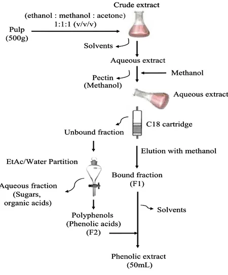
Figure 4: Procedure used for extraction of mango polyphenols... 41
Figure 5: Representative chromatograms at 280 nm of Ataulfo (A) and Haden polyphenols (B). (1) gallotannins; (2) gallic acid. Representative chromatograms at 366 nm of Ataulfo (A1) and Haden (B1) (3) mangiferin ... 48
Figure 6: Cell proliferation of prostate LnCap, leukemia Molt-4, lung A-549, breast MDA-MB-231 and colon SW-480 cancer cells treated with Ataulfo (A) and Haden (B) polyphenols. Cells were treated with different concentrations of extracts and cell growth was assessed after 72 h incubation. Values are means ± SD, n=3 ... 50
growth was measured after 72 h incubation. Values are mean ± SD, n=3.. ... 52
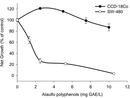
Figure 8: Cell-growth suppressive effects of Ataulfo polyphenols on the human SW-480 colon cancer cells and colonic myofibroblasts CCD-18Co cells. Cells were incubated with extracts and net growth was measured after 48 h. Values are mean ± SD, n=3. Asterisk indicates a significant difference compared to untreated control (*) p ≤ 0.01 ... 55
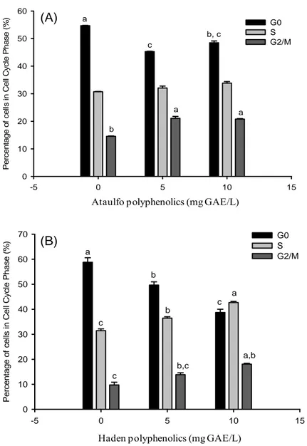
Figure 9: Cell cycle analysis of SW-480 colon cancer cells teated with Ataulfo and Haden polyphenols for 24 h. Values are means ± SE (n=3), different letters indicate significance at p < 0.05... 56
Figure 10: mRNA expression of SW-480 colon cancer cells treated with Ataulfo (A) and Haden (B) polyphenols after 24 h and analyzed by real time PCR as ratio to TATA-binding protein (TBP) mRNA. Values are means ± SE (n=3). Different letters indicate significance at p < 0.05. ... 58
Figure 11: Protective effects of Ataulfo and Haden polyphenols against H2O2- induced ROS production on SW-480 cancer cells (A) and CCD-18Co non-cancer cells (B). Cells were pretreated with extract for 24h and
exposed to 200μM H2O2 for 2h. Values are means ± SD (n=6), different letters indicate significance at p < 0.05.. ... 62
Figure 12: Procedure used for extraction of mango polyphenols... 75
Figure 13: Procedure used for fractionation of mango phenolic extract. ... 77
Figure 15: Representative chromatograms at 280 nm (A) and 340 nm (B) of Ataulfo mango pulp polyphenols from hydrolysed phenolic extracts. Peak assignments: (1) gallic acid; (2) p-OH-benzoic acid; (3) gallotannin; (4) vanillic acid; (5) caffeic acid; (6) mangiferin; (7) p-coumaric acid; (8) ferulic acid. ... 87
Figure 16: Chromatograms (280 nm) from samples taken from the basolateral compartment of Caco-2 cells following 2h of incubation with HBSS (pH 6.0) (A), control (B) and hydrolyzed (C) Ataulfo phenolic extracts. Peak assignments: (1) gallic acid; (2) p-OH-benzoic acid; (3) vanillic acid; (4) caffeic acid; (5) p-coumaric acid; (6) ferulic acid... 90
Figure 17: Cell-growth suppressive effects of mango pulp polyphenols on estrogen independent MDA-MB-231 breast (A) and HT-29 colon (B) human cancer cells. Cells were incubated for 48h with control (c) and hydrolyzed (h) phenolic extracts. Values are mean ± SD, n=3... 94
Figure 18: Chromatograms at 280 nm from HMW (A) and LMW (B) fractions from Ataulfo hydrolyzed extract. Peak assignments: (1) gallic acid; (2) p-OH-benzoic acid; (3) vanillic acid; (4) caffeic acid; (5) mangiferin; (6) p-coumaric acid; (7) ferulic acid... 99
Figure 19: Cell-growth suppressive effects of mango pulp polyphenols on estrogen independent MDA-MB-231 breast (A) and human HT-29 colon (B) cancer cells. Cells were incubated for 48h with HMW and LMW fractions from Ataulfo hydrolyzed extract. Error bars represent the standard error of the mean (n=3)... 101
RESUMO
BERTOLDI, Michele Corrêa, D.Sc.,Universidade Federal de Viçosa, dezembro, 2009. Capacidade antioxidante, efeitos anticarcinogênicos eabsorção de polifenóis de de manga (Mangifera indica L.) in vitro. Orientador: Paulo Cesar Stringheta. Co-orientadores: Susanne U. Mertens-Talcott e Tânia Toledo de Oliveira.
igualmente maior sensibilidade ao tratamento com polifenóis de Ataulfo, enquanto SW-480 e MOLT-4 mostraram-se mais sensíveis ao tratamento com polifenóis de Haden, segundo resultados obtidos por contagem de células. O efeito antiproliferativo dos extratos fenólicos de todas as variedades de manga foi avaliado em células cancerosas de colón humano (SW-480). As variedades Ataulfo e Haden demonstraram maior efeito supressor, seguidas de Kent, Francis e Tommy Atkins. Quando células cancerosas de colón SW-480 foram tratadas com 5 mg GAE/L de polifenóis de Ataulfo, o crescimento celular foi inibido em ~79%, enquanto a proliferação de miofibroblastos não cancerosos CCD-18Co não foi inibida. A supressão do crescimento celular pelo tratamento com polifenóis de Ataulfo e Haden em células de câncer de colón SW-480 foi associada com o aumento na expressão gênica de biomarcadores de apoptose (caspase 8, Bax e Bim) e reguladores do ciclo celular (PKMYT1), atraso do ciclo celular e alteração na produção de espécies reativas de oxigênio. Os extratos fenólicos da polpa de manga continham ácido gálico, mangiferina, derivados de ácidos fenólicos e galotaninos, os quais foram caracterizados por análises em HPLC-DAD e HPLC-ESI-MSn antes e após a hidrólise enzimática (0.17 mg β-glicosidase 1000 KU/g polpa de manga / 4 h / 35°C). Ácidos fenólicos incluindo ácido gálico, caféico, ferúlico, p-coumárico e p-hidroxibenzóico consistiram os principais compostos derivados da hidrólise enzimática. Monocamadas de células Caco-2 foram incubadas por 2h no compartimento apical com extratos controle e hidrolisado. Quando incubadas com o extrato hidrolisado, ácido gálico, caféico, ferúlico, p-coumárico, vanílico e p-hidroxibenzóico foram detectados no compartimento basolateral, enquanto apenas ácido gálico foi detectado quando as células foram tratadas com o extrato controle. Polifenóis de elevado peso molecular, incluindo mangiferina e galotaninos, não foram transportados. Polifenóis de polpa (controle) de todas as variedades inibiram a proliferação de células humanas de câncer de colón HT-29 (0-27 μg de ácido gálico equiv./mL) e de mama MDA-MB-231 (0-24
ABSTRACT
BERTOLDI, Michele Corrêa, D.Sc.,Universidade Federal de Viçosa, December, 2009. Antioxidant capacity, anticancer effects and absorption of mango (Mangifera indica L.) polyphenols in vitro. Adviser: Paulo Cesar Stringheta. Co-advisers: Susanne U. Mertens-Talcott and Tânia Toledo de Oliveira.
Polyphenols found in mango pulp, including gallotannins, flavonol glycosides, gallic acid, benzophenone derivatives and mangiferin have shown anticancer activity. Biological activities of polyphenols have been related to their bioavailability. Deglycosylation by β-glucosidases is a critical step in the metabolism and absorption of dietary polyphenols in humans, which might influence their anticancer properties. The objective of this study was to elucidate the anti-cancer effects of mango polyphenols of several varieties (Francis, Kent, Ataulfo, Tommy Atkins and Haden) in different types of cancer. The antiproliferative effects of mango polyphenols were studied in vitro
INTRODUCTION
Carcinogenesis
Cancer is one of the leading chronic diseases and causes of death worldwide (1). In 2008, 12 million of new cases of cancer and around 7 million of deaths were estimated, with the highest incidence for lung, breast and colon cancer (1). Colon cancer is the third most common cancer in USA for both men and women, with 108,070 estimated cases of colon and 40,740 cases of rectal cancer diagnosed in 2008 (2). According to INCA (National Institute of Cancer) (3), the occurrence of 489.270 new cases of cancer is expected to 2010-2011 in Brazil, with the highest incidence for skin (114,000 cases), prostate (52,000), breast (18,000) and colon (28,000).
Neoplasia consists in an abnormal, uncontrolled and exaggerated proliferation of cells as a result of damage in mechanisms of cell cycle regulation or alteration in genes that regulate the growth and differentiation of cells, which may be benign or malignant (4-5). Cancer is a term used for diseases in which abnormal cells divide without control and are able to spread through the blood and lymph systems and invade others tissues in the body. The process is initially characterized in cell mutation (initiation) as a result of a change in the genetic material of the cell primes. This alteration may occur spontaneously or by an agent that causes cancer (carcinogen). Development of cancer includes invasion, which refers to the direct migration and penetration by cancer cells into neighboring tissues, and metastasis, which is the ability of cancer cells to penetrate into lymphatic and blood vessels, circulate through the bloodstream, and then invade normal tissues elsewhere in the body. Cancerous cells present uncontrolled growth, evasion of apoptosis, self-sufficiency in growth signals, insensitivity to growth-inhibitory signals, limitless replicative potential, sustained angiogenesis, invasion of adjacent tissues, and sometimes metastasis, process by which cells spread to other locations in the body via lymph or blood. These malignant properties are hallmarks of cancer and differentiate cancer cells from begin tumors, which are self-limited, and do not invade or metastasize (6-10).
process that may involve the sequential activation of oncogenes as well as the inactivation of tumor suppressor genes often in the same clone of cells. These genetic changes cause phenotypic alterations in tumor cells that allow them to continue to survive and expand. Both tumor suppressor genes and oncogenes are responsible for proliferation control or differentiation after mutation, which may result in an overexpression of proteins and tumor formation (11). In normal cells, oncogenes often are underexpressed or inactivated (12). Cancer cells are generated, when DNA is damaged, and cells do not undergo apoptosis but instead oncogenes are activated to prevent cell death, resulting in continued and disorderly proliferation (11). Tumor suppressor genes (anti-oncogenes) normally function to limit cell proliferation. Their loss of function d facilitates cancer development, usually in combination with other genetic changes (13). Tumor suppressor genes, or more precisely, the proteins they encode, either have a dampening or repressive effect on the regulation of the cell cycle or promote apoptosis, and sometimes both. The products of oncogenes and tumor suppressor genes have therefore become an important target for new anti-cancer drugs. Thus, knowledge regarding the cancer molecular biology, following the identification and functional characterization of many oncogenes and tumor suppressor genes are critical for therapeutic approach in targeting and eliminating cancer cells (11, 14).
Cell culture models have been extensively used as in vitro trials to evaluate the biological activities of several drugs and natural compounds, including polyphenols (15-19). Because the great availability of different types of human cancer cell lines, including colon, prostate, breast, and so on (20), the use of cell culture models permits evaluation of the potential anticancer effects with higher specificity, reduced cost, and in a shorter period of time.
Polyphenols in cancer prevention
beta-carotene, and phenolic compounds, which comprise the major antioxidant compounds derived from diet (24).
Phenolic compounds include a complex mixture of secondary plant metabolites, with high diversity in chemical structure and reactivity. Chemically, they are composed by aromatic rings with one or more hydroxyl groups, including their functional derivatives (25). Currently, more than 8000 polyphenols have been identified (17), including phenolic acids, flavonoids, lignans, estilbens, cumarins and tannins, and their consumption has been estimated in 1 g/day (26).
Polyphenols are associated not only with color and sensory properties, but also are increasingly being considered as natural cancer chemopreventive compounds based on safety and efficacy assessments (27-29). These compounds may prevent cancer by interfering in enzymatic expression (30), improving intestinal health (31), reducing oxidative stress (32-33), interfering in synthesis and repair of DNA (34), as well as inducing apoptosis (35).
Although many studies have been shown in vitro and in vivo cancer-inhibitory activities from individual compounds, the combination of a variety of phytochemicals in fruits and vegetables may be increased in cancer prevention due to synergistic effects (16, 35-36). Therefore, studies with complex mixtures of these compounds might represent the health-benefits derived from food comsumption.
Bioactive effects of mango phytochemicals
Mangoes (Mangifera indica L.) are amongthe most important tropical fruits marked in the world, with a global production exceeding 33 million tons in 2007. Moreover, mangoes consist one of the most consumed fruits in Brazil due to sensorial characteristics and nutritional value, ranking the seventh position worldwide in terms of production in 2007 (37).
kg/person/year (39). Furthermore, there has been increasing interest in understanding the mango phytochemicals biological properties due to their health-promoting characteristics such as antioxidant, antitumoral, anti-inflammatory, and immunomodulatory activities (40-45). Recently, mango fruit has been listed as a nutrient-rich fruit into the unofficial classification and so-called superfruit due to its considerable content of carotenoids, specially β-carotene, vitamin C and polyphenols, including gallic acid, gallotannins, quercetin and kaempferol glycosides as well as xanthone-C-glucosides (46-48). Gallotannins (Figure 1) represent the major high molecular weight polyphenols found in mango pulp, with molecular weights ranging from 332 (mono-O-galloyl-glucose) to over 1852 Da (undeca-O-galloyl-glucose) (47).
Figure 1. Chemical structure of 1,2,3,4,6-penta-O-galloyl-β-D-glucose (PGG), the precursor of gallotannins.
In traditional medicine, the use of mango extracts as herbal drugs is widespread.
Vimang, a mango stem bark extract, have been used at least 10 years in Cuban medicine with effectiveness against several diseases, like cancer (45). Moreover, studies in vitro
demonstrated chemopreventive action against lung carcinogenesis induced by benzo(a)pyrene in Swiss albino mice (59), which might be related to induction of mitochondria permeability (60). Lupeol and related terpenoids exerted varied cytotoxicity against several cancer cell types, including B16 2F2 mouse melanoma, leukemia HL60, U937, K562 melanoma G36, human lung carcinoma (A-549) and human colon adenocarcinoma (DLD-1) (61). Gallic acid showed to inhibit human cancer cell lines including esophageal, gastric, breast, cervix and colon cancer cells HT-29, Colo201 and colon 26 (mouse colon cancer). In esophageal cancer cells, apoptosis-related molecular mechanism included up-regulation of the pro-apoptotic protein Bax, PARP cleavage induction and caspase-cascade activity, down-regulation of antiapoptotic proteins Bcl-2 and Xiap and the survival Akt/mTOR pathway (62). The effect of gallic acid and its alkyl esters is known to induce cell death or cell cycle arrest in a variety of cancer cells, including colon cancer (63). This phenolic acid (100μM) significantly increased the number of cells in G2/M phase, with a consequent reduction of cells in G1 and S phases on human colon adenocarcinoma cells (Caco2) (64). Additionally, several studies have been shown the anticancer-related activities of gallotannins. 1,2,3,4,6-penta-O-galloyl-beta-d-glucose (PGG), which is the precursor of gallotannins, showed to induce cell cycle arrest, cell proliferation and apoptosis in a cell type-dependent manner (51-53), and to suppress tumor growth in vivo via angiogenesis inhibition and stimulation of apoptosis (65).
The overall anti-cancer potential of mango polyphenols might be in part associated to their antioxidant properties (72), which may contribute to enhance cell defense capacity and modulate molecular pathways in target cells. These compounds might also inhibit promotion and progression stages of cancer by interfering in cell cycle regulation, signal transduction pathways, transcription and activating apoptosis (activation of pro-apoptotic genes and pro-apoptotic proteins) in neoplastic cells (73).
Oxidative stress and cancer
Free radicals are highly reactive chemical species which contains one or more unpaired electrons. These species comprise reactive oxygen species (ROS) and reactive nitrogen species (RNS), which may attack molecules in vivo including proteins, lipids and DNA (74). Superoxide, hydroxyl radical, hydrogen peroxide, hypochlorite and singlet oxygen includes the most important reactive oxygen species (75), whereas reactive nitrogen species constitute the molecules derived from nitric oxide (76).
Reactive oxygen species are produced as natural byproducts of oxygen reduction during the normal metabolism and play an important role in cell signaling (77-78). However, during times of environmental oxidative stress, ROS may cumulated and result in significant damage to cell structures (DNA, RNA and proteins), which is caused by an imbalance between the production of ROS and a biological system's ability to readily detoxify the reactive intermediates or repair the resulting damage. ROS can potentially cause DNA damage and promote tumor progression. Thus, oxidative stress has been considered a hallmark of many tumors (79). In addition to causing genomic instability, ROS are know to increase tumorigenesis by activating signaling pathways that regulate cellular proliferation, angiogenesis, and metastasis (78, 80-81). Low or transient levels of ROS can activate cellular proliferation or survival signaling pathways, while high levels of ROS can initiate damage or cell death (78).
signal transduction (72). Interestingly, the induction in ROS generation by some dietary anticancer agents is tumor cell specific, which may not occur in normal cells (85-87).
Bioavailability of polyphenols
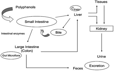
Polyphenols are extensively metabolized either in tissues, once they are absorbed by small intestine, or by the colonic microflora, that transform the non-absorbed fraction or the fraction re-excreted in the bile (Figure 2). Once non-absorbed, polyphenols are conjugated to form O-glucuronides, sulphate esters and O-methyl ether in the small intestine and later in the liver, and no free aglycones are found in the plasma. Conjugation (methylation, sulfation and glucuronidation) reduces polyphenols potential toxicity, enhances their hidrophilicity and facilitates their biliary and urinary elimination. Trough enterophatic recirculation, conjugated compounds are excreted by the liver as components of bile into the intestine, while deconjugated compounds are transformed by microbial enzymes before being absorbed (31, 88-90).
Figure 2. Routes of absorption and metabolism of dietary polyphenols in human body.
Most of polyphenols are found in food in the form of esters, glycosides, or polymers that cannot directly be absorbed in their intact form through small intestine.
Polyphenols C
P450
Tissues
Large Intestine (Colon)
Feces Liver
Kidney
Urine
Gut Microflora Intestinal enzymes
Small Intestine
Bile
Excretion
Polyphenols C
P450
Tissues
Large Intestine (Colon)
Feces Liver
Kidney
Urine
Gut Microflora Intestinal enzymes
Small Intestine
Bile
Thus, exogenous (luminal) deglycosylation by several β-glucosidases in small intestine, or coordination between epithelial transportes and intracellular β-glucosidases facilitate the absorption of some phenolic compounds such as flavonoid glycosides (90-91). Because only aglycones and some glucosides can be absorbed in the small intestine, most of dietary polyphenols need to be firstly transformed in the colon by intestinal microflora before their metabolites may be absorbed (31, 88). Thus, gut bacteria can hydrolyze glycosides, glucuronides, sulfates, amides, esters and lactones. They also are responsible for reduction, descarboxilation, ring-cleavage, demethylation, and dehydroxylation reactions. However, when the microflora is involved, the efficiency of absorption is often reduced because the flora also degrades the aglycones into simple aromatic acids. In addition, polyphenols, which present antimicrobial activities, can interact specifically in a structure-dependent way with certain types of microorganisms in the gut, also modulating the microbial population of the gastrointestinal tract. This has effects on gastrointestinal health as well as in polyphenolic metabolism (92-93). Therefore, the biotransformation by gut bacteria may result in metabolites with higher or lower biological activity than the parent compounds (94-95).
In addition to molecular weight, glycosylation has a considerable impact in the intestinal absorption of dietary polyphenols, because it influences chemical, physical and biological properties of polyphenols and it directly affects its polarity and partition coefficient (26). The aglycones are able to be absorbed by the small intestine. Thus, a commonly accepted concept regarding polyphenols intestinal absorption is that glycosylated polyphenols need to be converted to the aglycone by glycosidases in the food or gastrointestinal mucosa, or from the colon microflora in order to be absorbed by passive diffusion (100). Esterification has also an impact in polyphenols absorption. Esterified polyphenols are, in general, lower absorbed than their non-esterified forms. Galloylated catechins, for example, were better recovered in human urine after black tea consumption than non-galloylated catechins, demonstrating their low availability (101).
water soluble ellagitannin found in pomegranates was hydrolyzed into ellagic acid in vitro across the mitochondrial membrane (109), and converted in vivo by gut microflora to 3,8-dihydroxy-6H-dibenzo[b,d]pyran-6-one (urolithin A, UA) derivatives (18).
Caco-2 cell monolayers are among the most functional in vitro models in the field of drug absorption and permeability, which has been used to predict the in vivo
intestinal absorption and transport of various polyphenols including phenolic acids (110), flavonoids (111), and procyanidins (103). Several studies have been shown that
β-glucosidase activity is related to the intestinal absorption of glycosides in vivo and in caco-2 cell monolayers (112-117). The β-glucosidases (β-glucan glucohydrolase; EC 3.2.1.21) are a widespread group of enzymes that hydrolyze a broad variety of glycosides including aryl- and alkyl- β -D-glycosides, where β-D-galactoside and β -D-glucoside substrates can be hydrolyzed with comparable efficiencies (118). In mammals, several β-glucosidases have been characterized, including the lysosomal β -glucosidase (also called acid β-glucosidase or glucocerebrosidase), lactase phlorizin hydrolase (LPH) and cytosolic (or broad-specificity) β-glucosidase (118). The human cytosolic β-glucosidase (hCBG) constitutes a group of nine enzymes with similarities in the amino-acid and structural features related to their substrate specificities. They are found in the cytosol of liver, spleen, kidney, small intestine, and lymphocytes of mammals and can catalyze the hydrolysis of O-linked β-glycosidic bonds at the non-reducing end of carbohydrates with retention of anomeric configuration (118).
their metabolites and test their respective biological activities, since the hydrolysis of glycosides and further gut bacterial transformation of aglycones may lead to the production of more or less biologically active compounds, which may protect against carcinogenesis (100, 119).
Apoptosis
Apoptosis or Programmed cell death is a physiological process that leads to cellular self-destruction, which is an essential process in development, maintenance of homeostasis and host defense in multicellular organisms. Apoptosis involves typical morphological characteristics including plasma membrane blebbing, cell shrinkage, nuclear chromatin condensation and fragmentation. In contrast to necrosis, apoptosis is a tightly regulated process, which requires the activation of the intracellular machinery by energy requiring (120). Dysregulation of this process contribute to various diseases, including cancer (121-122).
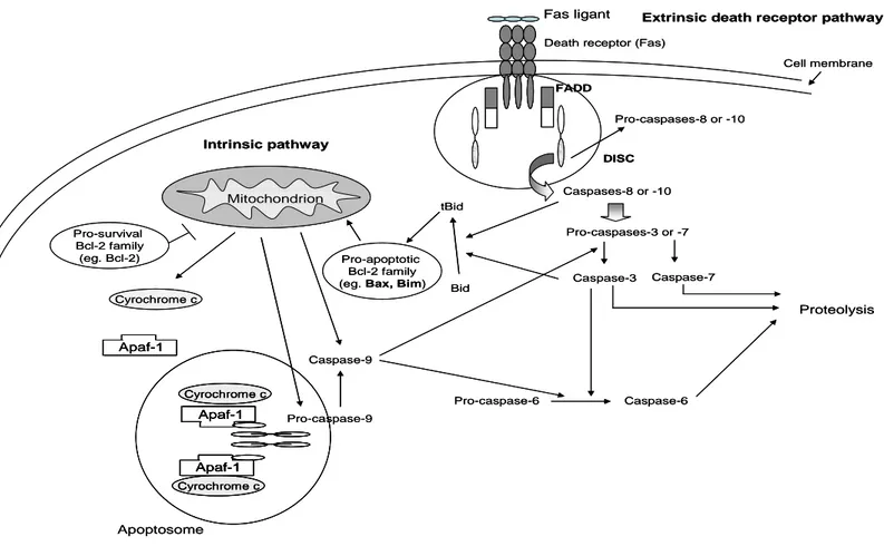
A family of cystein-dependent aspartate-directed proteases, called caspases, plays an essential role in apoptosis. Caspases are synthesized in normal cells as inactive proenzymes (123), and can be activated by apoptosis signaling, which includes autoproteolic cleavage or cleavage of other caspases at specific residues of aspartic acid (124). During apoptosis, initiator caspases function as upstream signal transducers and proteolytically activate downstream caspases (‘effector’ caspases). The initiator caspases-2, -8, -9 and -10 are involved in interactions with adaptor proteins, whereas effector caspases-3, -6 and -7 act on a variety of substrates resulting in proteolysis of cellular proteins, leading to the apoptotic features (internucleosomal DNA fragmentation, and culminating in cell death by apoptosis. Two pathways involved in the activation of caspases have been extensivelly studied: the extrinsic death receptor pathway, and the intrinsic or mitochondrial pathway (125-129).
Extrinsic death receptor pathway
tumour necrosis factor (TNF)-receptor superfamily. This family includes Fas (Apo-1 or CD95), TNF-receptor-1 (TNF-R1), death receptor-3, TNF-related apoptosis inducing ligand receptor-1, TRAIL-R2 and DR6.
Figure 3. Pathways involved in apoptosis signaling: Fas-mediated extrinsic death receptor pathway and mitochondrial pathway; ( ) inhibition; ( ) activation.
Extrinsic death receptor pathway
Intrinsic pathway
Death receptor (Fas)
Cell membrane
Mitochondrion
Fas ligant
FADD
DISC
Pro-caspases-8 or -10
Caspases-8 or -10
Pro-caspases-3 or -7
Caspase-3 Caspase-7
Proteolysis Caspase-6 Pro-caspase-6 Apoptosome Apaf-1 Cyrochrome c Apaf-1 Apaf-1 Cyrochrome c Cyrochrome c Bid Pro-caspase-9 tBid Caspase-9 Pro-apoptotic Bcl-2 family (eg. Bax, Bim) Pro-survival
Bcl-2 family (eg. Bcl-2)
Extrinsic death receptor pathway
Intrinsic pathway
Death receptor (Fas)
Cell membrane Mitochondrion Mitochondrion Fas ligant FADD DISC
Pro-caspases-8 or -10
Caspases-8 or -10
Pro-caspases-3 or -7
Caspase-3 Caspase-7
Proteolysis Caspase-6 Pro-caspase-6 Apoptosome Apaf-1 Apaf-1 Cyrochrome c Apaf-1 Apaf-1 Cyrochrome c Cyrochrome c Apaf-1 Apaf-1 Apaf-1 Apaf-1 Cyrochrome c Cyrochrome c Bid Pro-caspase-9 tBid Caspase-9 Pro-apoptotic Bcl-2 family (eg. Bax, Bim) Pro-survival
Mitochondrial pathway
The second pathway involves the participation of mitochondria in apoptosis, which release caspase-activating proteins into the cytosol (Figure 3). The release of cytochrome c from the mitochondria results in the activation of the apoptotic protease activating factor-1 (Apaf-1). In the presence of cytochrome c and ATP, Apaf-1 bind and activate procaspase-9, thus forming the mitochondrial DISC, also designated as ‘apoptosome’ (133). Following activation, the apoptosome-associated caspase-9 will in turn activate downstream caspases like caspase- 3, -6 and -7 (132).
Intrinsic or mitochondria pathway is mainly regulated by proteins members of the B cell lymphoma (Bcl-2) family, which plays multiple roles in carcinogenesis process. These proteins normally reside in the intermembrane space of mitochondria and, in response to a variety of apoptotic stimuli, can be released to the cytosol. It has been suggested that changes in the concentration or function of Bcl-2 protein family may lead to the cytochrome c release by modulating the permeabilization of the inner and/or outer mitochondria membranes (134-135).
Bcl-2 family includes pro-apoptotic proteins, which facilitate this physiological process of cell death, and pro-survival proteins, that inhibit apoptosis (136). The homology between members of the Bcl-2 family is greatest within four small segments, designated Bcl-2 homology (BH) regions, some of which contribute to the interactions between Bcl-2 family members. Even though conclusion regarding whether the ability to associate with other family members is central to apoptosis regulation, previous studies suggested that the ability of Bcl-2 in inhibiting cell death requires binding to a pro-apoptotic family member. Bcl-2 family can be divided into three groups according to BH domains, mitochondrial anchorage (MA) and pro or anti-apoptotic action of their proteins. They consist in anti-apoptotic (Bcl-2 and Bcl-xL) members, which contain
BH1, BH2, BH3 and BH4 domains; pro-apoptotic members with multidomains including BH1, BH2 and BH3 (Bax and Bak), and citosolic BH3-only pro-apoptotic members (Bid, Bad and Bim) (137-138).
membrane, thus releasing caspase-activating proteins into the cytosol (139-140). Likewise, Bim acts as sensor for cytoskeleton integrity and induce apoptosis by neutralizing certain pro-survival Bcl-2 sub-family of proteins (141). Recently, it was demonstrated the direct interaction between Bim and Bax domains, which suggest a possible Bax activation by Bim (142).
Cell cycle regulation
Cell cycle consists in the series of events which occur in a cell that lead to its division and replication. In eukaryotes, cell cycle is divided in interphase, process of high expression of cell growth proteins which are required to mitosis; and mitosis, the process of nuclear division. Mitosis includes four stages: prophase, metaphase, anaphase and telophase, whereas interphase includes G1, S and G2 phases. DNA Replication occurs in S phase, which is preceded by a gap called G1, a phase characterized by cell growth and high biosynthetic activity of enzymes that are required in S phase. Likewise, RNA transcription and protein synthesis rates are high in G2 phase, during which the cell prepares for mitosis (143-144). Cells in G1 can, before commitment to DNA replication, enter a resting state called G0. The major part of the non-growing, non-proliferating cells in the human body is found in G0 phase Although the duration of cell cycle in tumor cells is equal to or longer than that of normal cell cycle, the proportion of cells that are in active cell division (versus quiescent cells in G0
phase) in tumors is much higher than that in normal tissue. Thus there is a net increase in cell number, whereas the number of cells that die by apoptosis or senescence may remain the same (145).
Cell cycle, a highly conserved and regulated process, is essential to cell survival because includes the repair of genetic damage as well as the prevention of uncontrolled cell division. The mutation in some genes is the main cause of cell cycle dysregulation, which may lead to an uncontrolled cellular proliferation, tumor formation and carcinogenesis development (143, 146).
regulation mechanisms involve controlled expression and destruction of cyclins, activation and inhibitory phosphorylation and dephosphorylation of the cyclin-dependent kinases (Cdks), cyclins, and other proteins; the binding of a number of Cdk inhibitory proteins; and the expression and destruction of inhibitory proteins associated with Cdks, or Cdk/cyclin complexes (143, 146, 148-150).
Cyclins are targets for extracellular signaling and frequently are dysregulated during oncogenesis. These proteins are produced or degraded as needed during interphase in order to drive the cell through the different stages of cell cycle. They control the progression of cell cycle by activating cyclin-dependent kinase (Cdk) enzymes. Cyclins consist in cell cycle regulators which are activated by Cdks at specific points: at the G1 phase (Cyclin D), at G1/S transition (Cyclin E), at S phase (Cyclin A), at G2/M phase transition ( Cyclin A) and in mitosis (Cyclin B) (145, 151).
Likewise, cyclin-dependent kinases are cell cycle regulatory proteins from a serine/threonine protein kinases family. Some of these proteins are known to induce downstream processes by phosphorylating specific proteins upon becoming active by forming clyclin complexes with other proteins, or by phosphorylation during different cell cycle phases (148). In humans, Cdk4, Cdk6, Cdk2 are know to regulate G1 phase, Cdk2 is active at S phase, while Cdk1 controls mitosis (152).
by the PKMYT1 gene in humans (157). Therefore, this enzyme plays an important role in carcinogenesis, because negatively regulates cell cycle G2/M transition by cdc-2 inactivation (157), which may leads to G2/M cell cycle arrest and cell growth suppression.
The activity of cyclin-dependent kinase (Cdks) can be counteracted by cell cycle inhibitory proteins, known as cyclin-dependent kinase inhibitors (CKI), which are produced by genes called tumor suppressor genes. CKIs can halt cell cycle and, consequently, the tumor progression, by binding Cdk alone or Cdk/cyclin complexes. CKIs are distributed in two major families: the INK4a/ARF (Inhibitor of Kinase 4/Alternative Reading Frame) family, composed of inhibitors (p15, p16, p18 and p19) of single Cdks enzyme before cyclin binding (158); and the cip/kip family, composed of CDKs (p21, p27 and p57) that inactivate CDK-cyclin complexes (159). In addition, they inhibit the CDK/cyclin complexes at G1 phase, and to a lesser extent, Cdk1/cyclin B complexes (160).
Anticarcinogenic effects of polyphenols in normal as compared to cancer
cells
Tumor cells present several aspects which make them different from normal cells. Cancerous cells do not depend on growth factors like normal cells do, because they are able to produce their own growth factors to stimulate cell proliferation. Normal cells need to keep in contact with the extracellular medium to growth, whereas cancer cells are not dependent on anchorage. Moreover, in cell culture, normal cells growth in a monolayer and may be inhibited by closed cells, whereas cancerous cells growth to form several monolayers. Tumoral cells present less adherence than normal cells. Additionally, normal cells interrupt cell proliferation as soon as they reach certain density, whereas tumoral cells keep growing. Normal cells remain in the area where they belong and do not spread to other parts of the body. Cancer cells, otherwise, may spread through the body (metastasize), by direct invasion and destruction of the organ of origin, or spread through the lymphatic system or bloodstream to distant organs such as the bone, lung, and liver (10).
Several in vitro studies have been demonstrated the chemopreventive effects of polyphenols in cancer cells but not in normal cells (169-173). Previous studies have reported that growth-inhibitory effects of polyphenols were greater in colon cancer as compared to non-tumorigenic colon cells (169-170). The isogenic human colon cancer cell lines HCT-116(p53 +/+) (IC50 value 45 μg/mL) and HCT-116 (p53 -/-) (IC50 value 30 μg/mL) showed to be more sensitive to the treatment with gallotannins than normal human intestinal epithelial cells FHs 74Int (IC50 value > 60 μg/mL) (173).
Objectives
The overall objective of this study was to elucidate the anti-cancer effects of mango polyphenols of several varieties in different types of cancer.
Based on previous published data which state that individual polyphenols found in mango pulp present anticancer properties including antiproliferative effects, induction of apoptosis, regulation of cell cycle and inhibition of angiogenesis, we hypothesize that mango phenolic extracts present chemopreventive properties, and might represent the health benefits derived from the consumption of the entire fruit instead of isolated compounds. The antiproliferative effects in vitro of polyphenols from different mango varieties (Francis, Kent, Ataulfo, Tommy Atkins and Haden) were compared on different cancer cell lines (Molt-4 leukemia, A-549 lung, MDA-MB-231 breast, LnCap prostate, SW-480 colon cancer cells and the non-cancer cell line CCD-18Co). Molecular mechanisms involved on the anti-cancer activities of mango polyphenols on colon cancer cells were assessed by studying the effect of mango polyphenols on gene expression, cell cycle regulation and reactive oxygen species production.
Based on previous publications indicating the increased bioavailability of polyphenolics after metabolism by β-glucosidase, we hypothesize that the enzymatic hydrolysis of mango polyphenols could enhance their absorption in a selected cell culture model. This hypothesis was tested using Caco-2 human coloncarcinoma cell monolayers in vitro model. In order to elucidate the in vivo intestinal absorption, mango polyphenols were prior hydrolyzed using the enzyme β-glucosidase, commonly found in gut microflora. The effect of enzymatic hydrolysis on antioxidant activity, phenolic content and cell-growth suppressive activity of mango polyphenols was evaluated on MDA-MB-231 breast and HT-29 colon human cancer cells. The antiproliferative activity of high molecular weight polyphenols-rich fraction, characterized by mangiferin (422.35 Da) and gallotannins (788-1851 Da), and low molecular weight polyphenols-rich fraction, comprising mainly phenolic acids (138-194 Da), was studied using cell culture models.
polyphenols contributes to the ongoing evaluation of health benefits derived from fruit consumption.
REFERENCES
1. WHO, World Health Statistics 2008, World Health Organization: Geneva Switzerland, 2008.
2. American Cancer Society Cancer Facts & Figures 2008
http://www.cancer.org/downloads/STT/2008CAFFfinalsecured.pdf (03/13/09).
3. INCA, Estimativas 2010: Incidência de Câncer no Brasil. 2009, National Institutto of Cancer http://www1.inca.gov.br/vigilancia/incidencia.html (28-11-09). 4. Brasilerio Fillho, G., Patologia Geral 3. ed. Rio de Janeiro: Guanabara Koogan, 2004.
5. Harnden, D. G.; Mcgee, J. O. D., Textbook of Patology 1992, Oxford University Press v.1, 571-717.
6. Soussi, T., Cancer epidemiology: from the description to molecular biology. M S-Med Sci 2000, 16, (12), 1397-1404.
7. Bogliolo, L.; Pereira, F. E. L.; Guimaraes, R. C., Bogliolo Patologia 1987, 4. ed. Rio de Janeiro: Guanabara Koogan, 180-205.
8. Cotran, R. S.; Kumar, V.; Collins, T. R., Patologia Estrutural e Funcional.
2000, 6. ed. Rio de Janeiro: Guanabara Koogan, 233-293.
9. Coussens, L. M.; Werb, Z., Inflammation and cancer. Nature 2002, 20, 860-867. 10. Hanahan, D.; Weinberg, R. A., The hallmarks of cancer. Cell 2000, 100, (1), 57-70.
11. Shay, J. W.; Roninson, I. B., Hallmarks of senescence in carcinogenesis and cancer therapy. Oncogene 2004, 23, (16), 2919-2933.
12. Vattemi, E.; Claudio, P. P., Tumor suppressor genes as cancer therapeutics.
Drug News Perspect 2007, 20, (8), 511-520.
13. Burzynski, S. R., Aging: gene silencing or gene activation? Med Hypotheses
2005, 64, (1), 201-208.
14. Ross, D. W., Cancer: The emerging molecular biology. Hosp Pract 2000, 35, (1), 63-+.
MAP-kinases without affecting intracellular generation of reactive oxygen species in vitro. J Nutr 2005, 135, (3), 609-614.
16. Mertens-Talcott, S. U.; Bomser, J. A.; Talcott, S. T.; Percival, S. S., Quercetin and ellagic acid act synergistically in the induction of apoptosis and p21(WAF1/C1P1)-involved signal transduction in human MOLT-4 leukemia cells. Faseb J 2004, 18, (4), A379-A379.
17. Mertens-Talcott, S. U.; Chintharlapalli, S.; Li, M. R.; Safe, S., The oncogenic microRNA-27a targets genes that regulate specificity protein transcription factors and the G(2)-M checkpoint in MDA-MB-231 breast cancer cells. Cancer Res 2007, 67, (22), 11001-11011.
18. Mertens-Talcott, S. U.; Jilma-Stohlawetz, P.; Rios, J.; Hingorani, L.; Derendorf, H., Absorption, metabolism, and antioxidant effects of pomegranate (Punica granatum L.) polyphenols after ingestion of a standardized extract in healthy human volunteers. J Agr Food Chem 2006, 54, (23), 8956-8961.
19. De Castro, W. V.; Mertens-Talcott, S.; Derendorf, H.; Butterweck, V., Grapefruit juice-drug interactions: Grapefruit juice and its components inhibit p-glycoprotein (ABCB1) mediated transport of talinolol in caco-2 cells. J Pharm Sci-Us
2007, 96, (10), 2808-2817.
20. Ross, D. T.; Scherf, U.; Eisen, M. B.; Perou, C. M.; Rees, C.; Spellman, P.; Iyer, V.; Jeffrey, S. S.; Van de Rijn, M.; Waltham, M.; Pergamenschikov, A.; Lee, J. C. E.; Lashkari, D.; Shalon, D.; Myers, T. G.; Weinstein, J. N.; Botstein, D.; Brown, P. O., Systematic variation in gene expression patterns in human cancer cell lines. Nat Genet
2000, 24, (3), 227-235.
21. Block, K. I.; Koch, A. C.; Mead, M. N.; Tothy, P. K.; Newman, R. A.; Gyllenhaal, C., Impact of antioxidant supplementation on chemotherapeutic efficacy: A systematic review of the evidence from randomized controlled trials. Cancer Treat Rev
2007, 33, (5), 407-418.
22. Abdulla, M.; Gruber, P., Role of diet modification in cancer prevention.
Biofactors 2000, 12, (1-4), 45-51.
24. Moure, A.; Cruz, J. M.; Franco, D.; Dominguez, J. M.; Sineiro, J.; Dominguez, H.; Nunez, M. J.; Parajo, J. C., Natural antioxidants from residual sources. Food Chem
2001, 72, (2), 145-171.
25. Shahidi, F.; Naczk, M., Food phenolics: souces, chemistry, effects and applications. 1. ed. Lancaster: Technomic Publishing Co, Inc. 1995, 331p.
26. Scalbert, A.; Williamson, G., Dietary intake and bioavailability of polyphenols.
J Nutr 2000, 130, (8), 2073s-2085s.
27. Amin, A. R. M. R.; Kucuk, O.; Khuri, F. R.; Shin, D. M., Perspectives for Cancer Prevention With Natural Compounds. J Clin Oncol 2009, 27, (16), 2712-2725. 28. Korkina, L. G.; De Luca, C.; Kostyuk, V. A.; Pastore, S., Plant Polyphenols and Tumors: From Mechanisms to Therapies, Prevention, and Protection Against Toxicity of Anti-Cancer Treatments. Curr Med Chem 2009, 16, (30), 3943-3965.
29. Brisdelli, F.; D'Andrea, G.; Bozzi, A., Resveratrol: A Natural Polyphenol with Multiple Chemopreventive Properties (Review). Curr Drug Metab 2009, 10, (6), 530-546.
30. Magrone, T.; Candore, G.; Caruso, C.; Jirillo, E.; Covelli, V., Polyphenols from Red Wine Modulate Immune Responsiveness: Biological and Clinical Significance.
Curr Pharm Design 2008, 14, (26), 2733-2748.
31. Selma, M. V.; Espin, J. C.; Tomas-Barberan, F. A., Interaction between Phenolics and Gut Microbiota: Role in Human Health. J Agr Food Chem 2009, 57, (15), 6485-6501.
32. Parvez, S.; Tabassum, H.; Rehman, H.; Banerjee, B. D.; Athar, M.; Raisuddin, S., Catechin prevents tamoxifen-induced oxidative stress and biochemical perturbations in mice. Toxicology 2006, 225, (2-3), 109-118.
33. Yamamoto, M.; Miyamoto, S.; Moon, J. H.; Murota, K.; Hara, Y.; Terao, J., Effect of dietary green tea catechin preparation on oxidative stress parameters in large intestinal mucosa of rats. Biosci Biotech Bioch 2006, 70, (1), 286-289.
35. Mertens-Talcott, S. U.; Talcott, S. T.; Percival, S. S., Low concentrations of quercetin and ellagic acid synergistically influence proliferation, cytotoxicity and apoptosis in MOLT-4 human leukemia cells. J Nutr 2003, 133, (8), 2669-2674.
36. Mertens-Talcott, S. U.; Percival, S. S., Ellagic acid and quercetin interact synergistically with resveratrol in the induction of apoptosis and cause transient cell cycle arrest in human leukemia cells. Cancer Lett 2005, 218, (2), 141-151.
37. FAOSTAT, FAO Statistical Database – Agriculture. http://apps.fao.org ( 11-09-2009) 2009.
38. Masibo, M.; He, Q., Major mango polyphenols and their potential significance to human health. Compr Rev Food Sci F 2008, 7, (4), 309-319.
39. Camargo Filho, W. P. d.; Alves, H. S.; Mazzei, A. R., Mercado de manga no Brasil: contexto mundial, variedades e estacionalidade Informações Econômicas, SP
<http://www.iea.sp.gov.br/OUT/publicacoes/pdf/tec4-0504.pdf> (11-11-2009). 2004,
34, (5).
40. Garrido, G.; Gonzalez, D.; Lemus, Y.; Garcia, D.; Lodeiro, L.; Quintero, G.; Delporte, C.; Nunez-Selles, A. J.; Delgado, R., In vivo and in vitro anti-inflammatory activity of Mangifera indica L. extract (VIMANG (R)). Pharmacol Res 2004, 50, (2), 143-149.
41. Garrido, G.; Delgado, R.; Lemus, Y.; Rodriguez, J.; Garcia, D.; Nunez-Selles, A. J., Protection against septic shock and suppression of tumor necrosis factor alpha and nitric oxide production on macrophages and microglia by a standard aqueous extract of Mangifera indica L. (VIMANG (R)) - Role of mangiferin isolated from the extract.
Pharmacol Res 2004, 50, (2), 165-172.
42. Garrido, G.; Gonzalez, D.; Delporte, C.; Backhouse, N.; Quintero, G.; Nunez-Selles, A. J.; Morales, M. A., Analgesic and anti-inflammatory effects of Mangifera indica L. extract (Vimang). Phytother Res 2001, 15, (1), 18-21.
43. Martinez, G.; Delgado, R.; Perez, G.; Garrido, G.; Selles, A. J. N.; Leon, O. S., Evaluation of the in vitro antioxidant activity of Mangifera indica L. extract (Vimang).
Phytother Res 2000, 14, (6), 424-427.
as pharmaceuticals - Experimental evidences of a mango stem bark extract. Pharmacol Res 2007, 55, (5), 351-358.
45. Nunez-Selles, A. J., Antioxidant therapy: Myth or reality? J Brazil Chem Soc
2005, 16, (4), 699-710.
46. Schieber, A.; Ullrich, W.; Carle, R., Characterization of polyphenols in mango puree concentrate by HPLC with diode array and mass spectrometric detection.
Innovative Food Science and Emerging Technologies 2000, 1, (2), 161-166.
47. Berardini, N.; Carle, R.; Schieber, A., Characterization of gallotannins and benzophenone derivatives from mango (Mangifera indica L. cv. 'Tommy Atkins') peels, pulp and kernels by high-performance liquid chromatography electrospray ionization mass spectrometry. Rapid Commun Mass Sp 2004, 18, (19), 2208-2216.
48. Barreto, J. C.; Trevisan, M. T. S.; Hull, W. E.; Erben, G.; de Brito, E. S.; Pfundstein, B.; Wurtele, G.; Spiegelhalder, B.; Owen, R. W., Characterization and quantitation of polyphenolic compounds in bark, kernel, leaves, and peel of mango (Mangifera indica L.). J Agr Food Chem 2008, 56, (14), 5599-5610.
49. Huh, J. E.; Lee, E. O.; Kim, M. S.; Kang, K. S.; Kim, C. H.; Cha, B. C.; Surh, Y. J.; Kim, S. H., Penta-O-galloyl-beta-D-glucose suppresses tumor growth via inhibition of angiogenesis and stimulation of apoptosis: Roles of cyclooxygenase-2 and mitogen-activated protein kinase pathways. J Vasc Res 2006, 43, 75-75.
50. Zhang, J. H.; Li, L.; Kim, S. H.; Hagerman, A. E.; Lu, J. X., Cancer, Anti-Diabetic and Other Pharmacologic and Biological Activities of Penta-Galloyl-Glucose.
Pharm Res-Dord 2009, 26, (9), 2066-2080.
51. Chen, W. J.; Chang, C. Y.; Lin, J. K., Induction of G1 phase arrest in MCF human breast cancer cells by pentagalloylglucose through the down-regulation of CDK4 and CDK2 activities and up-regulation of the CDK inhibitors p27(Kip) and p21(Cip).
Biochem Pharmacol 2003, 65, (11), 1777-1785.
52. Chen, W. J.; Lin, J. K., Induction of G1 arrest and apoptosis in human jurkat T cells by pentagalloylglucose through inhibiting proteasome activity and elevating p27Kip1, p21Cip1/WAF1, and Bax proteins. . J Biol Chem 2004 2004, 279, (14), 496-505.
1,2,3,4,6-penta-O-galloyl-beta-D-glucose on human hepatocellular carcinoma cell line, SK-HEP-1 cells.
Cancer Lett 2001, 174, (1), 17-24.
54. Liu, Z. J.; Schwimer, J.; Liu, D.; Lewis, J.; Greenway, F. L.; York, D. A.; Woltering, E. A., Gallic acid is partially responsible for the antiangiogenic activities of Rubus leaf extract. Phytother Res 2006, 20, (9), 806-813.
55. Jagetia, G. C.; Venkatesha, V. A., Effect of mangiferin on radiation-induced micronucleus formation in cultured human peripheral blood lymphocytes. Environ Mol Mutagen 2005, 46, (1), 12-21.
56. Jagetia, G. C.; Baliga, M. S., Radioprotection by mangiferin in DBAxC(57)BL mice: a preliminary study. Phytomedicine 2005, 12, (3), 209-215.
57. Leiro, J. M.; Alvarez, E.; Arranz, J. A.; Siso, I. G.; Orallo, F., In vitro effects of mangiferin on superoxide concentrations and expression of the inducible nitric oxide synthase, tumour necrosis factor-alpha and transforming growth factor-beta genes.
Biochem Pharmacol 2003, 65, (8), 1361-1371.
58. Yoshimi, N.; Matsunaga, K.; Katayama, M.; Yamada, Y.; Kuno, T.; Qiao, Z.; Hara, A.; Yamahara, J.; Mori, H., The inhibitory effects of mangiferin, a naturally occurring glucosylxanthone, in bowel carcinogenesis of male F344 rats. Cancer Lett
2001, 163, (2), 163-170.
59. Rajendran, P.; Ekambaram, G.; Sakthisekaran, D., Effect of mangiferin on benzo(a)pyrene induced lung carcinogenesis in experimental Swiss albino mice. Nat Prod Res 2008, 22, (8), 672-680.
60. Pardo-Andreu, G. L.; Dorta, D. J.; Delgado, R.; Cavalheiro, R. A.; Santos, A. C.; Vercesi, A. E.; Curti, C., Vimang (Mangifera indica L. extract) induces permeability transition in isolated mitochondria, closely reproducing the effect of mangiferin, Vimang's main component. Chem-Biol Interact 2006, 159, (2), 141-148.
61. Chaturvedi, P. K.; Bhui, K.; Shukla, Y., Lupeol: Connotations for chemoprevention. Cancer Lett 2008, 263, (1), 1-13.
63. Serrano, A.; Palacios, C.; Roy, G.; Cespon, C.; Villar, M. L.; Nocito, M.; Gonzalez-Porque, P., Derivatives of gallic acid induce apoptosis in tumoral cell lines and inhibit lymphocyte proliferation. Arch Biochem Biophys 1998, 350, (1), 49-54. 64. Salucci, M.; Stivala, L. A.; Maiani, G.; Bugianesi, R.; Vannini, V., Flavonoids uptake and their effect on cell cycle of human colon adenocarcinoma cells (Caco2). Brit J Cancer 2002, 86, (10), 1645-1651.
65. Huh, J. E.; Lee, E. O.; Kim, M. S.; Kang, K. S.; Kim, C. H.; Cha, B. C.; Surh, Y. J.; Kim, S. H., Penta-O-galloyl-beta-D-glucose suppresses tumor growth via inhibition of angiogenesis and stimulation of apoptosis: roles of cyclooxygenase-2 and mitogen-activated protein kinase pathways. Carcinogenesis 2005, 26, (8), 1436-1445.
66. Percival, S. S.; Talcott, S. T.; Chin, S. T.; Mallak, A. C.; Lounds-Singleton, A.; Pettit-Moore, J., Neoplastic transformation of BALB/3T3 cells and cell cycle of HL-60 cells are inhibited by mango (Mangifera indica L.) juice and mango juice extracts. J Nutr 2006, 136, (5), 1300-1304.
67. Garcia-Solis, P.; Yahia, E. M.; Morales-Tlalpan, V.; Diaz-Munoz, M., Screening of antiproliferative effect of aqueous extracts of plant foods consumed in Mexico on the breast cancer cell line MCF-7. . Int J Food Sci Nutr 2009, 1-15.
68. Garcia-Solis, P.; Yahia, E. M.; Aceves, C., Study of the effect of 'Ataulfo' mango (Mangifera indica L.) intake on mammary carcinogenesis and antioxidant capacity in plasma of N-methyl-N-nitrosourea (MNU)-treated rats. Food Chem 2008, 111, (2), 309-315.
69. Prasad, S.; Kalra, N.; Shukla, Y., Induction of apoptosis by lupeol and mango extract in mouse prostate and LNCaP cells. Nutr Cancer 2008, 60, (1), 120-130.
70. Prasad, S.; Kalra, N.; Singh, M.; Shukla, Y., Protective effects of lupeol and mango extract against androgen induced oxidative stress in Swiss albino mice. Asian J Androl 2008, 10, (2), 313-318.
71. Ribeiro, S. M. R., Caracterização e avaliação do potencial antioxidante de mangas (Mangifera indica L.) cultivadas no Estado de Minas Gerais. PhD Dissertation – Universidade Federal de Viçosa, Viçosa, M.G. 2006.
72. Balsano, C.; Alisi, A., Antioxidant Effects of Natural Bioactive Compounds.
73. Surh, Y. J., Cancer chemoprevention with dietary phytochemicals. Nat Rev Cancer 2003, 3, (10), 768-780.
74. Auten, R. L.; Davis, J. M., Oxygen Toxicity and Reactive Oxygen Species: The Devil Is in the Details. Pediatric Research 2009, 66, (2), 121-127.
75. Afonso, V.; Champy, R.; Mitrovic, D.; Collin, P.; Lomri, A., Reactive oxygen species and superoxide dismutases: Role in joint diseases. Joint Bone Spine 2007, 74, (4), 324-329.
76. Martinez, M. C.; Andriantsitohaina, R., Reactive Nitrogen Species: Molecular Mechanisms and Potential Significance in Health and Disease. Antioxid Redox Sign
2009, 11, (3), 669-702.
77. Datta, K.; Sinha, S.; Chattopadhyay, P., Reactive oxygen species in health and disease. National Medical Journal of India 2000, 13, (6), 304-310.
78. Weinberg, F.; Chandel, N. S., Reactive oxygen species-dependent signaling regulates cancer. Cell Mol Life Sci 2009, 66, (23), 3663-3673.
79. Behrend, L.; Henderson, G.; Zwacka, R. M., Reactive oxygen species in oncogenic transformation. Biochem Soc T 2003, 31, 1441-1444.
80. Feig, D. I.; Reid, T. M.; Loeb, L. A., Reactive Oxygen Species in Tumorigenesis. Cancer Res 1994, 54, (7), S1890-S1894.
81. Storz, P., Reactive oxygen species in tumor progression. Front Biosci 2005, 10, 1881-1896.
82. Raza, H.; John, A., In Vitro Effects of Tea Polyphenols on Redox Metabolism, Oxidative Stress, and Apoptosis in PC12 Cells. Recent Advances in Clinical Oncology
2008, 1138, 358-365.
83. Hong, J.; Kim, M. R.; Lee, N. H.; Lee, B. H., Inhibition of Oral Epithelial Cell Growth in vitro by Epigallocatechin-3-gallate; Its Modulation by Serum and Antioxidant Enzymes. Food Sci Biotechnol 2009, 18, (4), 971-977.
84. Hadi, S. M.; Bhat, S. H.; Azmi, A. S.; Hanif, S.; Shamim, U.; Ullah, M. F., Oxidative breakage of cellular DNA by plant polyphenols: A putative mechanism for anticancer properties. Semin Cancer Biol 2007, 17, (5), 370-376.
86. Antosiewicz, J.; Ziolkowski, W.; Kar, S.; Powolny, A. A.; Singh, S. V., Role of Reactive Oxygen Intermediates in Cellular Responses to Dietary Cancer Chemoprevention Agents. Planta Med 2008, 74, (13), 1570-1579.
87. Engel, R. H.; Evens, A. M., Oxidative stress and apoptosis: a new treatment paradigm in cancer. Front Biosci 2006, 11, 300-312.
88. Manach, C.; Scalbert, A.; Morand, C.; Remesy, C.; Jimenez, L., Polyphenols: food sources and bioavailability. Am J Clin Nutr 2004, 79, (5), 727-747.
89. Scalbert, A.; Manach, C.; Morand, C.; Remesy, C., Bioavailability of dietary polyphenols is a key issue to assess their impact on physiological functions. Free Radical Bio Med 2004, 36, S27-S27.
90. Scalbert, A.; Morand, C.; Manach, C.; Remesy, C., Absorption and metabolism of polyphenols in the gut and impact on health. Biomed Pharmacother 2002, 56, (6), 276-282.
91. Nemeth, K.; Plumb, G. W.; Berrin, J. G.; Juge, N.; Jacob, R.; Naim, H. Y.; Williamson, G.; Swallow, D. M.; Kroon, P. A., Deglycosylation by small intestinal epithelial cell beta-glucosidases is a critical step in the absorption and metabolism of dietary flavonoid glycosides in humans. Eur J Nutr 2003, 42, (1), 29-42.
92. Lee, H. C.; Jenner, A. M.; Low, C. S.; Lee, Y. K., Effect of tea phenolics and their aromatic fecal bacterial metabolites on intestinal microbiota. Res Microbiol 2006, 157, (9), 876-884.
93. Davis, C. D.; Milner, J. A., Gastrointestinal microflora, food components and colon cancer prevention. J Nutr Biochem 2009, 20, (10), 743-752.
94. Rechner, A. R.; Smith, M. A.; Kuhnle, G.; Gibson, G. R.; Debnam, E. S.; Srai, S. K. S.; Moore, K. P.; Rice-Evans, C. A., Colonic metabolism of dietary polyphenols: Influence of structure on microbial fermentation products. Free Radical Bio Med 2004, 36, (2), 212-225.
95. Lampe, J. W.; Chang, J. L., Interindividual differences in phytochemical metabolism and disposition. Semin Cancer Biol 2007, 17, (5), 347-353.
97. Lipinski, C. A., Drug-like properties and the causes of poor solubility and poor permeability. J Pharmacol Toxicol 2000, 44, (1), 235-249.
98. Lipinski, C. A.; Lombardo, F.; Dominy, B. W.; Feeney, P. J., Experimental and computational approaches to estimate solubility and permeability in drug discovery and development settings. Adv Drug Deliver Rev 2001, 46, (1-3), 3-26.
99. Lee, C. Y.; Cheng, H. M.; Sim, S. M., Bioavailability of dietary flavonoids and carotenoids. Curr Top Nutraceut R 2006, 4, (1), 33-51.
100. Yang, C. S.; Landau, J. M.; Huang, M. T.; Newmark, H. L., Inhibition of carcinogenesis by dietary polyphenolic compounds. Annu Rev Nutr 2001, 21, 381-406. 101. Warden, B. A.; Smith, L. S.; Beecher, G. R.; Balentine, D. A.; Clevidence, B. A., Catechins are bioavailable in men and women drinking black tea throughout the day.
J Nutr 2001, 131, (6), 1731-1737.
102. Santos-Buelga, C.; Scalbert, A., Proanthocyanidins and tannin-like compounds - nature, occurrence, dietary intake and effects on nutrition and health. J Sci Food Agr
2000, 80, (7), 1094-1117.
103. Deprez, S.; Mila, I.; Huneau, J. F.; Tome, D.; Scalbert, A., Transport of proanthocyanidin dimer, trimer, and polymer across monolayers of human intestinal epithelial Caco-2 cells. Antioxid Redox Sign 2001, 3, (6), 957-967.
104. Holt, R. R.; Lazarus, S. A.; Sullards, M. C.; Zhu, Q. Y.; Schramm, D. D.; Hammerstone, J. F.; Fraga, C. G.; Schmitz, H. H.; Keen, C. L., Procyanidin dimer B2 [epicatechin-(4 beta-8)-epicatechin] in human plasma after the consumption of a flavanol-rich cocoa. Am J Clin Nutr 2002, 76, (4), 798-804.
105. Prasain, J. K.; Peng, N.; Dai, Y. Y.; Moore, R.; Arabshahi, A.; Wilson, L.; Barnes, S.; Wyss, J. M.; Kim, H.; Watts, R. L., Liquid chromatography tandem mass spectrometry identification of proanthocyanidins in rat plasma after oral administration of grape seed extract. Phytomedicine 2009, 16, (2-3), 233-243.
106. Appeldoorn, M. M.; Vincken, J. P.; Gruppen, H.; Hollman, P. C. H., Procyanidin Dimers A1, A2, and B2 Are Absorbed without Conjugation or Methylation from the Small Intestine of Rats. J Nutr 2009, 139, (8), 1469-1473.