Article
Flavonoid Composition and Antitumor Activity of
Bee Bread Collected in Northeast Portugal
Filipa Sobral1, Ricardo C. Calhelha1, Lillian Barros1,2,*, Montserrat Dueñas3, Andreia Tomás1, Celestino Santos-Buelga3, Miguel Vilas-Boas1and Isabel C. F. R. Ferreira1,*
1 Mountain Research Centre (CIMO), ESA, Polytechnic Institute of Bragança, Campus de Santa Apolónia,
1172, 5300-253 Bragança, Portugal; [email protected] (F.S.); [email protected] (R.C.C.); [email protected] (A.T.); [email protected] (M.V.-B.)
2 Laboratory of Separation and Reaction Engineering—Laboratory of Catalysis and Materials (LSRE-LCM),
Polytechnic Institute of Bragança, Campus de Santa Apolónia, 1134, 5301-857 Bragança, Portugal 3 Grupo de Investigación en Polifenoles (GIP-USAL), Facultad de Farmacia, Universidad de Salamanca,
Campus Miguel de Unamuno s/n, 37007 Salamanca, Spain; [email protected] (M.D.); [email protected] (C.S.-B.) * Correspondence: [email protected] (L.B.); [email protected] (I.C.F.R.F.);
Tel.: +351-273-303-285 (L.B.); +351-273-303-219 (I.C.F.R.F.); Fax: +351-273-325-405 (L.B.); +351-273-325-405 (I.C.F.R.F.)
Academic Editor: Derek J. McPhee
Received: 1 January 2017; Accepted: 3 February 2017; Published: 7 February 2017
Abstract:Bee bread (BB) is a fermented mixture of plant pollen, honey, and bee saliva that worker bees use as food for larvae, and for young bees to produce royal jelly. In the present study, five BB samples, collected from Apis mellifera iberiensis hives located in different apiaries near Bragança, in the northeast region of Portugal, and one BB commercial sample were characterized by high-performance liquid chromatography coupled to a diode array detector and electrospray mass spectrometry (HPLC-DAD-ESI/MS) in terms of phenolic compounds, such as flavonoid glycoside derivatives. Furthermore, the samples were screened, using in vitro assays, against different human tumor cell lines, MCF-7 (breast adenocarcinoma), NCI-H460 (non-small cell lung cancer), HeLa (cervical carcinoma) and HepG2 (hepatocellular carcinoma), and also against non-tumor liver cells (porcine liver cells, PLP2). The main phenolic compounds found were flavonol derivatives, mainly quercetin, kaempferol, myricetin, isorhamnetin and herbacetrin glycoside derivatives. Thirty-two compounds were identified in the six BB samples, presenting BB1 and BB3 with the highest contents (6802 and 6480µg/g extract, respectively) and the highest number of identified compounds. Two isorhamnetin glycoside derivatives, isrohamnetin-O-hexosyl-O-rutinoside and isorhamnetin-O-pentosyl-hexoside, were the most abundant compounds present in BB1; on the other hand, quercetin-3-O-rhamnoside was the most abundant flavonol in BB3. However, it was not possible to establish a correlation between the flavonoids and the observed low to moderate cytotoxicity (ranging from >400 to 68µg/mL), in which HeLa and NCI-H460 cell lines were the most susceptible to the inhibition. To the authors’ knowledge, this is the first report characterizing glycosidic flavonoids in BB samples, contributing to the chemical knowledge of this less explored bee product.
Keywords:bee bread;Apis mellifera iberiensis; phenolic compounds; HPLC-DAD-ESI/MS; cytotoxicity
1. Introduction
The nutritional requirements of honeybees,Apis mellifera, are met by the collection of pollen, nectar, and water. Nectar is the primary source of carbohydrates, while pollen provides proteins, lipids, vitamins and minerals [1]. Bee bread (BB) is a fermented mixture of plant pollen, honey,
and bee saliva that worker bees use as food for larvae, and for young bees to produce royal jelly. Pollen collected by bees is mixed with a small amount of honey and saliva, and packed into the cells of the honeycomb where it undergoes a chemical change to form a product called bee bread [2]. This mixture undergoes different chemical processes due to the action of distinct enzymes from glandular secretions, microorganisms, moisture and temperature (35–36◦C chamber temperature
offspring), allowing the transformation, improvement and preservation of the stored pollen, which is called bee bread after two weeks of initial storage [3,4].
Despite the role of BB as the main source of protein for the bees, its functional properties have been correlated, as well as its flavonoid content, with the BB’s floral origin [4]. In particular, BB has demonstrated in vitro antibacterial [5,6], antioxidant [3], and antitumor [2,7] properties. For the last activity, ethanolic extracts were screened against tumor cell lines (human glioblastoma cell line U87MG) and the normal human astroglia cell line SVGp12 (CRL-08621) using in vitro assays [2,7].
The BB composition varies according to the origin of the pollen but is mainly composed of water, proteins, carbohydrates, lipids, inorganic elements and various other minor components such as decanoic acid, gamma globulin, nucleic acids, vitamins B and C, pantothenic acid, biopterin, neopterin, acetylcholine, and reproductive hormones, among others [8].
The quality information available on the literature for bee bread remains limited, with few reports on the phenolic composition of this mixture. Some phenolic compounds were previously identified in BB samples from Poland, Russia, Latvia and Georgia [9,10]. Other reports on BB samples from Spain and Poland mentioned only total phenolics measured by the Folin-Ciocalteu colorimetric assay [2,4] and did not provide detailed characterization in terms of individual phenolic compounds.
In the present study, five BB samples, collected fromApis mellifera iberiensis hives located in different apiaries near Bragança, in the northeast region of Portugal, and one sample of commercial BB were characterized by HPLC-DAD-ESI/MS in terms of their phenolic profile. Furthermore, the samples were screened against different human tumor cell lines, as well as against non-tumor liver cells.
2. Results and Discussion
2.1. Chromatographic Profile of the BB
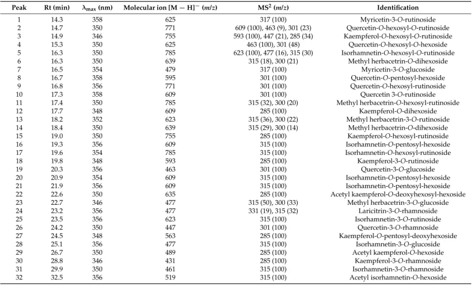
The chromatographic profile of BB1 recorded at 370 nm can be observed in Figure1; the peak characteristics, tentative identities and quantification of all the samples are presented in Table1and the quantification results are presented in Table2. The main phenolic compounds found in bee bread were flavonol derivatives, mainly quercetin, kaempferol, myricetin, isorhamnetin and herbacetrin glycosides. The phenolic composition of bee bread has hardly been explored, only having been reported by a few authors [2,9,10], but using different analytical approaches. Tavdidishvili et al. [10] used HPLC-UV-Vis to study Georgian bee bread samples, reporting the presence of three flavonoids, naringin, rutin and quercetin. Isidorou et al. [9], using GC-MS, identified four phenolic acids (4-hydroxybenzoic, p-coumaric, ferulic and caffeic acids) and six flavonoids (chrysin, naringenin, kaempferol, isorhamnetin, apigenin and quercetin), whereas Markiewicz-Zukowska et al. [2], also using GC-MS, reported the presence of just two flavonoids, kaempferol and apigenin.
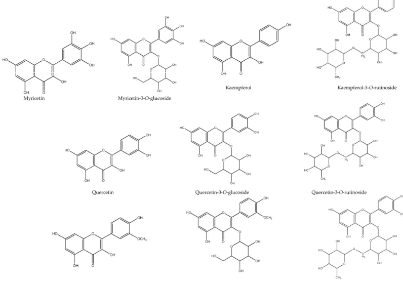
In our samples, up to 32 different flavonoids were detected (Table1). Myricetin-3-O-glucoside (peak 7), quercetin-3-O-rutinoside (peak 10), kaempferol-3-O-rutinoside (peak 18), quercetin-3-O-glucoside (peak 19), isorhamnetin-3-O-rutinoside (peak 25) and isorhamnetin-3-O-glucoside (peak 28) were positively identified according to their retention, mass and UV-vis characteristics in comparison with commercial standards (Figure2). Among them, peaks 10, 18 and 19 were found in all the studied samples, while compounds7(BB3),25(BB1) and28(BB1, BB3, BB5 and BBC) were only detected in some bee bread samples (Table2).
of quercetin, kaempferol and isorhamnetin 3-O-rutinosides may point to peak 1 also a 3-O-rutinoside, and thus it was tentatively assigned as myricetin-3-O-rutinoside.
λ − − − − − − − − − − − − Time (min)
0 5 10 15 20 25 30 35 40 45
mAU 0 50 100 150 200 250 300 350 1 2 3 5 81013 16 18 19 20 2123 25 27 28 32 4 15 26
Figure 1.Individual phenolic compound profile of BB1 recorded at 370 nm. Peak numbering is the same as in Tables1and2.
Peaks 2, 4, 8, 9 and 26 were identified as quercetin derivatives owing to the product ion observed at m/z301 and UV spectra (λmaxaround 350–358 nm). Peaks 2 and 9 presented the same pseudomolecular
ion [M−H]−atm/z771. Peak 2’s MS2fragments revealed the alternative loss of hexosyl (m/zat 609;
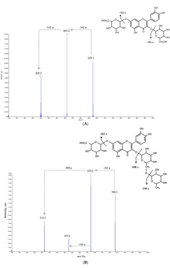
−162 u) and deoxyhexosyl-hexoside (m/zat 463;−308 u) residues, indicating the location of each residue on different positions of the aglycone. Nevertheless, for peak 9 the observation of only one MS2 fragment suggested that the three sugars were linked together. For both peaks 2 and 9, no information about the identity of the sugar moieties and the location on the aglycone could be obtained, so the compounds were tentatively identified as quercetin-O-hexosyl-O-(deoxyhexosyl-hexoside) and quercetin-O-hexosyl-deoxyhexosyl-hexoside, respectively. Nevertheless, the positive identification of different rutinosides, including quercetin-3-O-rutinoside, in the analyzed samples may suggest a rutinose identity for the deoxyhexosyl-hexose residue present in peaks 2 and 9. The mass characteristics of peak 4 ([M −H]− atm/z625) indicated that it corresponds to a quercetin derivative bearing two hexosyl residues. The observation of MS2 fragments at m/z 463 (−162 u) and 301 (−162 u) also indicated the alternative loss of each of the hexosyl moieties, respectively, pointing to their location on different positions of the aglycone. Thus, this compound was tentatively identified as quercetin-O-hexosyl-O-hexoside. Figure3a exemplifies the fragmentation pattern of these types of compounds, and gives a tentative identification for peak 4. This compound was the majority flavonoid in the commercial sample and it was present in all the samples, with the exception of BB3 (Table2).
Peaks 8 ([M−H]−atm/z595) and 26 ([M−H]−atm/z447) showed a similar fragmentation pattern
Table 1.Retention time (Rt), wavelengths of maximum absorption in the visible region (λmax), mass spectral data and identification of phenolic compounds in bee bread samples.
Peak Rt (min) λmax(nm) Molecular ion [M
−H]−(m/z) MS2(m/z) Identification
1 14.3 358 625 317 (100) Myricetin-3-O-rutinoside
2 14.7 350 771 609 (100), 463 (9), 301 (23) Quercetin-O-hexosyl-O-rutinoside 3 14.9 346 755 593 (100), 447 (21), 285 (34) Kaempferol-O-hexosyl-O-rutinoside
4 15.3 350 625 463 (100), 301 (48) Quercetin-O-hexosyl-O-hexoside
5 16.3 350 785 623 (100), 477 (16), 315 (30) Isorhamnetin-O-hexosyl-O-rutinoside
6 16.3 350 639 315 (18), 300 (21) Methyl herbacetrin-O-dihexoside
7 16.5 354 479 317 (100) Myricetin-3-O-glucoside
8 16.7 358 595 301 (100) Quercetin-O-pentosyl-hexoside
9 16.8 356 771 301 (100) Quercetin-O-hexosyl-rutinoside
10 17.3 358 609 301 (100) Quercetin 3-O-rutinoside
11 17.4 350 785 315 (32), 300 (20) Methyl herbacetrin-O-hexosyl-rutinoside
12 17.7 348 609 285 (100) Kaempferol-O-dihexoside
13 18.2 352 623 315 (36), 300 (22) Methyl herbacetrin-3-O-rutinoside
14 18.4 350 639 315 (29), 300 (14) Methyl herbacetrin-O-dihexoside
15 19.0 350 755 285 (100) Kaempferol-O-hexosyl-rutinoside
16 19.3 356 609 315 (100) Isorhamnetin-O-pentosyl-hexoside
17 19.6 354 785 315 (100) Isorhamnetin-O-hexosyl-rutinoside
18 19.8 348 593 285 (100) Kaempferol-3-O-rutinoside
19 20.3 356 463 301 (100) Quercetin-3-O-glucoside
20 20.9 354 609 315 (100) Isorhamnetin-O-pentosyl-hexoside
21 21.9 356 609 315 (100) Isorhamnetin-O-pentosyl-hexoside
22 22.6 350 635 285 (100) Acetyl kaempferol-O-deoxyhexosyl-hexoside
23 22.7 346 477 315 (50), 300 (33) Methyl herbacetrin-3-O-glucoside
24 23.2 356 477 331 (19), 315 (32) Laricitrin-3-O-rhamnoside
25 23.5 356 623 315 (100) Isorhamnetin-3-O-rutinoside
26 24.2 350 447 301 (100) Quercetin-3-O-rhamnoside
27 24.5 348 563 285 (100) Kaempferol-O-pentosyl-deoxyhexoside
28 25.1 356 477 315 (100) Isorhamnetin-3-O-glucoside
29 26.7 350 489 285 (100) Acetyl kaempferol-O-hexoside
30 28.8 346 431 285 (100) Kaempferol-3-O-rhamnoside
31 29.9 350 461 315 (100) Isorhamnetin-3-O-rhamnoside
Table 2.Quantification of the phenolic compounds (µg/g of extract) present in the bee bread samples.
BB1 BB2 BB3 BB4 BB5 BBC Normal
Distribution1 Homoscedasticity2 among MeansDifferences3
Myricetin-3-O-rutinoside 41±3c nd 322±7a nd 38±4c 118±4b 0.002 0.719 <0.001
Quercetin-O-hexosyl-O-rutinoside 156±8 nd nd nd nd nd - -
-Kaempferol-O-hexosyl-O-rutinoside 69±1 nd nd nd nd nd - -
-Quercetin-O-hexosyl-O-hexoside 129±5c 211±6b nd 127±4c 74±3d 1580±31a <0.001 0.089 <0.001
Isorhamnetin-O-hexosyl-O-rutinoside 2615±54 nd nd nd nd nd - -
-Methyl herbacetrin-O-dihexoside nd 622±25a 70±3d 460±3b 192±1c nd 0.046 0.084 <0.001
Myricetin-3-O-glucoside nd nd 36±2 nd nd nd - -
-Quercetin-O-pentosyl-hexoside 100±5 nd nd nd nd 139±1 0.023 0.148 <0.001
Quercetin-O-hexosyl-rutinoside nd 314±6b 106±9c 367±1a nd nd 0.006 0.290 <0.001
Quercetin 3-O-rutinoside 158±3c 88±5e 312±5b 105±6d 91±2e 377±7a 0.001 0.688 <0.001
Methyl herbacetrin-O-hexosyl-rutinoside nd nd nd nd 217±1 nd - -
-Kaempferol-O-dihexoside nd 91±6c 246±8b 83±4c 108±1c 1167±30a <0.001 0.094 <0.001 Methyl herbacetrin-3-O-rutinoside 186±25c nd 71±1d 435±5a 225±7b nd 0.037 0.119 <0.001 Methyl herbacetrin-O-dihexoside nd nd 39±3d 164±13b 268±11a 105±5c 0.152 0.424 <0.001 Kaempferol-O-hexosyl-rutinoside 212±10d 3597±69b 403±1c 3755±46a nd 130±2e <0.001 0.095 <0.001
Isorhamnetin-O-pentosyl-hexoside 1448±37 nd nd nd nd nd - -
-Isorhamnetin-O-hexosyl-rutinoside nd 103±1 nd 43±3 nd nd 0.009 0.208 <0.001
Kaempferol-3-O-rutinoside 62±7de 94±21d 355±10c 56±4e 815±16b 1627±32a <0.001 0.300 <0.001 Quercetin-3-O-glucoside 248±5a 52±8e 236±1b 53±3e 177±3c 72±1d 0.001 0.249 <0.001
Isorhamnetin-O-pentosyl-hexoside 94±8 nd nd nd nd nd - -
-Isorhamnetin-O-pentosyl-hexoside 30±2 nd 47±3 nd nd nd 0.081 0.743 <0.001
Acetyl kaempferol-O-deoxyhexosyl-hexoside nd nd 20±1 nd 11±1 nd 0.037 0.639 <0.001
Methyl herbacetrin-3-O-glucoside tr 32±4d 53±6c 224±10a 138±17b nd 0.033 0.383 <0.001
Laricitrin-3-O-rhamnoside nd nd 125±5 nd nd nd - -
-Isorhamnetin-3-O-rutinoside 836±35 nd nd nd nd nd - -
-Quercetin-3-O-rhamnoside tr 280±22c 3029±72a 168±19d 2001±17b 190±7d 0.001 0.236 <0.001
Kaempferol-O-pentosyl-deoxyhexoside 82±6 nd nd nd nd nd - -
-Isorhamnetin-3-O-glucoside 140±1b nd 199±1a nd 118±4c 64±2d 0.103 0.269 <0.001
Acetyl kaempferol-O-hexoside nd nd nd nd nd 22±3 - -
-Kaempferol-3-O-rhamnoside nd nd 141±12 nd 29±10 nd 0.038 0.747 <0.001
Isorhamnetin-3-O-rhamnoside nd 73±10c 670±44a nd 232±14b nd 0.022 0.234 <0.001
Acetyl isorhamnetin-O-hexoside 197±12 nd nd nd nd nd - -
-Total flavonoids 6802±204a 5557±179d 6480±128b 6040±76c 4733±106e 5593±118d 0.417 0.804 <0.001
nd: not detected; tr: traces.1Normal distribution of the residuals was evaluated using Shapiro-Wilk test (p> 0.05 indicates normal distribution).2Homoscedasticity among bread
formulations was tested by Levene’s test: homoscedasticity,p> 0.05; heteroscedasticity,p< 0.05.3p< 0.05 indicates that the mean value of the corresponding phenolic compound of at least
(A)
(B)
Peaks 3, 12, 15, 27 and 30 were identified as kaempferol glycosides based on their UV spectra (λmaxaround 348 nm) and the production of an MS2fragment ion atm/z285. Similarly, peaks 5, 16,
17, 20, 21 and 31 were identified as isorhamnetin (λmaxaround 356 nm, MS2fragment atm/z315)
glycosides. Tentative identities of these compounds were assigned based on their pseudomolecular ions using similar reasoning as for the quercetin derivatives. Thus, peaks 3 ([M−H]−atm/z755)
and 5 ([M−H]−atm/z785) could correspond to kaempferol-O-hexosyl-O-(deoxyhexosyl-hexoside)
and isorhamnetin-O-hexosyl-O-(deoxyhexosyl-hexoside), whereas peaks 15 and 17, with the same pseudomolecular ions as 3 and 5, could correspond to kaempferol-O-hexosyl-deoxyhexosyl-hexoside (peak 15) and isorhamnetin-O-hexosyl-deoxyhexosyl-hexoside (peak 17). As assumed for the quercetin derivatives, the positive identification of kaempferol and isorhamnetin 3-O-rutinoside allows us to speculate that peaks 3 and 5 correspond to kaempferol-O-hexosyl-O-rutinoside and isorhamnetin-O-hexosyl-O-rutinoside, respectively. Compound5was the majority flavonoid in sample BB1, and compound15in BB2 and BB4 samples. Figure3b exemplifies the fragmentation pattern of these types of compounds, and gives a tentative identification for peak 5. Peaks 12 ([M−H]−
atm/z 609), 27 ([M − H]− atm/z563) and 30 ([M − H]− at m/z 431) could be assumed as a kaempferol-O-dihexoside, kaempferol-O-pentosyl-deoxyhexoside and kaempferol-O-deoxyhexoside, respectively. This latter might be supposed to be kaempferol-3-O-rhamnoside, based on the same considerations as for peak 26.
Peaks 16, 20 and 21, mostly identified in sample BB1, showed the same pseudomolecular ion ([M−H]− atm/z609), pointing out that they might correspond to different isorhamnetin-O-pentosyl-hexoside isomers. Peak 31 ([M − H]− at m/z 461), also bearing −146 u (loss of a
deoxyhexosyl moiety), can be speculated to correspond to an isorhamnetin-O-deoxyhexoside, possibly isorhamnetin-3-O-rhamnoside.
Peaks 22 ([M − H]− at m/z 635) and 32 ([M − H]− at m/z 519) possessed molecular
weights 42 u higher than peaks 18 and 28, pointing to the existence of an additional acetyl residue, thus being tentatively identified as acetyl kaempferol-O-deoxyhexosyl-hexoside and acetyl isorhamnetin-O-hexoside, respectively. Similarly, peak 29 ([M−H]− atm/z489) was tentatively identified as acetyl kaempferol-O-hexoside.
Peaks 6, 11, 13, 14 and 23 were tentatively identified as methyl-herbacetin glycosides based on their UV spectra and the production of two MS2fragments atm/z315 and 300. This assignment was supported by the previous identification of similar compounds in bee pollen samples [11–13]. Compound identities were assigned based on their pseudomolecular ions, as methyl-herbacetin-O-deoxyhexosyl-hexoside (peak 13), methyl-herbacetin-O-methyl-herbacetin-O-deoxyhexosyl-hexoside (peak 23), methyl-herbacetin-O-hexosyl-deoxyhexosyl-hexoside (peak 11) and two methyl-herbacetin-O-dimethyl-herbacetin-O-hexosyl-deoxyhexosyl-hexoside isomers (peaks 6 and 14). Peaks 13 and 23 can be speculated to correspond respectively to herbacetin-3-O-rutinoside and methyl-herbacetin-3-O-glucoside, taking into account the presence in the samples of equivalent glycosides derived from other flavonols. With the exception of peak 11, which was only found in sample BB5, the rest of the methyl-herbacetin derivatives were detected in most of the analyzed samples (Table2).
Finally, peak 24 ([M − H]− at m/z 477), only detected in sample BB3, showed a similar
fragmentation pattern as the methyl-herbacetin derivatives, releasing in this case two MS2 fragment ions atm/z331 and 315, and was tentatively identified as a methylmyricetin derivative, possibly laricitrin (i.e., 3′-O-methylmyricetin). According to its pseudomolecular ion, it would be a laricitrin-O-deoxyhexoside, which can also be speculated to correspond to laricitrin-3-O-rhamnoside owing to the detection of other laricitrin-3-O-rhamnosides in the sample (peaks 26 and 30). To the authors' knowledge, this is the first report on the presence of flavonol glycoside derivatives in bee bread.
2.2. Antitumoral Activity of the BB Samples
less than 50% of the growth of the tumor cells, the results being expressed in terms of GI25values
(sample concentration providing 25% of growth inhibition) (Table3). BBC was selective for the HeLa cell line, while BB3 inhibited the growth of all the tested human tumor cell lines, being the only one that was able to inhibit HepG2 growth. Besides the mentioned data, BB1 and mostly BB2 were also active against MCF-7, BB4 and BB5 against NCI-H460, and BB1, BB5 and principally BB4 against HeLa. It should be highlighted that up to 400µg/mL, none of the BB samples showed toxicity for normal cells (non-tumor porcine liver primary cells).
Table 3.Cytotoxic activity (GI25values,µg/mL) of the bee bread (BB) samples.
Human Tumor Cell Lines Non-Tumor Porcine Liver Cells
MCF-7 NCI-H460 HeLa HepG2 PLP2
BB1 186±6a >400 345±13a >400 >400 BB2 84±3c >400 >400 >400 >400 BB3 164±4b 253±10a 225±12bc 67±1 >400 BB4 >400 85±5b 209±21c >400 >400 BB5 >400 68±8b 276±18b >400 >400 BBC >400 >400 366±7a >400 >400 Ellipticine 0.45±0.02 0.74±0.01 0.55±0.03 1.61±0.07 1.06±0.02
GI25values: sample concentration providing 25% of growth inhibition in human tumor cell lines or in liver primary
culture PLP2. In each column different letters mean significant statistical differences (p< 0.05).
Despite the reports on the antitumor properties of different phenolic compounds, including flavonoids [14], it was not possible to establish a positive correlation between the concentration of flavonoids in each sample and the corresponding cytotoxicity. Therefore, these properties could be attributed to specific individual flavonoids, synergism/antagonism dynamics in the samples and, moreover, the presence of other compounds rather than flavonoids.
3. Materials and Methods
3.1. Samples Collection
The samples of BB were collected in 2012 fromApis melliferaiberiensis hives located in different apiaries near Bragança, in the northeast region of Portugal; specifically located in Bragança (BB1), Montesinho (BB2), Rio de Onor (BB3), Vinhais (BB4), and Castrelos (BB5). For extraction, the frames next to the bee brood were removed from the hives, freeze and the wax crushed mechanically to collect the bee bread. An additional commercial sample (BBC), collected fromApis dorsataon the Himalayan region, was courtesy of Bee Healthy Farms (Springfield, MO, USA).
Once extracted, the samples were lyophilized (FreeZone 4.5 model 7750031, Labconco, Kansas City, MO, USA), reduced to a fine dried powder (20 mesh), mixed to obtain homogenous samples and stored in a desiccator, protected from light, until further analysis.
3.2. Standards and Reagents
3.3. Extracts Preparation
A methanol:water (80:20,v/v) extract was obtained from the lyophilized material. Each sample (1 g) was extracted twice by stirring (25◦C at 150 rpm) with 30 mL of methanol:water (80:20,v/v) for
1 h and subsequently filtered through a Whatman No. 4 paper. The combined methanol:water extracts were evaporated at 40◦C (rotary evaporator Büchi R-210, Flawil, Switzerland) to remove the methanol and further frozen and lyophilized.
3.4. Characterization of the Extracts by HPLC-DAD-ESI/MS
The phenolic compounds were analysed using a Hewlett-Packard 1100 chromatograph (Hewlett-Packard 1100, Agilent Technologies, Santa Clara, CA, USA) with a quaternary pump and a diode array detector (DAD) coupled to an HP Chem Station (rev. A.05.04) data-processing station. A Waters Spherisorb S3 ODS-2 C18, 3µm (4.6 mm×150 mm) column thermostatted at 35◦C was
used and 10µL of each sample was injected. The solvents used were: (A) 0.1% formic acid in water, (B) acetonitrile. The elution gradient established was 15% B for 5 min, 15% B to 20% B over 5 min, 20%–25% B over 10 min, 25%–35% B over 10 min, 35%–50% B for 10 min, and re-equilibration of the column, using a flow rate of 0.5 mL/min. Double online detection was carried out in the DAD using 280 nm and 370 nm as preferred wavelengths and in a mass spectrometer (MS) connected to HPLC system via the DAD cell outlet.
MS detection was performed in an API 3200 Qtrap (Applied Biosystems, Darmstadt, Germany) equipped with an ESI source and a triple quadrupole-ion trap mass analyser that was controlled by the Analyst 5.1 software. Zero grade air served as the nebulizer gas (30 psi) and turbo gas for solvent drying (400◦C, 40 psi). Nitrogen served as the curtain (20 psi) and collision gas (medium). The quadrupoles were set at unit resolution. The ion spray voltage was set at−4500 V in the negative mode. The MS detector was programmed for recording in two consecutive modes: Enhanced MS (EMS) and enhanced product ion (EPI) analysis. EMS was employed to show full scan spectra, so as to obtain an overview of all of the ions in sample. Settings used were: declustering potential (DP)−450 V, entrance potential (EP)−6 V, collision energy (CE)−10 V. EPI mode was performed in order to obtain the fragmentation pattern of the parent ion(s) in the previous scan using the following parameters: DP−50 V, EP−6 V, CE−25 V, and collision energy spread (CES) 0 V. Spectra were recorded in negative ion mode betweenm/z100 and 1000.
The phenolic compounds present in the samples were characterised according to their UV and mass spectra and retention times compared with standards, when available. For the quantitative analysis of phenolic compounds, a fiev-level calibration curve was obtained by injection of known concentrations (2.5–100 µg/mL) of different standards compounds: isorahmetin-3-O-glucoside (y = 218.26x − 0.98; R2 = 0.999); isorahmetin-3-O-rutinoside (y = 284.12x + 67.055; R2 = 0.999); kaempferol-3-O-glucoside (y= 288.55x−4.05;R2= 1); kaempferol-3-O-rutinoside (y= 182.94x+ 96.644;
R2 = 1); quercetin-3-O-glucoside (y= 236.33x + 70.006; R2 = 0.999) and quercetin-3-O-rutinoside (y= 280.87x+ 0.37373;R2= 1). The results were expressed inµg per g of extract.
3.5. Evaluation of In Vitro Cytotoxic Properties of the Extracts
The BB extracts were dissolved in water at 8 mg/mL and then submitted to further dilutions from 400 to 1.56µg/mL. Four human tumor cell lines were used: MCF-7 (breast adenocarcinoma), NCI-H460 (non-small cell lung cancer), HeLa (cervical carcinoma) and HepG2 (hepatocellular carcinoma). Cells were routinely maintained as adherent cell cultures in RPMI-1640 medium containing 10% heat-inactivated FBS and 2 mM glutamine (MCF-7, NCI-H460 HeLa and HepG2 cells), at 37 ◦C,
in a humidified air incubator containing 5% CO2. Each cell line was plated at an appropriate density
For evaluation of the cytotoxicity in non-tumor cells, a cell culture was prepared from a freshly harvested porcine liver obtained from a local slaughterhouse, according to a procedure established by the authors [16]; it was designed as PLP2. Cultivation of the cells was continued with direct monitoring every two to three days using a phase contrast microscope. Before confluence was reached, cells were subcultured and plated in 96-well plates at a density of 1.0×10⁴cells/well, and commercial in DMEM medium with 10% FBS, 100 U/mL penicillin and 100µg/mL streptomycin. Ellipticine was used as positive control and all the results were expressed in GI25 values (concentration that inhibited 25% of the net cell growth).
3.6. Statistical Analysis
Data on phenolic compounds quantification were expressed as mean ± standard deviation. Differences in the phenolic compounds levels among each of the assayed bee bread samples were analyzed through one-way analysis of variance (ANOVA). The fulfilment of the one-way ANOVA requirements, specifically the normal distribution of the residuals and the homogeneity of variance, was tested by means of the Shapiro Wilk’s and the Levene’s tests, respectively. All dependent variables were compared using Tukey’s honestly significant difference (HSD) or Tamhane’s T2 multiple comparison tests, when homoscedasticity was verified or not, respectively. For all compounds detected in only two bee bread samples, statistically significant differences among means were classified according tot-student test. All statistical tests were performed at a 5% significance level using SPSS software, version 23.0 (IBM Corp., Armonk, NY, USA).
4. Conclusions
Overall, bee bread is a very recent bee product and at this stage it can be collected and consumed as a food supplement named “bee bread”. To the authors’ knowledge, this is the first report characterizing glycosidic flavonoids in bee bread samples, contributing to the chemical knowledge of this less explored bee product. Thirty-two flavonol glycoside derivatives, such as quercetin, kaempferol, myricetin, isorhamnetin and herbacetrin derivatives, were identified in the six samples of bee bread. BB1 and BB3 were the bee bread samples that presented the highest content and the highest number of identified compounds (19 compounds). Two isorhamnetin glycoside derivatives, isrohamnetin-O-hexosyl-O-rutinoside and isorhamnetin-O-pentosyl-hexoside, were the most abundant compounds present in BB1; on the other hand, quercetin-3-O-rhamnoside was the most abundant flavonol in BB3. BB samples showed moderate antitumor activity; however, none of the BB samples have shown toxicity for normal cells.
Acknowledgments:The authors are grateful to the Foundation for Science and Technology (FCT, Portugal) for financial support to CIMO (Pest-OE/AGR/UI0690/2013) and L. Barros grant (SFRH/BPD/107855/2015). Also, to POCI-01-0145-FEDER-006984 (LA LSRE-LCM), funded by FEDER, through POCI-COMPETE2020 and FCT. The GIP-USAL is financially supported by the Spanish Government through the project AGL2015-64522-C2-2-R.
Author Contributions:I.C.F.R.F. and M.V.-B. designed the experiments; F.S. and A.T. performed all the experimental assays with the collaboration of R.C.C. in the cytotoxicity assays and L.B. in chemical analyses; M.D. and C.S.-B. supported the chemical characterization of the extracts. F.S., L.B. and I.C.F.R.F. wrote the manuscript; C.S.-B. and M.V.-B. revised the manuscript.
Conflicts of Interest:The authors declare no conflict of interest.
References
1. Gilliam, M. Microbiology of pollen and bee: The yeasts.Apidologie1979,10, 43–53. [CrossRef]
2. Markiewicz- ˙Zukowska, R.; Naliwajko, S.K.; Bartosiuk, E.; Moskwa, J.; Isidorov, V.; Soroczy ´nska, J.; Borawska, M.H. Chemical composition and antioxidant activity of beebread, and its influence on the glioblastoma cell line (U87MG).J. Apic. Sci.2013,57, 147–157. [CrossRef]
4. Durán, X.A.; Mardones, I.Q.; Gutiérrez, M.M.; Ulloa, D.M. Polifenoles totales en pan de abeja (Apis melliferaL.) de colmenas de la Región de La Araucanía.Idesia (Arica)2014,32, 107–111. [CrossRef]
5. Abouda, Z.; Zerdani, I.; Kalalou, I.; Faid, M.; Ahami, M.T. The antibacterial activity of Moroccan bee bread and bee-pollen (fresh and dried) against pathogenic bacteria.Res. J. Microbiol.2011,6, 376–384.
6. Baltrušaityt ˙e, V.; Venskutonis, P.R.; ˇCeksteryt ˙e, V. Antibacterial activity of honey and beebread of different origin againstS. aureusandS. epidermidis.Food Technol. Biotechnol.2007,45, 201–208.
7. Borawska, M.H.; Markiewicz- ˙Zukowska, R.; Naliwajko, S.K.; Moskwa, J.; Bartosiuk, E.; Socha, K.; Sura ˙zy ´nsk, A.; Kochanowicz, J.; Mariak, Z. The Interaction of Bee Products With Temozolomide in Human Diffuse Astrocytoma, Glioblastoma Multiforme and Astroglia Cell Lines.Nutr. Cancer2014,66, 1247–1256. [CrossRef] [PubMed]
8. Giroud, B.; Vauchez, A.; Vulliet, E.; Wiest, L.; Buleté, A. Trace level determination of pyrethroid and neonicotinoid insecticides in beebread using acetonitrile-based extraction followed by analysis with ultra-high-performance liquid chromatography-tandem mass spectrometry. J. Chromatogr. A2013,1316, 53–61. [CrossRef] [PubMed]
9. Isidorov, V.A.; Isidorova, A.G.; Sczczepaniak, L.; Czy ˙zewska, U. Gas chromatographic-mass spectrometric investigation of volatile and extractable compounds of crude royal jelly.Food Chem.2009,115, 1056–1063. [CrossRef]
10. Tavdidishvili, D.; Khutsidze, T.; Pkhakadze, M.; Vanidze, M.; Kalandia, A. Flavonoids in Georgian bee bread and bee pollen.J. Chem.2014,8, 676–681.
11. Markham, K.R.; Mitchell, K.A.; Campos, M. An unusually lipophilic flavonol glycoside fromRanunculus sardous pollen.Phytochemistry1997,45, 203–204. [CrossRef]
12. Markham, K.R.; Campos, M. 7- and 8-O-Methylherbacetin-3-O-sophorosides from bee pollens and some structure/activity observations.Phytochemistry1996,43, 763–767. [CrossRef]
13. Campos, M.; Markham, K.R.; Mitchell, K.A.; Cunha, A.P. An approach to the characterization of bee pollens via their flavonoid/phenolic profiles.Phytochem. Anal.1997,8, 181–185. [CrossRef]
14. Carocho, M.; Ferreira, I.C.F.R. A review on antioxidants, prooxidants and related controversy: Natural and synthetic compounds, screening and analysis methodologies and future perspectives.Anti Cancer Agents Med. Chem.2013,13, 1236–1258. [CrossRef]
15. Barros, L.; Pereira, E.; Calhelha, R.C.; Dueñas, M.; Carvalho, A.M.; Santos-Buelga, C.; Ferreira, I.C.F.R. Bioactivity and chemical characterization in hydrophilic and lipophilic compounds ofChenopodium ambrosioidesL. J. Funct. Food2013,5, 1732–1740. [CrossRef]
16. Abreu, R.M.; Ferreira, I.C.F.R.; Calhelha, R.C.; Lima, R.T.; Vasconcelos, M.H.; Adega, F.; Chaves, R.; Queiroz, M.J.R. Anti-hepatocellular carcinoma activity using human HepG2 cells and hepatotoxicity of 6-substituted methyl 3-aminothieno [3,2-b]pyridine-2-carboxylate derivatives: In vitro evaluation, cell cycle analysis and QSAR studies.Eur. J. Med. Chem.2011,46, 5800–5806. [CrossRef] [PubMed]
Sample Availability:BB samples are available from the authors.