doi: 10.15171/bi.2014.007 http://bi.tbzmed.ac.ir/
Development and characterization of solid dispersion of piroxicam for
improvement of dissolution rate using hydrophilic carriers
Mohammad Barzegar-Jalali1,2, Saeed Ghanbarzadeh2,3,4, Khosro Adibkia1,2,4, Hadi Valizadeh2,4, Siamak Bibak2,
Ghobad Mohammadi5, Mohammad Reza Siahi-Shadbad2*
1 Drug Applied Research Center, Tabriz University of Medical Science, Tabriz, Iran 2 Faculty of Pharmacy, Tabriz University of Medical Science, Tabriz, Iran
3 Student Research Committee, Tabriz University of Medical Sciences, Tabriz, Iran
4 Research Center for Pharmaceutical Nanotechnology, Tabriz University of Medical Sciences, Tabriz, Iran 5 School of Pharmacy, Kermanshah University of Medical Science, Kermanshah, Iran
Article info
Article History: Received: 17 Nov. 2013 Revised: 09 Mar. 2014 Accepted: 14 Apr. 2014 ePublished: 31 Aug. 2014
Keywords: Piroxicam Solid dispersion Dissolution rate Cogrinding
Abstract
Introduction:The main objective of this study was preparation and characterization of solid dispersion of piroxicam to enhance its dissolution rate.
Methods: Solid dispersion formulations with different carriers including crospovidone, microcrystalline cellulose, Elaeagnus angustifolia fruit powder, with different drug to carrier ratios were prepared employing cogrinding method. Dissolution study of the piroxicam powders, physical mixtures and solid dispersions was performed in simulated gastric fluid and simulated intestinal fluid using USP Apparatus type II. The physical characterization of formulations were analyzed using powder X ray diffraction (PXRD), particle size analyzer and differential scanning calorimetry (DSC). Interactions between the drug and carriers were evaluated by Fourier transform infrared (FT-IR) spectroscopic method.
Results: It was revealed that all of three carriers increase the dissolution rate of piroxicam from physical mixtures and especially in solid dispersions compared to piroxicam pure and treated powders. PXRD and DSC results confirmed the reduction of crystalline form of piroxicam. FT-IR analysis did not show any physicochemical interaction between drug and carriers in the solid dispersion formulations.
Conclusion: Dissolution rate was dependent on the type and ratio of drug to carrier as well as pH of dissolution medium. Dissolution data of formulations were fitted well into the linear Weibull as well as non-linear logistic and suggested models.
Introduction
The rate or extent of dissolution of drug from any solid dosage form is a rate limiting step in the poor process of water soluble drug absorption. Potential absorption problem is due to erratic and incomplete absorption from gastrointestinal tract and occurs if the aqueous solubility is less than 1mg/mL. Therefore, for better and quick
absorption, dissolution rate enhancement is critical.1, 2
Several techniques have been developed to enhance the solubility and overcome the problems associated with poorly water soluble drugs such as solid dispersion, inclusion complex, ultra rapid freezing process, melt sono crystallization, solvent change method, melt granulation technique, supercritical solvent, supercritical and cryogenic technique, micronization, cosolvent approach,
salt formation, use of surfactant and use of pro-drug.3-8
Solid dispersion technique has been widely used to
increase the solubility of a poorly water soluble drug. Based on this method, a drug is utterly dispersed in a hydrophilic carrier by appropriate methods of preparation. The main mechanism for increasing the solubility and the dissolution rate of the drug is the included reduction of particle size of drug to submicron size or molecular size. The particle size reduction is attributed to increased wettability within the dispersions which generally increases the rate of dissolution. Furthermore, the drug is changed from amorphous to crystalline form, the high energetic state, which is highly soluble. At last, wettability
of drug particle is improved by the hydrophilic carrier.1,9-12
Solid dispersion, which was introduced in the early 1970s, is basically a multicomponent system, having drug dispersed in and around hydrophilic carrier (s) and can be prepared by various methods such as solvent evaporation,
cogrinding and melting method.13-17 A cogrinding
*Corresponding author: Mohammad Reza Siahi-Shadbad, Email: [email protected]
© 2014 The Author(s). This work is published by BioImpacts as an open access article distributed under the terms of the Creative Commons Attribution License (http://creativecommons.org/licenses/by-nc/4.0/). Non-commercial uses of the work are permitted, provided the original work is properly cited.
Research Article
BioImpacts PublishingGroup
et al.
technique has been employed to prepare the formulations. The method has some advantages over other techniques of solid dispersion formulations in that it does not require the use of toxic solvent therefore environmentally
favorable and economically less expensive.18
Different hydrophilic carriers, such as polyethylene glycols, polyvinylpyrrolidone, hydroxypropyl methylcellulose, gelucires, poloxamers, gums, sugar, mannitol and urea have been examined for improvement of dissolution characteristics and bioavailability of poorly water soluble
drugs.10,11,19-27
Piroxicam (PRX) is one of the most potent non-steroidal anti-inflammatory drugs which is employed in musculoskeletal and joint disorders such as ankylosing spondylitis, osteoarthritis, and rheumatoid arthritis. Based on the Biopharmaceutical Classification System, PRX belongs to the class II, characterized by low solubility. A brief review of literature showed that many different carriers and methods were used to formulate solid dispersion formulations of PRX. However, Elaeagnus angustifolia fruit powder was not used in the formulation of solid dispersions before our
investigation in 2010.1,18,28-30 Considering the abundance
of Elaeagnus angustifolia in vast areas of central and western Asia including Iran, the anti-nociceptive and anti-inflammatory effects could potentiate the effects of ibuprofen. The major constituents of oleaster powder are glucose and fructose (more than 55% w/w) which confer
a high degree of hydrophilicity to it.18 In this study, solid
dispersions were formulated with three water soluble polymers including microcrystalline cellulose (MCC), crospovidone and Elaeagnus angustifolia fruit powder by cogrinding technique.
Materials and methods Materials
PRX was a gift from Zahravi Pharmaceutical Company (Iran). Microcrystalline cellulose (Avicel PH-101) and crospovidone were supplied from Blanver (Korea) and BASF (Germany) Companies, respectively. Elaeagnus angustifolia fruit (vernacular name is senjed which is native to central and western Asia including Iran) powder (Tabriz product, Iran) was used as received. HCl, NaOH
and KH2PO4 were obtained from Merck Company
(Germany).
Methods
Preparation of physical mixtures
To prepare physical mixtures of drug and carriers with different ratios (1:0.5, 1:1, 1:2, 1:5 and 1:10), the calculated amounts of PRX and carriers were weighed and mixed by tumbling method in a glass bottle.
Preparation of solid dispersions
Solid dispersions of PRX with different carriers were prepared by cogrinding method. Accurately weighed quantities (10 g) of PRX and the respective carrier were located into a Ball Mill (Fritsch, Germany). The mixtures were then rotated (rpm=360) at room temperature for 3 h
at different angles mostly in 45 º.
Characterization of solid dispersion Powder X-Ray Diffraction (PXRD) studies
The powder X-ray diffraction analysis was conducted for pure and treated drug, polymers, physical mixtures and solid dispersions. X-ray diffractograms were obtained using the Siemens (Germany) X- ray diffractometer with Cu-Kα (λ= 1.54 Å) radiation at 40 kV and 30 mA. All samples were scanned over a 2θ range of 30-80 ° at a step size of 0.02 °.
Differential scanning calorimetry (DSC)
Accurately weighed samples (pure PRX, treated PRX, carriers, physical mixtures and solid dispersions) were placed into the standard aluminum pans with lids. Subsequently, the physical status of PRX of the formulations was monitored using the differential scanning calorimetry
(DSC60, Shimadzu, Japan). The heating rate was 20 ºC/
min and the heat flow was recorded from 25 ºC to 250 ºC.
The aluminum oxide was used as reference.
Fourier Transform Infrared (FT-IR) spectroscopic analysis
FTIR spectra of the treated and pure powder of PRX and all carriers as well as physical mixtures and solid dispersions with drug to carrier ratio of 1:2 were obtained using a spectrophotometer (Bomem, USA) by potassium bromide (KBR, 150 bar) pellet method. The scanning
range was 450–4000 cm−1, and the resolution was 4 cm−1.
Particle size analysis
The mean particle size and size distribution of the pure and treated powder of PRX were determined (n=3) using the laser diffraction particle size analyzer (Shimadzu, Japan) equipped with the Wing SALD software (version 2101).
In vitro release study
In vitro dissolution study was conducted using USP apparatus No. II (paddle method) in a speed of 100 rpm
and a fixed amount ofsolid dispersions, physical mixtures
and PRX powders (treated and nontreated) containing
20 mg equivalent of PRX. Nine hundred milliliters of
simulated gastric fluid (SGF, pH 1.2) and simulated
intestinal fluid (SIF, pH 6.8) at temperature of 37 ± 0.5 °C
wereused as dissolution media. The dissolution test was
carried outfor 60 min and 5 mL sample was withdrawn at
predetermined intervals of 5, 10, 15, 20, 30, 45 and 60min.
The dissolution samples were then analyzed by UV-VIS spectrophotometer (SHIMADZU, Japan) at λ max =234
nm (linear in the range of 2.5-20 µg/mL, R2= 0.9996 at pH
1.2 and R2= 0.9997 at pH 6.8).
Release kinetic analysis
The release data obtained from in vitro dissolution studies
were fitted to ten linear and seven non-linear kinetic equations to find out the best model of drug release (Table 1). The precision and prediction power of the models were evaluated by calculation of mean percent error (MPE) for each set as well as overall mean percent error (OMPE) for all sets using following equations:
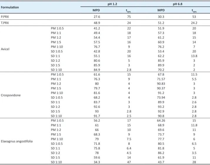
Table 2. Mean percent dissolution (MPD) and time needed to release 50% of incorporated drug (t50%) of formulations in pH 1.2 and 6.8.
Formulation pH 1.2 pH 6.8
MPD t
50% MPD t50%
P.PRX 27.6 75 30.3 53
T.PRX 48.9 24 51.2 24.2
Avicel
PM 1:0.5 41.2 22 51.9 20
PM 1:1 49.4 18 57.3 18
PM 1:2 54.4 17 61.2 15
PM 1:5 57.5 16 60.9 14
PM 1:10 76.7 9 76.2 7
SD 1:0.5 42.8 20 53.4 20
SD 1:1 55.1 16 62.2 13.8
SD 1:2 80.6 5 85.9 3
SD 1:5 85.9 3 89.9 2
SD 1:10 84.9 2.8 70.2 2
Crospovidone
PM 1:0.5 61.6 15 67.8 11.5
PM 1:1 76.3 9 71.57 5.5
PM 1:2 80 4 90.83 3
PM 1:5 79.7 4 90.37 3
PM 1:10 81.6 4 91.2 3
SD 1:0.5 68.2 4 73.94 2.8
SD 1:1 83.7 3 89.9 2.6
SD 1:2 92.6 3 93.2 2.8
SD 1:5 93 2.8 92.9 2.8
SD 1:10 91.7 2.5 90.8 2.8
Elaeagnus angostifolia
PM 1:0.5 56.2 17 64.26 15
PM 1:1 61 15 68.9 11.8
PM 1:2 66 10 69.6 11
PM 1:5 68.3 9 73.7 9
PM 1:10 79 7.5 77.7 6
SD 1:0.5 71.8 8 80.5 6.5
SD 1:1 75.8 6.4 81.8 5
SD 1:2 78 4.5 86.2 3.5
SD 1:5 59.6 14 61.9 11
SD 1:10 34.3 47 43.7 28
P=Pure, T=Treated, PM=Physical mixture, SD=Solid dispersion
Model Equation MRSQ MPE
Zero order F = k0t 0.878 17.116
First order ln (1-F) = -kf t 0.965 33.131
Higuchi F = kH t 0.937 8.947
Pepas ln F = ln kp+ plnt 0.988 19.555
Hixon-Crowell 1-
31F = k1/3t 0.964 30.229
Square root of mass 1- 1F = k1/2t 0.95 10.005 Three second root of
mass 1 - 3
2
1F = k2/3t 0.93 13.023
Weibull ln[-ln(1-F)] = -βln td + βln t 0.981 4.718
Wagner Linear
probability Z = Z0 + qt 0.945 8.222
Wagner Log
probability Z' = Z0' + q'lnt 0.963 9.696
Gompertz 𝐹𝐹 = 𝑒𝑒(−𝑎𝑎×𝑒𝑒
−𝑏𝑏𝑏𝑏𝑏𝑏𝑏𝑏 𝑡𝑡)
0.962 12.132
Logistic 𝐹𝐹 = 𝑒𝑒( 𝑎𝑎−𝑏𝑏𝑏𝑏𝑏𝑏𝑏𝑏𝑏𝑏1+𝑒𝑒(𝑎𝑎+𝑏𝑏𝑏𝑏𝑏𝑏𝑏𝑏𝑡𝑡)) 0.979 6.771
Skrdla-homogen 𝐹𝐹 = 1 − 𝑒𝑒(𝑎𝑎𝑏𝑏𝑎𝑎−𝑏𝑏𝑡𝑡2−1) 0.946 13.154
Skrdla-hetrogen
𝐹𝐹 = 1 − 𝑒𝑒(
−𝑎𝑎
𝑏𝑏 (𝑒𝑒𝑏𝑏𝑡𝑡2−1)) 0.966 23.963
Reciprocal powered time
(Suggested 1)* (1𝐹𝐹 − 1) =𝑚𝑚𝑡𝑡𝑏𝑏
0.927 14.147
Suggested 2* 𝑛𝑛 < 1 1 − (1 − 𝐹𝐹)1−𝑛𝑛= 𝐾𝐾 0.977 5.154
Suggested 3*
𝑛𝑛′> 1 1
(1 − 𝐹𝐹)𝑛𝑛′−1− 1 = (𝑛𝑛′− 1)𝐾𝐾𝑛𝑛′−1𝑡𝑡
0.966 31.957
Table 1. Mean squared correlation coefficients (MRSQ), mean percent error (MPE) and percent of total number of error (NE) of the kinetic
et al.
Where Fobs and Fcal are the measured and calculated
fraction of the drug released in each sampling time, and N
is the number of sampling times.2, 31
32
1 D
OMPE 32
=
∑
(2)Where, 32 is the number of formulations.
Dissolution profile of different formulations were compared using calculation of mean percent dissolution (MPD) and time needed to release 50% of incorporated drug (t50%) in pH 1.2 and 6.8.
MPD was calculated according to the following
equation:23,32
n i=1
MPD=
∑
D (3)Where D is the percent of drug dissolved at different sampling times.
Results
Characterization of the solid dispersions Particle size analysis
The particle size analysis results indicated that the mean particle size diameter of treated PRX powder was
significantly (p<0.05) lower than pure drug (Fig. 1).
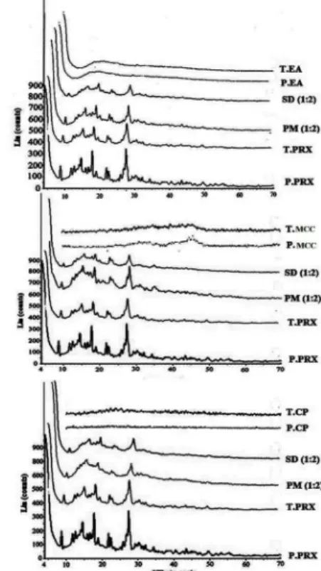
Powder X-Ray Diffraction studies
PXRD spectrum of pure PRX showed distinctive peaks in the 2θ= 8.99, 14.46, 17.67, 21.83 and 27.36 which indicate the crystalline form β of PRX (Fig. 2).
Differential Scanning Calorimetry
Differential scanning calorimetry pattern displayed a
sharp endothermic peak at 206 ºC, corresponding to the
melting point of PRX (Fig. 3).
Fourier Transform Infrared spectroscopy
The FTIR studies were performed in order to find out the probable intermolecular interactions between the drug and carriers. The FTIR spectroscopy pattern of pure and treated PRX and carriers, as well as physical mixtures and solid dispersions (drug: carrier, 1:2) are presented in Fig .4.
Fig 1. Particle size distribution of pure (Top) and treated PRX
powder (Below)
Fig 2. Powder X-Ray Diffraction patterns of pure PRX (P. PRX),
treated FZM (T. PRX), physical mixtures (PM) 1:2, solid disper -sions (SD) 1:2, pure microcrystalline cellulose (P. MCC) and treat-ed microcrystalline cellulose (T. MCC), pure crospovidone (P.CP), treated crospovidone (T.CP), pure Elaeagnus angostifolia (P.EA) and treated Elaeagnus angostifolia (T.EA)
Fig. 3. DSC thermogram of pure PRX (P. PRX), treated FZM (T.
Fig. 4. FT-IR patterns of pure PRX (P. PRX), treated FZM (T.
PRX), physical mixtures (PM) 1:2, solid dispersions (SD) 1:2, pure microcrystalline cellulose (P. MCC) and treated microcrys-talline cellulose (T. MCC), pure crospovidone (P.CP), treated crospovidone (T.CP), pure Elaeagnus angostifolia (P.EA) and treated Elaeagnus angostifolia (T.EA)
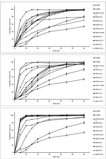
In vitro drug release
Dissolution profiles of physical mixtures and solid dispersions of PRX prepared with various drug to carrier ratios as well as treated and pure PRX powders at pH 1.2 and 6.8 are presented in Fig. 5 and 6, respectively. Dissolution rate of all physical mixtures were much higher than the pure PRX which may be because of high hydrophilicity of the polymers. Table 2 illustrates the time needed to release 50% of incorporated drug and the mean percent of dissolution from pure and treated PRX as well as physical mixtures and solid dispersions of PRX. Dissolution rate
is considered faster, when the value of t50% is lower and
MPD value is higher. In solid dispersions, drug release rate was enhanced as a consequence of increasing carrier concentration up to ratio of 1:2, while MCC containing solid dispersions showed the maximum release rate at the drug to carrier ratio of 1:2 (Table 2 and Fig. 5).
Fig. 6 shows that the dissolution of PRX is remarkably influenced by pH of the dissolution media in the presence or absence of a carrier.
Solid dispersions prepared by crospovidone with ratios of 1:0.5 and 1:1 showed a higher dissolution rate compared to MCC and Elaeagnus angustifolia fruit powder. Increasing the ratios of crospovidone from 1:1, Elaeagnus angustifolia fruit powder from 2:2 and MCC from 1:5 did not result in further increasing in dissolution rate. Therefore the best ratios for drug to carriers were as follows: crospovidone; 1:1, EA; 1:2 and MCC; 1:5.
0 20 40 60 80 100
0 10 20 30 40 50 60
C
u
m
u
la
ti
v
e r
el
ea
se (
%
)
Time (hrs)
P.PRX T.PRX PM.EA1:0.0 5 PM.EA1:1 PM.EA1:2 PM.EA1:5 PM.EA1:10 SD.EA1:0.0 5 SD.EA1:1 SD.EAG1:2 SD.EA1:5 SD.EA1:10
0 20 40 60 80 100
0 10 20 30 40 50 60
C
u
m
u
la
ti
v
e r
el
ea
se (
%
)
Time (hrs)
P.PRX T.PRX PM.Av1:0.0 5 PM.Av1:1 PM.Av1:2 PM.Av1:5 PM.Av1:10 SD.Av1:0.05 SD.Av1:1 SD.Av1:2 SD.Av1:5 SD.Av1:10
0 20 40 60 80 100
0 10 20 30 40 50 60
C
u
m
u
la
ti
v
e r
el
ea
se (
%
)
Time (hrs)
P.PRX T.PRX PM.CP1:0.0 5 PM.CP1:1 PM.CP1:2 PM.CP1:5 PM.CP1:10 SD.CP1:0.05 SD.CP1:1 SD.CP1:2 SD.CP1:5 SD.CP1:10
Fig. 5. Dissolution profiles of pure PRX (P. PRX), treated PRX (T.
PRX) physical mixtures (PM) 1:2 and solid dispersions (SD) 1:1 containing microcrystalline cellulose (MCC), crospovidone (CP) and Elaeagnus angostifolia (EA) fruit powder in pH 1.2. (mean ± SD, n=3)
The percentage of drug released at pH 6.8 is obviously higher than the amount of drug dissolved at pH 1.2. This could be due to the better solubility of the weak acid PRX because of a greater ionization at higher pH values.
Release kinetics
The in vitro release data of prepared solid dispersions were fitted to 10 linear and 7 non-linear kinetic models to clarify the mechanism of drug release. Results are presented in Table 1.
Discussion
The treated powder had a reduced geometric diameter and as a result, a higher surface area than that of pure PRX. According to the Noyes–Whitney equation, the amount of solid drug per unit time, dM/dt, is related to the surface area of the solid (S).
(4) dM DS( )
dt = h Cs−C
Where D is the diffusion coefficient of the solute in solution, h stands for the thickness of the diffusion layer,
Cs and C are the solubility and the concentration of the
et al.
main reason of higher dissolution rate of the treated PRX compared to pure PRX may be described by particle size reduction during grinding process.
The treated PRX showed a different PXRD pattern with lower crystallinity indicating the effect of grinding on crystallinity. Comparing peaks height in the physical mixtures (drug: carrier, 1:2) indicated a reduction in the magnitude of peaks due to the dilution effect of the carriers. Reduction in the height of peaks and absence of some major peaks seen in PXRD patterns of the solid dispersions containing MCC and Elaeagnus angustifolia powder indicated a decrease of PRX crystallinity in these formulations (Fig. 2). Conversely, the PXRD patterns of physical mixtures and solid dispersions containing crospovidone did not show any reduction in crystallinity. The DSC spectrum of the treated PRX also exhibited endothermic melting peak at 206 ºC. The lower intensity of peaks in physical mixtures of carriers and PRX compared to the pure PRX, and reduction of molar melting enthalpy could be explained by lower amount of PRX in PMs and dissolving of PRX in melted carriers. The reduction of molar melting enthalpy value and melting point in DSC spectra of the solid dispersions indicated the lower crystallinity of PRX in the solid dispersions. These
0 20 40 60 80 100
0 10 20 30 40 50 60
C u m u la ti v e r el ea se ( % ) Time (hrs) P.PRX T.PRX PM.EA1:0.0 5 PM.EA1:1 PM.EA1:2 PM.EA1:5 PM.EA1:10 SD.EA1:0.05 SD.EA1:1 SD.EAG1:2 SD.EA1:5 SD.EA1:10 0 20 40 60 80 100
0 10 20 30 40 50 60
C u m u la ti v e r el ea se ( % ) Time (hrs) P.PRX T.PRX PM.Av1:0.0 5 PM.Av1:1 PM.Av1:2 PM.Av1:5 PM.Av1:10 SD.Av1:0.05 SD.Av1:1 SD.Av1:2 SD.Av1:5 SD.Av1:10 0 20 40 60 80 100
0 10 20 30 40 50 60
C u m u la ti v e r el ea se ( % ) Time (hrs) P.PRX T.PRX PM.CP1:0.0 5 PM.CP1:1 PM.CP1:2 PM.CP1:5 PM.CP1:10 SD.CP1:0.05 SD.CP1:1 SD.CP1:2 SD.CP1:5 SD.CP1:10
Fig. 6. Dissolution profiles of pure PRX (P. PRX), treated PRX (T.
PRX) physical mixtures (PM) 1:2 and solid dispersions (SD) 1:1 containing microcrystalline cellulose (MCC), crospovidone (CP) and Elaeagnus angostifolia (EA) fruit powder in pH 6.8. (mean ± SD, n=3)
results together with PXRD results confirmed that the majority of crystalline PRX was altered to the amorphous state in the solid dispersions prepared with all carriers by cogrinding methods.
Absorption peaks of 3333 cm-1 and 3338 cm-1 inFTIR
spectroscopy pattern are related to the OH and NH bond, respectively. Absence of any other new peaks in the physical mixture and solid dispersions, and lack of elimination of these peaks and change in the positions of the absorption bands pointed to the absence of any significant interaction between PRX and carriers during cogrinding process.
Hydrophilic polymers cause aggregation reduction, wettability improvement and local solubilization by the carrier in the diffusion layer and hence increasing the dissolution rate. Although a direct relationship between the amount of carrier and PRX dissolution rate was found, increasing the carrier ratio more than 1:5 did not significantly affect the dissolution rate of different physical mixtures. On the other hand, the solid dispersion particles with different drug to polymer ratios showed the higher drug release rate compared to the related physical mixtures and pure drug.
Both of model independent parameters verified that the release rate is faster from the solid dispersions.
Piroxicam occurs as a white crystalline solid, sparingly soluble in water, dilute acid, and a most organic solvent. It is slightly soluble in alcohol and in aqueous solutions. It exhibits a weakly acidic 4-hydroxy proton (pKa 5.1) and a weakly basic pyridyl nitrogen (pKa 1.8). The improved drug release rate could be attributed to the drug crystallinity reduction in the PRX solid dispersions prepared by MCC, crospovidone and Elaeagnus angustifolia fruit powder. The amorphous form of a drug has a higher thermodynamic activity than its crystalline form, resulting in the rapid dissolution of the drug. It is usually believed that a drug in a solid dispersion system commonly exists in an amorphous form. Furthermore, the reduced particle size and accordingly elevated surface area could elevate the dissolution rate of PRX in the solid
dispersions.33-35 Plus latter evidences, increasing drug
wettability and solubility besides deaggregation of the drug particles caused by the polymers could be the reasons for enhanced drug release rate from the solid dispersions. The accuracy and prediction of the models were assessed by calculation of mean squared correlation coefficients
(MRSQ) and mean percent error (MPE).2,18,31 With the
MRSQ and MPE values in mind, release data of all the formulations were fitted best to the Weibull, suggested No.1 and logistic models with MPE values of 4.74, 5.15 and 6.77.
Conclusion
Solid dispersions of piroxicam were prepared with crospovidone, MCC and Elaeagnus angustifolia fruit powder with different ratios employing cogrinding method and characterized by PXRD, FT-IR spectroscopy,
solid state studies employing DSC and PXRD confirmed that solid dispersion of PRX with all carriers can decrease crystallinity or increase amorphousness of the drug. Results of FT-IR spectroscopy indicated the absence of any interaction between drug and carriers. Obtained solid dispersions had reduced size and were dissolved quickly in simulated gastric fluid and simulated intestinal fluid compared to the pure PRX, treated PRX and physical mixtures. The dissolution study results showed that solid dispersions can be beneficially applied to enhance the dissolution rate of the PRX. Furthermore, drug to carrier ratio, type of carriers and dissolution medium pH had the major roles in the dissolution rate in the solid dispersion. The maximum dissolution rate was obtained in the solid dispersions with drug to crospovidone ratio of 1:1, drug to Elaeagnus angustifolia fruit powder ratio of 1:2 and drug to MCC ratio of 1:5, where further increase of carriers did not increase the dissolution rate of PRX. Generally, solid dispersion formulations can increase the dissolution rate by several factors including the increasing of surface area, increase in drug wettability, prevention of drug aggregation, and creation of amorphous polymorph of the drug in the solid dispersions.
Acknowledgments
The authors would like to thank Drug Applied Research Center, Tabriz University of Medical Sciences, Tabriz, Iran. This article is based on a thesis submitted for Pharm.D degree (No. 3450) in Faculty of Pharmacy, Tabriz University of Medical Sciences, Tabriz, Iran.
Ethical issues
There is none to be declared.
Competinginterests
The authors declare no conflict of interests.
References
1. Adibkia K, Barzegar-Jalali M, Maheri-Esfanjani H, Ghanbarzadeh S, Shokri J, Sabzevari A, et al. Physicochemical characterization of naproxen solid dispersions prepared via spray drying technology. Powder Technology 2013; 246: 448-55. doi: 10.1016/j.powtec.2013.05.044
2. Adibkia K, Barzegar-Jalali M, Mohammadi G, Ebrahimnejhad H, Alaei-Beirami M. Effect of sodium alginate chain length and Ca2+and Al 3+on the release of diltiazem from matrices. Pharmaceutical Sciences 2011; 16: 221-8.
3. De Zordi N, Moneghini M, Kikic I, Grassi M, Del Rio Castillo AE, Solinas D, et al. Applications of supercritical fluids to enhance the dissolution behaviors of Furosemide by generation of microparticles and solid dispersions.
Eur J Pharm Biopharm2012; 81: 131-41. doi: 10.1016/j. ejpb.2012.01.002
4. Moneghini M, Kikic I, Voinovich D, Perissutti B, Filipović-Grčić J. Processing of carbamazepine–PEG 4000 solid dispersions with supercritical carbon dioxide: preparation, characterisation, and in vitro dissolution. Int J Pharm2001; 222: 129-38. doi: 10.1016/S0378-5173(01)00711-6
5. Reverchon E, Adami R, Caputo G, De Marco I. Spherical
microparticles production by supercritical antisolvent precipitation: Interpretation of results. J Supercrit Fluids
2008; 47: 70-84. doi: 10.1016/j.supflu.2008.06.002
6. Rogers TL, Overhoff KA, Shah P, Santiago P, Yacaman MJ, Johnston KP, et al. Micronized powders of a poorly water soluble drug produced by a spray-freezing into liquid-emulsion process. Eur J Pharm Biopharm 2003; 55: 161-72. doi: 10.1016/S0939-6411(02)00193-5
7. Sarkari M, Brown J, Chen X, Swinnea S, Williams Iii RO, Johnston KP. Enhanced drug dissolution using evaporative precipitation into aqueous solution. Int J Pharm2002; 243: 17-31. doi: 10.1016/S0378-5173(02)00072-8
8. Vo CL, Park C, Lee BJ. Current trends and future perspectives of solid dispersions containing poorly water-soluble drugs. Eur J Pharm Biopharm 2013;85:799-813. doi: 10.1016/j.ejpb.2013.09.007
9. Barmpalexis P, Koutsidis I, Karavas E, Louka D, Papadimitriou SA, Bikiaris DN. Development of PVP/ PEG mixtures as appropriate carriers for the preparation of drug solid dispersions by melt mixing technique and optimization of dissolution using artificial neural networks.
Eur J Pharm Biopharm 2013; 85:1219-31. doi: 10.1016/j. ejpb.2013.03.013
10. Karavas E, Georgarakis E, Sigalas MP, Avgoustakis K, Bikiaris D. Investigation of the release mechanism of a sparingly water-soluble drug from solid dispersions in hydrophilic carriers based on physical state of drug, particle size distribution and drug–polymer interactions.
Eur J Pharm Biopharm 2007; 66: 334-47. doi: 10.1016/j. ejpb.2006.11.020
11. Paudel A, Worku ZA, Meeus J, Guns S, Van den Mooter G. Manufacturing of solid dispersions of poorly water soluble drugs by spray drying: Formulation and process considerations. Int J Pharm 2013; 453: 253-84. doi: 10.1016/j.ijpharm.2012.07.015
12. Sjökvist E, Nyström C. Physicochemical aspects of drug release. VI. Drug dissolution rate from solid particulate dispersions and the importance of carrier and drug particle properties. Int J Pharm 1988; 47: 51-66. doi: 10.1016/0378-5173(88)90215-3
13. Broman E, Khoo C, Taylor LS. A comparison of alternative polymer excipients and processing methods for making solid dispersions of a poorly water soluble drug. Int J Pharm
2001; 222: 139-51. doi: 10.1016/S0378-5173(01)00709-8 14. Cui F, Yang M, Jiang Y, Cun D, Lin W, Fan Y, et al. Design
of sustained-release nitrendipine microspheres having solid dispersion structure by quasi-emulsion solvent diffusion method. J Control Release2003; 91: 375-84. doi: 10.1016/ S0168-3659(03)00275-X
15. Joe JH, Lee WM, Park Y-J, Joe KH, Oh DH, Seo YG, et al. Effect of the solid-dispersion method on the solubility and crystalline property of tacrolimus. Int J Pharm2010; 395: 161-6. doi: 10.1016/j.ijpharm.2010.05.023
16. Kim E-J, Chun M-K, Jang J-S, Lee I-H, Lee K-R, Choi H-K. Preparation of a solid dispersion of felodipine using a solvent wetting method. Eur J Pharm Biopharm 2006; 64: 200-5. doi: 10.1016/j.ejpb.2006.04.001
17. Zajc N, Obreza A, Bele M, Srčič S. Physical properties and dissolution behaviour of nifedipine/mannitol solid dispersions prepared by hot melt method. Int J Pharm 2005; 291: 51-8. doi: 10.1016/j.ijpharm.2004.07.042
et al.
solid dispersions in oleaster powder, microcrystalline cellulose and crospovidone. J Pharm Pharm Sci 2010; 13: 152-61.
19. Al-Hamidi H, Edwards AA, Mohammad MA, Nokhodchi A. To enhance dissolution rate of poorly water-soluble drugs: Glucosamine hydrochloride as a potential carrier in solid dispersion formulations. Colloids and Surfaces B: Biointerfaces 2010; 76: 170-8. doi: 10.1016/j. colsurfb.2009.10.030
20. Guns S, Kayaert P, Martens JA, Van Humbeeck J, Mathot V, Pijpers T, et al. Characterization of the copolymer poly(ethyleneglycol-g-vinylalcohol) as a potential carrier in the formulation of solid dispersions. Eur J Pharm Biopharm
2010; 74: 239-47. doi: 10.1016/j.ejpb.2009.09.009
21. Papadimitriou SA, Barmpalexis P, Karavas E, Bikiaris DN. Optimizing the ability of PVP/PEG mixtures to be used as appropriate carriers for the preparation of drug solid dispersions by melt mixing technique using artificial neural networks: I. Eur J Pharm Biopharm2012; 82: 175-86. doi: 10.1016/j.ejpb.2012.06.003
22. Watanabe T, Hasegawa S, Wakiyama N, Kusai A, Senna M. Comparison between polyvinylpyrrolidone and silica nanoparticles as carriers for indomethacin in a solid state dispersion. Int J Pharm 2003; 250: 283-6. doi: 10.1016/ S0378-5173(02)00549-5
23. Barzegar Jalali M, Dastmalchi S. Kinetic Analysis of Chlorpropamide Dissolution from Solid Dispersions. Drug Development and Industrial Pharmacy2007; 33: 63-70. 24. Mohammadi G, Barzegar-Jalali M, Siahi Shadbad
MR, Azarmi S, Barzegar-Jalali A, Rasekhian M, et al. The effect of inorganic cations Ca2+ and Al3+ on the release rate of propranolol hydrochloride from sodium carboxymethylcellulose matrices. Daru2009; 17: 131. 25. Barzegar-Jalali M, Adibkia K, Mohammadi G, Zeraati
M, Bolagh B, Nokhodchi A. Propranolol hydrochloride osmotic capsule with controlled onset of release. Drug Deliv
2007; 14: 461-8.
26. Barzegar-Jalali M, Nayebi A, Valizadeh H, Hanaee J, Barzegar-Jalali A, Adibkia K, et al. Evaluation of in vitro-in
vivo correlation and anticonvulsive effect of carbamazepine after cogrinding with microcrystalline cellulose. J Pharm Pharm Sci2006; 9: 307-16.
27. Dastmalchi S, Garjani A, Maleki N, Sheikhee G, Baghchevan V, Jafari-Azad P, et al. Enhancing dissolution, serum concentrations and hypoglycemic effect of glibenclamide using solvent deposition technique. J Pharm Pharm Sci
2005; 8: 175-81.
28. Aquino RP, Auriemma G, d’Amore M, D’Ursi AM, Mencherini T, Del Gaudio P. Piroxicam loaded alginate beads obtained by prilling/microwave tandem technique: Morphology and drug release. Carbohydr Polym2012; 89: 740-8. doi: 10.1016/j.carbpol.2012.04.003
29. Rasetti-Escargueil C, Grangé V. Pharmacokinetic profiles of two tablet formulations of piroxicam. Int J Pharm2005; 295: 129-34. doi: 10.1016/j.ijpharm.2005.02.006
30. Shojaee SA, Rajaei H, Hezave AZ, Lashkarbolooki M, Esmaeilzadeh F. Experimental measurement and correlation for solubility of piroxicam (a non-steroidal anti-inflammatory drugs (NSAIDs)) in supercritical carbon dioxide. J Supercrit Fluids2013; 80: 38-43. doi: 10.1016/j. supflu.2013.03.015
31. Barzegar-Jalali M, Adibkia K, Valizadeh H, Shadbad MRS, Nokhodchi A, Omidi Y, et al. Kinetic analysis of drug release from nanoparticles. J Pharm Pharm Sci2008; 11: 167-77. 32. Barzegar-Jalali M. A model for linearizing drug dissolution
data. Int J Pharm1990; 63: 9-11.
33. Guo Y, Shalaev E, Smith S. Physical stability of pharmaceutical formulations: solid-state characterization of amorphous dispersions. Trends Analyt Chem2013; 49: 137-44. doi: 10.1016/j.trac.2013.06.002
34. Gurunath S, Pradeep Kumar S, Basavaraj NK, Patil PA. Amorphous solid dispersion method for improving oral bioavailability of poorly water-soluble drugs. J Pharm Res
2013; 6: 476-80. doi: 10.1016/j.jopr.2013.04.008