Molecular Properties and Functional
Divergence of the Dehydroascorbate
Reductase Gene Family in Lower and Higher
Plants
Yuan-Jie Zhang, Wei Wang, Hai-Ling Yang, Yue Li, Xiang-Yang Kang, Xiao-Ru Wang, Zhi-Ling Yang*
National Engineering Laboratory for Tree Breeding, College of Biological Sciences and Technology, Beijing Forestry University, Beijing, China
Abstract
Dehydroascorbate reductase (DHAR), which reduces oxidized ascorbate, is important for maintaining an appropriate ascorbate redox state in plant cells. To date, genome-wide molecular characterization of DHARs has only been conducted in bryophytes ( Physcomi-trella patens) and eudicots (e.g.Arabidopsis thaliana). In this study, to gain a general under-standing of the molecular properties and functional divergence of the DHARs in land plants, we further conducted a comprehensive analysis of DHARs from the lycophyteSelaginella moellendorffii, gymnospermPicea abiesand monocotZea mays. DHARs were present as a small gene family in all of the land plants we examined, with gene numbers ranging from two to four. All the plants contained cytosolic and chloroplastic DHARs, indicating dehy-droascorbate (DHA) can be directly reduced in the cytoplasm and chloroplast by DHARs in all the plants. A novel vacuolar DHAR was found inZ.mays, indicating DHA may also be reduced in the vacuole by DHARs inZ.mays. The DHARs within each species showed extensive functional divergence in their gene structures, subcellular localizations, and enzy-matic characteristics. This study provides new insights into the molecular characteristics and functional divergence of DHARs in land plants.
Introduction
As a major antioxidant, ascorbic acid (Asc) provides crucial protection against oxidative dam-age in plants [1]. Besides its antioxidant role, Asc functions as a cofactor for a large number of key enzymes, and a regulator of cell division and growth in plants [2,3]. Once used, Asc is oxi-dized to monodehydroascorbate, a short-lived radical, which is reduced to Asc by monodehy-droascorbate reductase (MDHAR) or disproportionates to Asc and dehymonodehy-droascorbate (DHA) [4]. Dehydroascorbate reductase (DHAR) catalyzes the reduction of DHA to Asc using gluta-thione (GSH) as a reducing substrate [2].
OPEN ACCESS
Citation:Zhang Y-J, Wang W, Yang H-L, Li Y, Kang X-Y, Wang X-R, et al. (2015) Molecular Properties and Functional Divergence of the Dehydroascorbate Reductase Gene Family in Lower and Higher Plants. PLoS ONE 10(12): e0145038. doi:10.1371/journal. pone.0145038
Editor:Jin-Song Zhang, Institute of Genetics and Developmental Biology, Chinese Academy of Sciences, CHINA
Received:June 14, 2015
Accepted:November 29, 2015
Published:December 18, 2015
Copyright:© 2015 Zhang et al. This is an open access article distributed under the terms of the
Creative Commons Attribution License, which permits unrestricted use, distribution, and reproduction in any medium, provided the original author and source are credited.
Data Availability Statement:All relevant data are within the paper.
Funding:This study was supported by grants from the Natural Science Foundation of China (NSFC 31270641). The funders had no role in study design, data collection and analysis, decision to publish, or preparation of the manuscript.
DHAR plays important roles in regulating the cellular Asc redox state. For example, overex-pression of DHAR in tobacco leaves resulted in a more reduced Asc redox state, while suppres-sion of DHAR had the opposite effect [5]. Much evidence has shown that DHAR is crucial for protecting plants against oxidative injury. A tropical fig lacking heat-stable DHAR activity in its leaves exhibited high light sensitivity, indicating a role for DHAR in protecting plants against photoinhibition [6]. AnArabidopsismutant lacking cytosolic DHAR activity had lower apoplastic Asc content and was highly ozone sensitive [7]. Overexpression of a rice cytosolic DHAR gene conferred enhanced salt stress tolerance to rice plants by maintaining the Asc pool, ion homeostasis and redox homeostasis [8]. Transgenic tobacco overexpressing the Ara-bidopsiscytosolic DHAR gene showed enhanced tolerance to ozone and drought stresses [9].
DHARs usually exist as small gene families in plants. For example, the rice genome contains two DHARs, and thePopulus trichocarpa,S.moellendorffiiandP.patensgenomes contain three DHARs each [10]. Of the threeP.patensDHARs,PpDHAR1was expressed under all growth conditions tested,PpDHAR2was selectively expressed in response to specific treat-ments, andPpDHAR3expression was not detected by PCR in any of the samples tested [10]. Although the threePopulus tomentosaDHARs were expressed in all tissues examined, they showed different subcellular localizations. PtoDHAR1 was localized to the chloroplast, while PtoDHAR2 and PtoDHAR3 showed typical cytosolic localization [11]. Three of the four Arabi-dopsisDHARs were examined for their catalytic activities, which differed towards the DHA substrate [12]. These results show that the DHAR members in plants might have functionally diverged.
Previous genome-wide analyses of the DHAR gene family integrating sequence analysis, gene expression, protein subcellular localization and biochemical characterization have been conducted on the bryophyteP.patensand eudicots such asArabidopsis[10,12]. However, the molecular characteristics and functional divergence of DHAR families in other land plants have not been investigated. In this study, we conducted a comprehensive analysis of the gene sequences, gene structures, gene expression patterns, subcellular localization and biochemical characteristics of the DHARs in the lycophyteS.moellendorffii, gymnospermP.abiesand monocotZ.mays. By systematic analysis of the DHAR gene families in different land plants, we have gained a general understanding of the functional divergence of DHARs in land plants.
Results
Sequence characteristics of the DHAR genes in land plants
To investigate the sequence characteristics of DHARs in land plants, we conducted a joint sequence analysis of DHAR genes from a bryophyte (P.patens), a lycophyte (S.moellendorffii), gymnosperms (P.abiesandPinus taeda), and angiosperm eudicots (A.thalianaandP. tricho-carpa) and monocots (Z.maysandBrachypodium distachyon).P.abies,P.taedaandB. distach-yoneach contained two DHAR gene copies.P.patens,S.moellendorffiiandP.trichocarpaeach contained three copies. Four DHAR genes existed in theZ.maysandA.thalianagenomes, respectively. The DHARs fromS.moellendorffii,P.abies, andZ.mayswere cloned from the cDNAs of the above three species. Although the predictedSmDHAR3gene encodes protein with complete DHAR domain, no DNA fragments amplified fromS.moellendorffiicDNA for
SmDHAR3can be translated into protein containing complete DHAR domain. SoSmDHAR3
was considered to be a pseudogene. The predictedSmDHAR3splice variant encoding protein with complete DHAR domain was used for subsequent sequence analysis.
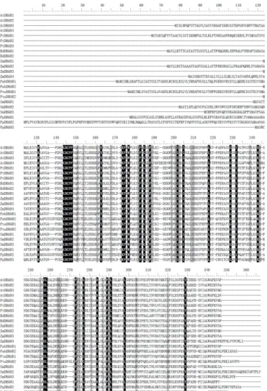
The land plant DHAR genes examined encoded proteins of different sizes, ranging from 212 to 349 amino acids. Each species contained DHARs that were more than 25 residues longer
than one of the others within the species. Multiple protein sequence alignment showed that the protein length differences were mainly due to excess peptides at the N-terminus (Fig 1). These excess peptides were predicted to be putative signal peptides that targeted the DHARs to spe-cific subcellular locations. After removing the highly variable peptides, we conducted pairwise comparison of the DHAR domain sequences in the above eight species. The pairwise sequence identity of DHARs within each species was>32%, and all land plant DHARs showed>29%
pairwise sequence identity in their DHAR domains.
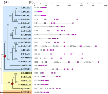
Phylogenetic relationships among the land plant DHARs were reconstructed using a neigh-bor-joining procedure. The 23 DHARs from the above eight species formed three clades in the phylogenetic tree with 100% bootstrap values. The three clades were termed clade I, II and III DHARs. Clade I contained DHARs from all eight species assessed in this study. Clade II con-tained DHARs from a bryophyte (P.patens), a lycophyte (S.moellendorffii), and gymnosperms (P.abiesandP.taeda), but not from angiosperms. Clade III only contained one member from
P.patens(Fig 2A). A previous study postulated that three ancestral DHAR copies might have existed in the common ancestor of land plants [10]. In this study, based on phylogenetic analy-sis, we also identified three ancestral DHAR copies (A, B and C, corresponding to clades I, II and III, respectively) in the common ancestor of land plants (Fig 2A). The ancestral DHAR A has been retained in all of the land plants, and expanded in angiosperms. The ancestral DHAR B has been lost in angiosperms, while the ancestral DHAR C has only been retained in bryophytes.
Except threeArabidopsisDHARs (AtDHAR1,2and4), the DHARs in clade I all had five introns, suggesting that the ancestral DHAR A might have five introns. The three-exon/two-intron structure ofAtDHAR1andAtDHAR2might result from intron loss events, and the one-exon structure ofAtDHAR4might result from a retrotransposition event. Highly variable gene structures were observed in clade II. The bryophyte DHAR genePpDHAR2and the lycophyte DHAR geneSmDHAR3each had seven introns.SmDHAR2had a seven-exon/six-intron gene structure. BothPabDHAR2andPtaDHAR2had five-exon/four-intron gene structures.
PpDHAR3in clade III had four introns (Fig 2B).
Expression of DHAR genes from
S
.
moellendorffii
,
P
.
abies
and
Z
.
mays
The expression patterns of the DHAR genes fromS.moellendorffii,P.abies, andZ.maysin three tissues including roots, stems and leaves were investigated. Some DHAR genes (e.g.
ZmDHAR1andPabDHAR1) were detected in all of the tested tissues by different numbers of amplification cycles (24, 26, 28 or 30), whileZmDHAR2was only detected in all of the tested tissues by 30 amplification cycles. All the DHAR genes from the above three species were expressed in all of the tissues examined by 30 PCR amplification cycles (Fig 3). The expression level of some DHARs was different in different tissues. For example, at 24 cycles,ZmDHAR1
andZmDHAR2showed much higher expression level in leaf than in root and stem. The two DHARs ofP.abiesalso showed the highest expression in leaf. At 28 cycles, the expression level ofSmDHAR3was higher in stem than in other two tissues.
Subcellular localization of DHARs from
S
.
moellendorffii
,
P
.
abies
and
Z
.
mays
We first predicted the subcellular localization of the DHARs fromS.moellendorffii,P.abies
species (SmDHAR2, PabDHAR1 and ZmDHAR2) was predicted to contain a cTP, ZmDHAR4 was predicted to contain a SP, and the other four (SmDHAR1, PabDHAR2, ZmDHAR1 and ZmDHAR3) were predicted not to contain any of the above three signal peptides. To experi-mentally verify the prediction results, translational fusions of the DHARs to the N-terminus of the enhanced green fluorescent protein (eGFP) were generated and transiently expressed in
Arabidopsisleaf protoplasts. Visualization by confocal microscopy showed that the GFP signals of three DHAR-eGFP fusions (SmDHAR2-, PabDHAR1- and ZmDHAR2-eGFP) were co-localized with the red autofluorescence signals of chlorophyll, demonstrating that they were localized to the chloroplast. In cells expressing the ZmDHAR4-eGFP fusion, the fluorescence was seen in the vacuole, indicating ZmDHAR4 was localized to the vacuole. The fluorescence patterns of other four DHAR-eGFP fusions (SmDHAR1, PabDHAR2, ZmDHAR1 and ZmDHAR3) were similar to that of eGFP alone, where the fluorescence was found throughout the cytosol (Fig 4).
Fig 1. Multiple sequence alignment of plant dehydroascorbate reductases (DHARs).Conserved residues in all plant DHARs are marked in black. At,A.thaliana; Pt,P.trichocarpa; Bd,B.distachyon; Zm,Z.
mays; Pab,P.abies; Pta,P.taeda; Sm,S.moellendorffii; Pp,P.patens.
doi:10.1371/journal.pone.0145038.g001
Expression and purification of DHAR proteins from
S
.
moellendorffii
,
P
.
abies
and
Z
.
mays
SmDHAR2, PabDHAR1, ZmDHAR2 and ZmDHAR4 all contained a signal peptide at their N-terminus. After removing the signal peptides, the eight DHARs fromS.moellendorffii,P.
abiesandZ.mayswere over-expressed inE.coli. Except PabDHAR1, all of the DHARs were expressed mainly as soluble proteins inE.coliat 37°C. After the induction temperature was lowered to 20°C, PabDHAR1 was also expressed as a soluble protein. The eight DHARs were purified using a Ni Sepharose High Performance column. In SDS-PAGE analysis, all of the purified recombinant proteins showed a single band (Fig 5).
Fig 2. Phylogenetic relationships among land plant dehydroascorbate reductases (DHARs) (A), and gene structures (B).Numbers on branches indicate the bootstrap values calculated from 100 replicates. Clades I, II and III DHARs are shaded blue, yellow and brown, respectively. The three ancestral genes of land plant DHARs are indicated by red circles. In (B), the GST N-terminal domain and C-terminal domain are highlighted by the blue and purple boxes, respectively, while introns are indicated as lines.
doi:10.1371/journal.pone.0145038.g002
Fig 3. Expression of the DHAR genes in various tissues ofZ.mays,P.abies, andS.moellendorffii.
Enzymatic activity and steady-state kinetics of the DHAR proteins from
S
.
moellendorffii
,
P
.
abies
and
Z
.
mays
The biochemical characteristics of the DHAR proteins fromS.moellendorffii,P.abiesandZ.
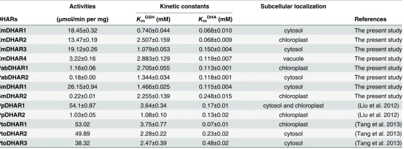
mayswere assayed using GSH and DHA as substrates. Among the eight DHARs, SmDHAR1 showed the highest catalytic activity (26.15 ± 0.94μmol min−1mg−1), while PabDHAR2
showed the lowest catalytic activity (0.18 ± 0.00μmol min−1mg−1). The highest catalytic activ-ity was 118.86-, 6.44- and 5.94-fold higher than the lowest catalytic activactiv-ity inS.moellendorffii,
P.abiesandZ.mays, respectively (Table 1).
The apparent kinetic constants of the DHAR proteins were assayed with various concentra-tions of GSH and DHA. The DHARs in the above three species showed apparentKmvalues for
GSH ranging from 0.74 to 2.88 mM, and for DHA ranging from 0.07 to 0.25 mM. ZmDHAR1 showed the highest affinity (lowestKmGSHandKmDHA) towards both the GSH and DHA
sub-strates (Table 1).
Fig 4. Subcellular localizations of DHAR proteins inArabidopsisprotoplasts.eGFP signal (green) and chlorophyll autofluorescence (red) were observed using confocal laser scanning microscopy, and an overlay is shown in yellow. Scale bars are 10μm.
doi:10.1371/journal.pone.0145038.g004
Fig 5. SDS-PAGE of purified DHAR proteins.Lane 1, molecular mass markers with the sizes shown on the left in kDa; lanes 2–9, SmDHAR1, SmDHAR2, PabDHAR1, PabDHAR2, ZmDHAR1, ZmDHAR2, ZmDHAR3 and ZmDHAR4, respectively.
doi:10.1371/journal.pone.0145038.g005
Temperature and pH profiles of the DHARs from
S
.
moellendorffii
,
P
.
abies
and
Z
.
mays
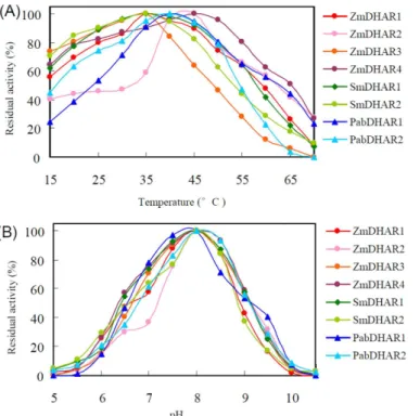
The eight DHARs fromS.moellendorffii,P.abiesandZ.maysshowed broad temperature opti-mums for enzymatic activity. All of the eight DHARs retained more than 45% of their maxi-mum enzymatic activity between 25 and 50°C. Optimaxi-mum catalytic temperature variation was also observed among the DHARs. At 60°C, SmDHAR2, PabDHAR2 and ZmDHAR3 retained no more than 29% of their maximum enzymatic activity, while the other DHARs retained more than 41% of their maximum enzymatic activity. At 25°C, PabDHAR1 and ZmDHAR2 retained no more than 54% of their maximum enzymatic activity, while all of the other DHARs retained more than 75% of their maximum enzymatic activity (Fig 6A).
A pH profile analysis showed that all eight DHARs of the three species had maximum enzy-matic activities at pH 8.0. They almost completely lost their enzyenzy-matic activity below pH 5.5 or above pH 10.0. ZmDHAR3, ZmDHAR4, SmDHAR1, PabDHAR1 and PabDHAR2 retained more than 53% of their maximum enzymatic activity between pH 7.0 and 9.0, while ZmDHAR1 and SmDHAR2 retained more than 57% of their maximum enzymatic activity between pH 7.0 and 8.5. ZmDHAR2 showed no more than 37% of its maximum enzymatic activity below pH 7.0 (Fig 6B).
Discussion
Dehydroascorbate reductase (DHAR), which reduces oxidized ascorbate, is important for maintaining an appropriate ascorbate redox state in plant cells. Two to four full-length DHAR copies have been found in each land plant species examined so far. A joint phylogenetic analy-sis of land plant DHARs showed that three ancestral DHAR copies (A, B and C) might have existed in the common ancestor of land plants, which is consistent with the previous study [10]. The ancestral DHAR gene C has only been retained in bryophytes and the ancestral DHAR B has been lost in angiosperms, while the ancestral DHAR A has been retained in all of the land plants and expanded in angiosperms. Protein catalytic activity analysis showed that the catalytic activities of all the DHARs examined in clade II were lower than those of clade III,
Table 1. Comparison of the specific activities and kinetic constants of land plant dehydroascorbate reductases (DHARs) towards the substrates dehydroascorbate (DHA) and glutathione (GSH).Pto,P.tomentosa.
Activities Kinetic constants Subcellular localization
DHARs (μmol/min per mg) KmGSH(mM) KmDHA(mM) References
ZmDHAR1 18.45±0.32 0.740±0.044 0.068±0.010 cytosol The present study
ZmDHAR2 13.47±0.19 2.507±0.159 0.068±0.009 chloroplast The present study
ZmDHAR3 19.12±0.26 1.079±0.053 0.150±0.004 cytosol The present study
ZmDHAR4 3.22±0.16 2.883±0.129 0.119±0.007 vacuole The present study
PabDHAR1 1.16±0.06 2.705±0.055 0.113±0.001 chloroplast The present study
PabDHAR2 0.18±0.00 1.344±0.034 0.118±0.001 cytosol The present study
SmDHAR1 26.15±0.94 1.466±0.025 0.115±0.004 cytosol The present study
SmDHAR2 0.22±0.01 2.255±0.139 0.248±0.015 chloroplast The present study PpDHAR1 54.1±0.87 3.64±0.34 0.17±0.01 cytosol and chloroplast (Liu et al. 2012)
PpDHAR2 1.03±0.05 1.08±0.10 0.13±0.02 chloroplast (Liu et al. 2012)
PtoDHAR1 53.02 3.75±0.77 0.07±0.01 chloroplast (Tang et al. 2013)
PtoDHAR2 49.89 2.28±0.22 0.23±0.02 cytosol (Tang et al. 2013)
PtoDHAR3 38.32 2.47±0.39 0.48±0.02 cytosol (Tang et al. 2013)
indicating that the ancestral DHAR copy with higher catalytic activity had a higher possibility to be retained and expand throughout its evolutionary history.
Ascorbate is mainly present in the cytosol and chloroplast [13]. After use, ascorbate is oxi-dized to MDHA and then disproportionated to DHA [4]. DHAR can reduce DHA to ascorbate [2]. The correct localization of DHAR proteins is essential for their function [14]. Both cyto-solic and chloroplastic DHARs were identified in plants, such as the bryophyteP.patens, and eudicotsA.thalianaandP.tomentosa[10,11,12]. In this study, we found that cytosolic and chloroplastic DHARs also existed in the lycophyteS.moellendorffii, gymnospermP.abiesand monocotZ.mays, indicating that all land plants might contain cytosolic and chloroplastic DHARs. Thus, DHA generated in the cytosol and chloroplast can be directly reduced to ascor-bate by corresponding DHARs in land plants.
Although the DHARs in each species shared high sequence identities in their DHAR domains, clear differences were observed in their gene structures, subcellular localizations, and enzymatic characteristics, which suggested functional divergence. For example, SmDHAR1, PabDHAR2, and ZmDHAR1/3 were localized to the cytoplasm, while SmDHAR2, PabDHAR1 and ZmDHAR2 were localized to the chloroplast. SmDHAR1 and PabDHAR1 had 118.86-and 6.44-fold higher catalytic activity to DHA than SmDHAR2 118.86-and PabDHAR2, respectively. Previous studies have shown that the patterns of functional divergence of DHARs differ inP.
tomentosaandA.thaliana[11]. The chloroplastic DHAR had the highest catalytic activity inP.
tomentosa, while the chloroplast-localized AtDHAR3 showed much lower catalytic activity than the cytosolic AtDHAR1 [11,12]. In this study, we found that the chloroplastic PabDHAR1 had much higher catalytic activity than the cytosolic PabDHAR2, whereas the catalytic activi-ties of chloroplastic DHARs were lower than those of cytosolic DHARs inS.moellendorffiiand
Z.mays. Our research supports that the patterns of functional divergence of DHARs are differ-ent in differdiffer-ent plant species.
Fig 6. Effects of temperature (A) and pH (B) on DHAR activity.
doi:10.1371/journal.pone.0145038.g006
Ascorbate appears to be synthesized in cytosol, and then translocated into the chloroplast, vacuole and apoplast following the concentration gradient [15]. As DHAR exists in cytosol and chloroplast, the ascorbate can be regenerated from DHA by DHAR in cytosol and chloroplast. No apoplastic and vacuolar DHARs were identified in previous studies. It has been postulated that DHA formed in the vacuole and apoplast may be transported to the cytoplasm to be reduced and then translocated back into the vacuole and apoplast [16]. In this study, a DHAR (ZmDHAR4) with dehydroascorbate reductase activity was found to localize to the vacuole in
Z.mays. So the DHA generated in vacuoles inZ.maysmay not need to be transported to cyto-sol for re-reduction, but be directly reduced in vacuole inZ.mays. We did not identify any vac-uolar DHAR in other species investigated in this study, indicating the reduction of DHA by DHAR in vacuole may only exist inZ.mays.
Of all the DHARs examined, only ZmDHAR4 was localized to vacuole. The vacuolar locali-zation of ZmDHAR4 may be the result of subcellular relocalilocali-zation. Signal peptide prediction showed that ZmDHAR4 contained a secretory pathway signal peptide (SP) which targets the ZmDHAR4 to vacuole. Except ZmDHAR4, none of the DHARs contained SP. The specific vacuolar localization of ZmDHAR4 may be the result of the gain of a vacuolar signal peptide. It has been proposed that protein subcellular relocalization is one source of retention and func-tional diversification of duplicate genes. Changes in the subcellular location of eukaryotic duplicate proteins can positively modify their function and therefore be beneficial to the organ-ism [17]. It has been shown that ascorbate may play important roles in detoxifying the hydro-peroxide generated in or diffused into vacuoles [16]. Direct reduction of DHA in the vacuole catalyzed by vacuolar DHAR avoids the transport of DHA to the cytoplasm and the transport of reduced ascorbate back to the vacuole, enabling faster reuse of ascorbate to detoxify reactive oxygen species in the vacuole. Thus, the vacuolar subcellular relocalization ofZ.maysDHAR may conferZ.maysspecific ability against oxidative stress.
Materials and Methods
DHAR gene identification
We conducted TBLASTN searches of the genome databases ofP.patens,S.moellendorffii,P.
abies,P.taeda,A.thaliana,P.trichocarpa,Z.maysandB.distachyonusing anArabidopsis
DHAR protein sequence (GenBank accession number NP_173387) as the template with default algorithm parameters. Of the two putative DHAR genes identified from theP.abies
genome, one was truncated at its N-terminus. As this truncated DHAR is located at a scaffold terminus in theP.abiesgenome database, the truncation may be due to the incomplete sequencing of theP.abiesgenome. To get its full-length sequence, we searched thePicea glauca
Phylogenetic analyses
Full-length DHAR protein sequences were aligned with the MUSCLE software (http://www. ebi.ac.uk/Tools/msa/muscle/) and manually adjusted using BioEdit [18]. The DHAR domain protein sequences were used to reconstruct phylogenetic relationships using a Neighbor-Join-ing (NJ) procedure in the MEGA software with the p-distance amino acid substitution model [19]. One hundred bootstrap replicates were performed in each analysis.
Detection of DHAR gene transcripts in various tissues
To investigate the expression patterns of the DHAR genes ofS.moellendorffii,P.abiesandZ.
maysin various tissues, total RNAs were isolated from roots, stems and leaves of the three spe-cies. The isolated RNAs were then treated with RNase-free DNase and reverse-transcribed into cDNA using an RNA PCR Kit (AMV) version 3.0 (TaKaRa, Dalian, China). TheActingenes of the three species were used as internal controls. PCR was performed in a volume of 25μL con-taining 3μL first-strand cDNA, 2.5μL TaKaRa 10× PCR buffer, 0.125μL TaKaRa Ex Taq (5 U/μL), 2μL dNTP (2.5 mM each), and 0.4 pmol primer. PCR conditions consisted of an initial denaturation of 3 min at 94°C, followed by cycles of 30 s at 94°C, 40 s at 60°C and 1 min at 72°C with a final extension of 3 min at 72°C. The number of cycles used for amplification with each primer pair was 24, 26, 28 or 30. The PCR products were each analyzed using a 1% aga-rose gel and were validated by DNA sequencing.
Subcellular localization of DHAR proteins
To investigate the subcellular localization of the DHAR proteins, the DHAR genes were sub-cloned into the pPSN transgenic vector to generate C-terminal green fluorescent protein (GFP) fusions driven by the 35S promoter [11]. Colonies containing potential appropriate recombi-nant plasmids were confirmed by sequencing. The pPSN vectors containing the DHAR genes were transiently transformed into protoplasts ofArabidopsis thalianaby polyethylene glycol—
calcium transformation [20]. The transformed protoplasts were then observed with a confocal laser microscope from 1 to 3 days after the transformation. GFP fluorescence was excited with a 488 nm laser, and chlorophyll autofluorescence was excited using a 543 nm laser.
DHAR protein expression and purification
To investigate the enzymatic activities of the DHAR proteins, the DHAR genes were subcloned into the pET-30a (Novagen, Madison, WI, USA) vector to express N-terminal 6×His-tagged fusion proteins. Recombinant plasmids containing the DHAR genes were then transformed into
Escherichia coliBL21 (DE3). Overnight cultures of theE.coliBL21 harboring the recombinant plasmids were diluted 1:100, and grown until the optical density (A600) reached 0.5. Synthesis
of the recombinant DHAR proteins was induced by isopropyl-beta-D-thiogalactopyranoside
(IPTG) at a final concentration of 0.1 mM. After incubation at 37 or 20°C overnight, the bacteria were harvested by centrifugation at 8000 × g for 3 min at 4°C, resuspended in binding buffer (20 mM sodium phosphate, 0.5 M NaCl, and 20 mM imidazole, pH 7.4), and disrupted by cold son-ication. The soluble fraction was separated from the insoluble fraction by centrifugation at 10,000 × g for 10 min at 4°C. The supernatant (soluble fraction) was then mixed with Ni Sephar-ose High Performance affinity media (Amersham Pharmacia Biotech, Piscataway, NJ, USA) that had been preequilibrated with binding buffer for 40 min at 25°C. After the unbound con-taminants were removed by washing with binding buffer, the bound proteins were eluted with elution buffer (20 mM sodium phosphate, 0.5 M NaCl, and 500 mM imidazole, pH 7.4).
Enzymatic activity characterization
The dehydroascorbate reductase activity was determined by measuring the increase inA265due
to the formation of ascorbate (ε= 14.0 mM−1cm−1). The reaction mixture contained 90 mM potassium phosphate buffer, pH 6.5, 0.42 mM GSH, 0.17 mM DHA, and various amounts of proteins. For a blank, the elution buffer was used instead of the purified DHAR proteins. All assays were carried out at 25°C. Sodium dodecyl sulfate-polyacrylamide gel electrophoresis (SDS-PAGE) was conducted on a 10% separating gel and a 5% stacking gel. Protein concentra-tions were determined by measuring absorbance at 280 nm.
The apparent kinetic constants of the DHAR proteins were studied in assays with various concentrations of GSH and DHA. The apparentKmvalues for GSH were measured using GSH
concentrations ranging from 0.125 to 2.0 mM in the presence of 0.17 mM DHA. The apparent
Kmvalues for DHA were measured using DHA concentrations ranging from 0.013 to 1.677
mM in the presence of 0.42 mM GSH. The kinetic parameters were derived from nonlinear regression analysis by the Hyper32 program available athttp://homepage.ntlworld.com/john. easterby/hyper32.html. The dependence of DHAR activity on temperature was examined in 90 mM potassium phosphate buffer (pH 6.5) at various temperatures from 15°C to 70°C at 5°C intervals. The dependence of DHAR activity on pH was measured according to the method described by Yuen and Ho [21].
Author Contributions
Conceived and designed the experiments: ZLY XRW. Performed the experiments: YJZ WW. Analyzed the data: HLY YL XYK. Wrote the paper: ZLY.
References
1. Noctor G, Foyer CH (1998) ASCORBATE AND GLUTATHIONE: keeping active oxygen under control. Annu Rev Plant Physiol Plant Mol Biol 49: 249–279. PMID:15012235
2. Chen Z, Gallie DR (2006) Dehydroascorbate reductase affects leaf growth, development, and function. Plant Physiol 142: 775–787. PMID:16891549
3. Kerk NM, Feldman N (1995) A biochemical model for the initiation and maintenance of the quiescent center: implications for organization of root meristems. Development 121: 2825–2833.
4. Kim YS, Kim IS, Bae MJ, Choe YH, Kim YH, Park HM, et al. (2013) Homologous expression of cytosolic dehydroascorbate reductase increases grain yield and biomass under paddy field conditions in trans-genic rice (Oryza sativa L. japonica). Planta 237: 1613–1625. doi:10.1007/s00425-013-1862-8PMID:
23519921
5. Chen Z, Gallie DR (2004) The ascorbic acid redox state controls guard cell signaling and stomatal movement. Plant Cell 16: 1143–1162. PMID:15084716
6. Yamasaki H, Takahashi S, Heshik R (1999) The tropical figFicus microcarpaL. f. cv. golden leaves lacks heat-stable dehydroascorbate reductase activity. Plant Cell Physiol 40: 640–646.
7. Yoshida S, Tamaoki M, Shikano T, Nakajima N, Ogawa D, Ioki M, et al. (2006) Cytosolic dehydroascor-bate reductase is important for ozone tolerance inArabidopsis thaliana. Plant Cell Physiol 47: 304– 308. PMID:16361320
8. Kim YS, Kim IS, Shin SY, Park TH, Park HM, Kim YH, et al. (2014) Overexpression of dehydroascor-bate reductase confers enhanced tolerance to salt stress in rice plants (Oryza sativa L. japonica). J Agron Crop Sci 200: 444–456.
9. Eltayeb AE, Kawano N, Badawi GH, Kaminaka H, Sanekata T, Morishima I, et al. (2006) Enhanced tol-erance to ozone and drought stresses in transgenic tobacco overexpressing dehydroascorbate reduc-tase in cytosol. Physiol Plant 127: 57–65.
10. Liu YJ, Han XM, Ren LL, Yang HL, Zeng QY (2012) Functional divergence of the glutathioneS -transfer-ase supergene family inPhyscomitrella patensreveals complex patterns of large gene family evolution in land plants. Plant Physiol 161: 773–786.
12. Dixon DP, Davis BG, Edwards R (2002) Functional divergence in the glutathione transferase superfam-ily in plants. Identification of two classes with putative functions in redox homeostasis inArabidopsis thaliana. J Biol Chem 277: 30859–30869. PMID:12077129
13. Horemans N, Foyer C, Potters G, Asard H (2000) Ascorbate function and associated transport systems in plants. Plant Physiol Biochem 38: 531–540.
14. Boruc J, Mylle E, Duda M, De Clercq R, Rombauts S, Geelen D, et al. (2010) Systematic localization of the Arabidopsis core cell cycle proteins reveals novel cell division complexes. Plant Physiol 152: 553– 565. doi:10.1104/pp.109.148643PMID:20018602
15. Rautenkranz A, Li L, Machler F, Martinoia E, Oertli JJ (1994) Transport of ascorbic and dehydroascor-bic acids across protoplast and vacuole membranes isolated from barley (Hordeum vulgare L. cv Ger-bel) leaves. Plant Physiol 106: 187–193. PMID:12232318
16. Takahama U (2004) Oxidation of vacuolar and apoplastic phenolic substrates by peroxidase: Physio-logical significance of the oxidation reactions. Phytochem Rev 3: 207–219.
17. Byun SA, Singh S (2013) Protein subcellular relocalization increases the retention of eukaryotic dupli-cate genes. Genome Biol Evol 5: 2402–2409. doi:10.1093/gbe/evt183PMID:24265504
18. Hall T (1999) BioEdit: a user-friendly biological sequence alignment editor and analysis program for Windows 95/98/NT. Nucleic Acids Symp Ser 41: 95–98.
19. Tamura K, Peterson D, Peterson N, Stecher G, Nei M, Kumar S. (2011) MEGA5: molecular evolution-ary genetics analysis using maximum likelihood, evolutionevolution-ary distance, and maximum parsimony meth-ods. Mol Biol Evol 28: 2731–2739. doi:10.1093/molbev/msr121PMID:21546353
20. Yoo SD, Cho YH, Sheen J (2007)Arabidopsismesophyll protoplasts: a versatile cell system for tran-sient gene expression analysis. Nat Protoc 2: 1565–1572. PMID:17585298
21. Yuen WK, Ho JW (2001) Purification and characterization of multiple glutathione S-transferase iso-zymes from Chironomidae larvae. Comp Biochem Physiol A Mol Integr Physiol 129: 631–640. PMID:
11423332