Genetic Modifiers of Duchenne Muscular
Dystrophy and Dilated Cardiomyopathy
Andrea Barp1☯, Luca Bello1☯, Luisa Politano2, Paola Melacini3, Chiara Calore3,
Angela Polo3, Sara Vianello1, Gianni Sorarù1, Claudio Semplicini1, Boris Pantic1, Antonella Taglia2, Ester Picillo2, Francesca Magri4, Ksenija Gorni5, Sonia Messina6, Gian
Luca Vita6, Giuseppe Vita6, Giacomo P. Comi4, Mario Ermani1, Vincenzo Calvo7, Corrado Angelini8, Eric P. Hoffman9, Elena Pegoraro1
*
1Neuromuscular Center, Department of Neuroscience, University of Padova, Padova, Italy,2Department of Experimental Medicine, Cardiomyology and Medical Genetics, Second University of Naples, Naples, Italy, 3Department of Cardiac, Thoracic and Vascular Sciences, Cardiology Section, University of Padova, Padova, Italy,4Dino Ferrari Centre, Department of Neurological Sciences, University of Milan, I.R.C.C.S. Foundation CàGranda, Ospedale Maggiore Policlinico, Milan, Italy,5NEuroMuscular Omnicentre (NEMO), Fondazione Serena Onlus, Ospedale Niguarda CàGranda, Milano, Italy,6Department of Neurosciences, Psychiatry and Anaesthesiology, University of Messina, Messina, Italy,7Department of Philosophy, Sociology, Pedagogy and Applied Psychology (FISPPA), University of Padova, Padova, Italy,8Istituto di Ricovero e Cura a Carattere Scientifico (IRCCS) San Camillo, Venice, Italy,9Research Center for Genetic Medicine, Children’s National Medical Center, 111 Michigan Avenue, NW, Washington, DC, 20010, United States of America
☯These authors contributed equally to this work. *[email protected].
Abstract
Objective
Dilated cardiomyopathy (DCM) is a major complication and leading cause of death in Duchenne muscular dystrophy (DMD). DCM onset is variable, suggesting modifier effects of genetic or environmental factors. We aimed to determine if polymorphisms previously associated with age at loss of independent ambulation (LoA) in DMD (rs28357094 in the
SPP1promoter, rs10880 and the VTTT/IAAM haplotype inLTBP4) also modify DCM onset.
Methods
A multicentric cohort of 178 DMD patients was genotyped by TaqMan assays. We per-formed a time-to-event analysis of DCM onset, with age as time variable, and finding of left ventricular ejection fraction<50% and/or end diastolic volume>70 mL/m2as event (con-firmed by a previous normal exam<12 months prior); DCM-free patients were censored at the age of last echocardiographic follow-up.
Results
Patients were followed up to an average age of 15.9±6.7 years. Seventy-one/178 patients developed DCM, and median age at onset was 20.0 years. Glucocorticoid corticosteroid treat-ment (n = 88 untreated; n = 75 treated; n = 15 unknown) did not have a significant indepen-dent effect on DCM onset. Cardiological medications were not administered before DCM
OPEN ACCESS
Citation:Barp A, Bello L, Politano L, Melacini P, Calore C, Polo A, et al. (2015) Genetic Modifiers of Duchenne Muscular Dystrophy and Dilated Cardiomyopathy. PLoS ONE 10(10): e0141240. doi:10.1371/journal.pone.0141240
Editor:Ashok Kumar, University of Louisville School of Medicine, UNITED STATES
Received:July 14, 2015
Accepted:October 5, 2015
Published:October 29, 2015
Copyright:© 2015 Barp et al. This is an open access article distributed under the terms of the
Creative Commons Attribution License, which permits unrestricted use, distribution, and reproduction in any medium, provided the original author and source are credited.
Data Availability Statement:Due to ethical restrictions, such as patient identifying information, anonymized datasets containing individual patients' sensitive data (e.g., genotype, onset of
cardiomyopathy) will be made available to other researchers (as per informed consent) upon motivated requests to the corresponding author.
onset in this population. We observed trends towards a protective effect of the dominant G allele atSPP1rs28357094 and recessive T allele atLTBP4rs10880, which was statistically significant in steroid-treated patients forLTBP4rs10880 (<50% T/T patients developing DCM during follow-up [n = 13]; median DCM onset 17.6 years for C/C-C/T, log-rank p = 0.027).
Conclusions
We report a putative protective effect of DMD genetic modifiers on the development of car-diac complications, that might aid in risk stratification if confirmed in independent cohorts.
Introduction
Duchenne muscular dystrophy (DMD) is a lethal, progressive neuromuscular disease due to
DMDgene mutations resulting in a complete lack of dystrophin in the skeletal muscle and myocardium[1]. Dilated cardiomyopathy (DCM) is a significant clinical feature of DMD, and increasing utilization of nocturnal ventilation has led to a greater proportion of DMD patients succumbing to DCM-related cardiac failure, in parallel to reduced mortality due to respiratory insufficiency[2]. DCM onset is variable: minor electrocardiographic alterations are usually detectable from the age of 10, evolving towards DCM with biventricular dilation and depres-sion of left ventricular ejection fraction. By the end of the second decade, most patients exhibit cardiac insufficiency[3]. DCM progression is also variable, with no obvious correlation to mus-cle weakness. Indeed, some authors argue that weaker patients show better preservation of myocardial function, due to less demand on the heart[3,4].
Glucocorticoid corticosteroids (hereafter“steroids”) are the only available pharmacological therapy able to slow the progression of muscle weakness in DMD[5], but there are contradic-tory reports on their effect on cardiac function. A protective effect in slowing DCM onset and progression has been reported by some authors[6–8], but denied by others[9,10]. Furthermore, steroids damage the myocardium in animal models of muscular dystrophy, exacerbating fibro-sis[11–13].
Osteopontin (OPN), encoded by theSecreted PhosphoProtein 1(SPP1) gene, is a cytokine involved in inflammation and tissue remodeling[14]. OPN is expressed by different cell types, including myoblasts, in themdxmouse muscle[15] and regulates inflammatory infiltration and muscle regeneration[16]. Moreover,SPP1genetic ablation in themdxmouse induces a milder disease course and a decrease in myocardial and diaphragmatic fibrosis through a reduction of TGFβ(Transforming Growth Factorβ)[17], which is itself a strong activator of theSPP1 pro-moter[18]. OPN is upregulated in dystrophic muscle[16,19–21], and, interestingly, is also a bio-marker and mediator of cardiovascular disease[22]. Its overexpression in the murine
myocardium causes myocarditis and DCM[23]. The G allele at the single nucleotide polymor-phism (SNP) rs28357094, in theSPP1promoter, was associated with more severe weakness in three independent DMD cohorts[19,24,25], although other authors failed to confirm this[26,27]. Underlying molecular mechanisms involve transcriptional regulation[28] and interactions with other pro-inflammatory factors, such as TGFβ[21]. Recent studies have shown the SNP to be ste-roid dependent, bothin vitro[29] andin vivo[25], suggesting a pharmacogenetic mechanism.
The latent TGFβbinding protein 4 (Ltbp4) locus showed linkage with disease severity in a mouse model of muscular dystrophy[30], and a common haplotype in the humanLTBP4gene, encoding different isoproteins, was found to modify age at loss of ambulation (LoA) in a cohort of patients with severe dystrophinopathy[26]. This finding was validated in independent cohorts [25,27]. As LTBP4 binds TGFβin a latent complex in the extracellular matrix, preventing it from
network (www.eurobiobank.org/). The funders had no role in study design, data collection and analysis, decision to publish, or preparation of the manuscript.
reaching its cell surface receptors, the proposed mechanism is that the protective haplotype ren-ders the complex more stable, preventing pro-fibrotic TGFβsignaling[31,32].
Here we test the hypothesis thatSPP1andLTBP4modify DCM onset in DMD.
Materials and Methods
Inclusion criteria
Inclusion criteria were as following: a) confirmed diagnosis of DMD (out-of-frame/nonsense
DMDgene mutations and/or absence of dystrophin by immunohistochemistry or western blot of muscle tissue); b) records of a regular (annual) cardiologic follow-up, including 2D-M-mode echocardiography; b) availability of a DNA sample.
Informed consent and ethics
All the patients or their guardians gave written informed consent to use of DNA samples and medical record data (including results of echocardiograms) for research purposes, at all partici-pating institutions which provided DNA samples (Universities of Padova, Naples, Messina and Milan; NEuroMuscular Omnicenter, Milan). The study was approved by the Ethics Committee at each institution where patients were recruited (Comitato Etico per la Sperimentazione dell'Azienda Ospedaliera di Padova, Comitato Etico dell'Azienda Ospedaliera Universitaria della Seconda Università di Napoli, Comitato Etico Interaziendale della Provincia di Messina, Comitato di Etica e Sperimentazione Farmacologica IRCCS Ca' Granda Ospedale Maggiore Policlinico, Comitato Etico Milano Area C), in compliance with the Declaration of Helsinki.
Steroid treatment and cardiological treatments
Patients were categorized as“steroid treated”if treated for at least one year with a standard dose of oral prednisone or equivalent dose of deflazacort (0.75 mg/kg/day; 0.9 mg/kg/day respectively) before events (DCM onset or LoA) or censoring. In this population, patients were not treated before DCM onset with prophylaptic cardiological medications such as angiotensin converting anzyme inhibitors (ACEi), angiotensin receptor blockers (ARB), or beta-blockers.
Echocardiographic studies
Echocardiographic studies were performed with Philips SONOS 5500 instruments with a 3 MHz transducer or equivalent instruments. Two-dimensional images and M-mode echocar-diograms of atrial and ventricular cavities were obtained in multiple cross-sectional planes, with the transducer in standard positions according to the recommendations of the American Society of Echocardiography[33,34]. Left ventricular (LV) ejection fraction (EF) was calculated from two-dimensional images with modified Simpson’s formula or area–length method[33].
Definition of DCM onset
DCM onset was defined as the age at the first echocardiographic finding of LV end diastolic volume (EDV)>70 mL/m2, and/or LV-EF<50%. We excluded patients whose first abnormal
echo was not preceded by a normal one<12 months prior, because age at DCM onset could
Genotyping and inheritance models
Genotypes at the SNPs rs28357094 (T/G nucleotide substitution at position -66 in the pro-moter region of theSPP1gene), rs2303729 (LTBP4 V194I), rs1131620 (LTBP4 T787A), and rs10880 (LTBP4 T1140M) were determined by Applied Biosystems TaqMan SNP genotyping assays and end-point allelic discrimination on an ABI-7000 SDS instrument. In the determina-tion of LTBP4 haplotypes, genotype at the fourth SNP rs1051303 (T820A) was imputed from rs1131620 genotype, assuming no recombination events due to very strong linkage disequilib-rium (LD). Haplotypes were phased by PLINK[35]. Patients were assigned to genotype groups according to previously described inheritance models: dominant for rs28357094[19], and recessive for rs10880[26]. Additionally, patients were grouped based on LTBP4 haplotype: homozygotes for the VTTT haplotype (rs2303729 G, rs1131620 A, rs10880 C), homozygotes for the IAAM haplotype (rs2303729 A, rs1131620 G, rs10880 T), or other.
Statistical analyses
The relation between age at LoA, age of DCM onset, steroid treatment and genotype at rs28357094, rs2303729, rs1131620, rs10880, andLTBP4haplotype was studied.
We used the Kaplan-Meier nonparametric method to estimate the survivor distribution functions of age at LoA and DCM onset. The log-rank test was used to test the significance of effects of genotype and steroid therapy. Linear correlations between 2 variables was tested by Pearson r. Concurrent effects of genotypes and age on LV-EF and LV-EDV (cross-sectional analysis) were evaluated by analysis of variance (ANOVA) in multiple linear regression models with EF or EDV as dependent variables, and age + genotype (dominant model for rs28357094 and recessive model for rs10880) as predictors. For all analyses, 2-tailed p values of less than 0.05 were considered significant. Analyses were done with SPSS version 18.0, R version 3.2.1, and Partek Genomics Suite 6.6. Based on Caucasian allele frequencies, we estimate that in our population statistical power for detection of SNP or haplotype effect is 0.8 with a median geno-type-related difference in age at onset of DCM of 10 years forSPP1rs28357094 (dominant model), and 12 years forLTBP4haplotype (recessive model).
Results
Patients
One hundred and seventy-eight patients selected according to inclusion criteria (seeMethods) were followed up to an average age of 15.9±6.7 years. Seventy-five/178 (42.1%) were steroid-treated, while 88/178 (49.4%) were untreated (or treated<1 year). For 15 patients (8.5%)
infor-mation about treatment was unavailable or insufficiently detailed (duration, dose adequacy, treatment before-after events etc).
SPP1
rs28357094 genotyping
There were 111 homozygotes for the T allele (62.4%), 59 T/G heterozygotes (33.1%) and 8 homozygotes for the G allele (4.5%). This distribution was close to expected minor allele fre-quency (MAF) in a Caucasian population (21.1%) and consistent with Hardy-Weinberg equi-librium (HWE).
LTBP4
genotyping
(MAF 39.0%, HWE p = 0.07). Total patient numbers do not coincide exactly because of limited DNA availability in a few patients. LTBP4 haplotype could be phased for 166 patients. Haplo-type frequencies were 50.1% VTTT, 27.3% IAAM, 8.8% IAAT, 6.7% VAAM, and 6.9% pooled rare haplotypes (including VTTM, ITTT and ITTM). Forty-nine patients (29.5%) were homo-zygotes for the VTTT haplotype, and 16 (9.6%) for the IAAM haplotype. These findings were close to the expected distribution for a Caucasian population.
DCM natural history
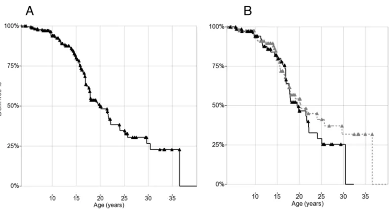
Seventy-one/178 patients (40%) developed DCM (as defined by echocardiographic criteria) during follow-up. Of these, 32 had both LV-EF and EDV available, 28 EF only, and 11 EDV only. Of the 32 patients who had both LV-EF and EDV available in the first pathological echocardiogram, both measures were altered in 15/32 (47%), EDV only with normal EF in 9/32 (28%), and EF only with normal EDV in 8/32 (25%). Mean age at onset in patients who presented with DCM was 16.5±6.0 years (range 5.4–40.1 years), with no significant differences between EF-defined and EDV-defined. In the 107 patients (60%) who did not develop DCM during the observation period (“censored”), mean age at last normal echocardiography was 15.5±7.1 years (range 3.6–36.5 years). Kaplan-Meier plots for DCM showed a median age onset at 20.0 years (95% confidence interval: 17.4–
22.6; first quartile 30.5 and third 16.0 years) (Fig 1A).
Steroid therapy and DCM
Kaplan-Meier plots comparing DCM-free survival between steroid-treated (n = 75) and untreated patients (n = 88) did not show significant differences (median onset 20.0 years vs. 20.5 years respectively,Fig 1B).
Fig 1. (A) Kaplan-Meier plot of DCM onset in 178 DMD patients. (B) Kaplan-Meier plot of DCM onset by steroid treatment: solid treated (>1 year before event/censoring), dashed untreated.Triangles indicate censoring.
Genotypes and DCM
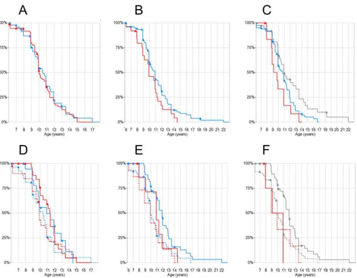
The cross-sectional analysis of LV-EF and LV-EDV values by age and genotype showed no sig-nificant correlations (ANOVA p = n.s. for both age and genotype;S1andS2Figs). Kaplan-Meier plots showed difference in estimated medians for DCM onset between rs28357094 geno-types: 24.1 years for T/G-G/G (n = 111) and 19.1 for T/T (n = 67), although not statistically sig-nificant (Fig 2A). There was a trend towards later onset of DCM in patients carrying the
LTBP4rs10880 T/T vs. C/C-C/T genotype (median 29.5 vs.19.0 years, n = 31 and 137 respec-tively, log-rank p = 0.13) (Fig 2B). Findings for otherLTBP4SNPs were similar due to LD, and also not significant (data not shown). The IAAM/IAAM haplotype showed a trend of associa-tion to later DCM onset (>50% DCM-free patients at last follow-up vs. 20.0 years at median
onset, n = 16 and 150 respectively, log-rank p = 0.15) (Fig 2C).
Steroid therapy and genotype concurrent effects
Kaplan-Meier plots with patient grouping by genotype x steroid treatment did not show signif-icant differences betweenSPP1genotypes in treated-untreated patients (Fig 2D). On the other hand, when grouping byLTBP4rs10880 x steroid treatment, within the steroid-treated group there was a significant difference between C/C-C/T genotype (median DCM onset 17.9 years,
Fig 2. Kaplan-Meier plots of DCM onset by genotypes and steroid treatment.Triangles indicate censoring. (A)SPP1rs28357094: T/T red, T/G-G/G blue; (B)LTBP4rs10880: T/T red, C/C-C/T blue; (C) LTBP4 haplotype: IAAM/IAAM red, VTTT/VTTT grey, other blue; (D)SPP1rs28357094 and steroid treatment: T/T red, T/G-G/G blue, solid treated (>1 year before event/censoring), dashed untreated; (E) rs10880 and steroid treatment: T/T red, C/C-C/T blue, solid treated, dashed untreated; (F) LTBP4 haplotype and steroid treatment: IAAM/IAAM red, other (including VTTT) grey, solid treated, dashed untreated.
n = 60) and T/T genotype (>50% of patients DCM-free at last follow-up, n = 13, log-rank
p = 0.027) (Fig 2E). When grouping byLTBP4haplotype, no DCM onset events were observed in 6 IAAM/IAAM steroid-treated patients, while median onset of DCM in 67 steroid-treated patients carrying other haplotypes was 19.0 years; however this difference was not significant (log-rank p = 0.26) (Fig 2F). Results regarding DCM onset are summarized inTable 1.
Loss of ambulation
Age at LoA was available for 163/178 patients, of whom 145 did not have severe cognitive impairment or other medical conditions potentially affecting age at LoA (e.g. bone fractures, prolonged immobilization). These patients were selected for association analysis between geno-types and LoA (“LoA cohort”). ForSPP1, we also excluded 22/145 patients who were previ-ously included in the original report of association betweenSPP1rs28357094 genotype and LoA in DMD[19].
Kaplan-Meier analysis showed no significant difference in median ages at LoA between
SPP1rs28357094 genotypes (T/T 10.0 years, n = 81, T/G-G/G 10.5 years, n = 55) (Fig 3A). Of these 123 patients, 47 (38.2%) had been treated with steroids at least 1 year before LoA, while 68 (55.3%) had not; for 8 patients (6.5%) steroid treatment status before LoA was not known with certainty. When performing Kaplan-Meier analysis grouping bySPP1genotype and ste-roid treatment, no significant differences were observed by the log-rank test, although the observed effect of steroid treatment on median age at LoA tended to be greater in T/T patients (9.9 years in 44 untreated vs. 11.3 years in 29 treated) than in T/G-G/G patients (10.3 years in 33 untreated vs. 10.9 in 19 treated), showing a trend towards greater efficacy of steroid treat-ment in T/T patients, as previously suggested[25] (Fig 3D).
LTBP4SNPs were successfully genotyped in 137/145 patients in the LoA cohort, and LTBP4 haplotype could be phased with certainty in 135. The rs10880 T/T genotype and the IAAM/IAAM haplotype, which are in close LD, were associated toearliermedian LoA: 9.9 years for T/T versus 10.9 years for C/C-C/T (n = 25 and 112 respectively, log-rank p = 0.058)
(Fig 3B); and 9.7 years for IAAM/IAAM, 10.8 years for other haplotypes, and 11.1 years for
VTTT/VTTT (n = 13, 83 and 39 respectively, log-rank test for IAAM/IAAM vs. all other haplotypes p = 0.037) (Fig 3C), in the opposite direction of association compared to Flanigan and colleagues’findings[26]. Findings for the other individualLTBP4SNPs showed similar trends (data not shown). When grouping for concurrent effects ofLTBP4genotypes and
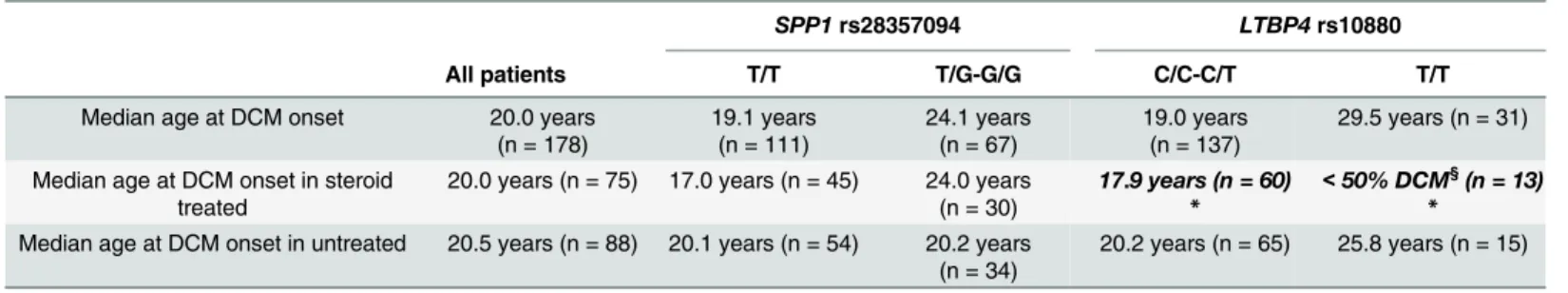
Table 1. Median age at DCM onset bySPP1andLTBP4genotype.
SPP1rs28357094 LTBP4rs10880
All patients T/T T/G-G/G C/C-C/T T/T
Median age at DCM onset 20.0 years (n = 178)
19.1 years (n = 111)
24.1 years (n = 67)
19.0 years (n = 137)
29.5 years (n = 31)
Median age at DCM onset in steroid treated
20.0 years (n = 75) 17.0 years (n = 45) 24.0 years (n = 30)
17.9 years (n = 60) *
<50% DCM§(n = 13) *
Median age at DCM onset in untreated 20.5 years (n = 88) 20.1 years (n = 54) 20.2 years (n = 34)
20.2 years (n = 65) 25.8 years (n = 15)
*Significant difference between genotypes (log-rank p<0.05).
§DCM onset was observed in less than 50% of patients, so no median value can be calculated.
Total n forLTBP4differs due to limited DNA availability in a few patients. ForSPP1, patients included in the previous report about loss of ambulation (Pegoraro et al, 2011) were also excluded.SPP1: Secreted PhosphoProtein 1.LTBP4: latent transforming growth factor beta binding protein 4. DCM: dilated cardiomyopathy.
steroid treatment before LoA, a bigger difference in median LoA was observed within treated and untreated patients in the rs10880 C/C-C/T genotype group (9.9 years untreated vs. 11.9 years treated, n = 55 and 47 respectively) than in the TT genotype (9.9 years untreated vs. 10.9 years treated, n = 15 and 8 respectively) (Fig 3E), although the log-rank test for geno-types between treated patients was not significant. Due to the strong LD between rs10880 and the IAAM haplortype, the same median values of age at LoA were observed in the haplo-type analysis (Fig 3F).
Loss of ambulation and cardiomyopathy
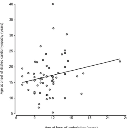
In 57 patients for whom both DCM onset and LoA were observed during follow-up, we did not observe a protective effect on the heart of early loss of ambulation (r = 0.199, p = ns) (Fig 4).
Fig 3. Kaplan-Meier plots of LoA by genotypes and steroid treatment.Triangles indicate censoring. (A)SPP1rs28357094 genotype: T/T red, T/G-G/G blue; (B)LTBP4rs10880: T/T red, C/C-C/T blue; (C) LTBP4 haplotype: IAAM/IAAM red, VTTT/VTTT grey, other blue; (D) rs28357094 genotype and steroid treatment: T/T red, T/G-G/G blue, solid treated (>1 year before LoA), dashed untreated; (E) rs10880 and steroid treatment: T/T red, C/C-C/T blue, solid treated, dashed untreated; (F) LTBP4 haplotype and steroid treatment: IAAM/IAAM red, other (including VTTT) grey, solid treated, dashed untreated.
Discussion
Known genetic modifiers of skeletal muscle function in DMD were tested for association with DCM onset in a DMD cohort. As DCM is often asymptomatic and underdiagnosed in DMD [10], we adopted a stringent phenotype definition and standardized echocardiographic param-eters, requiring a prior normal value in a narrow time window (1 year). Furthermore, pheno-type definition integrates both reduced LV-EF, an expression of ventricular hypokinesis, and increased LV-VTD, denoting ventricular dilation even in the absence of ventricular function depression. While LV-EF strictly correlates with fractional shortening (FS), a widely used mea-sure[6,8,10], including LV-VTD potentially increases diagnostic sensitivity.
A limit of this retrospective study is that only one of LV-EF or LV-EDV were available for some patients. However, average age at onset did not differ in these cases. Another limit is that age at DCM onset does not necessarily correlate with progression towards heart failure, which is variable and influenced by treatments. As treatment and follow-up were not standardized in this retrospective, multicentric study, capturing this variability in detail was not possible.
The natural history of thus-defined DCM in our population, depicted in the Kaplan-Meier plot (Fig 1A), places median onset at 20 years, 4~5 years later than a cohort similarly studied by Barber et al[8]. Mean age at onset in 71 patients developing DCM was also slightly later than in other studies: 16.5 years vs. 14.3 in Barber et al[8], and 15.4 in Jefferies et al[36] (also including Becker muscular dystrophy cases). The tendency to later onset cannot be ascribed to
Fig 4. Scatter plot of age at LoA and DCM onset in 57 patients shows no strong correlation (r = 0.31).
diagnostic delay, as we only included patients with normal echocardiography 1 year prior. Nor-mal LV-EF cut-off at 50%, on the other hand, might be considered strict in comparison to other authors using 55%[8,10]. While we chose the higher specificity of a lower LV-EF thresh-old, this difference might delay onset.
DCM onset was distributed over a wide range of 5 to 40 years. Individual patients showed surprisingly early echocardiographic alterations, or, on the contrary, preservation of ventricular size and function well into adult age. This leads to hypothesize the presence of other precipitat-ing or protective factors, includprecipitat-ing steroid treatment and genetic modifiers.
Our data do not confirm effectiveness of steroids in delaying DCM[6–8]; on the contrary, some“outlier”patients who never developed DCM, or developed it late in life, tended to be more frequently untreated (Fig 1B). This may reflect detrimental effects of steroids on the myo-cardium, promoting left ventricular fibrotic remodeling and dilation, as in prednisolone-treatedmdxmice[12,13]. Our definition of DCM onset, including indicators of left ventricular dilation, might have been sensitive to volume overload and mineralocorticoid activity second-ary to steroids, which might favor eccentric remodeling. Indeed, both ACEi[37] and aldoste-rone blockade[38] have been shown to improve DCM in DMD. Conversely, steroids might be effective in preventing progression to heart failure by other mechanisms.
When comparing DCM onset betweenSPP1genotypes, we observed a 5-year delay associ-ated with the T/G-G/G genotype, although not statistically significant. As the G allele is expected to reduceSPP1expression[28], this trend would be in the direction suggested by over-expression experiments ofSPP1in the murine myocardium, causing myocarditis and myocardial dilation[23]; but in the opposite direction compared to described effects ofSPP1
rs28357094 on skeletal muscle (greater weakness associated with the G allele)[19,24,25,29,39]. Perhaps relevant is thatSPP1genotype may represent a pharmacogenetic locus, influencing response to steroids[25,29]. As steroids seem to have little effect on heart involvement in the population studied here, the effect of this pharmacogenetic locus may be obscured in the heart. In fact, the difference in median age at DCM onset was more marked in steroid treated patients
(seeTable 1), although still not statistically significant. Further investigations of underlying
tis-sue-specific mechanisms would be warranted by stronger evidence of this genotype-phenotype association.
The rs10880 T/T genotype in theLTBP4gene, in LD with the IAAM haplotype, also showed a trend of association with delayed onset of cardiomyopathy, although not statistically signifi-cant (Fig 1B and 1C). If this trend were confirmed in independent populations, it could reflect reduced TGFβsignaling in homozygous T/T or IAAM patients, similar to what described by Flanigan and colleagues[26] in fibroblasts. This trend was clearer for rs10880 than other
LTBP4SNPs, and apparently stronger for the full haplotype, although rarity of homozygous IAAM/IAAM individuals hindered analyses, suggesting a biological effect of the T1140M ami-noacid change, within the TGFβ-binding domain of LTBP4.
risks. Further independent confirmations of this association are needed, beforeLTBP4 geno-typing is implemented in DMD clinical care with this purpose.
Survival analyses for LoA did not confirm expected associations in this cohort.SPP1
rs28357094 genotypes showed no significant differences in median delay of LoA, although the differences of median values between steroid treated and untreated were higher in the T/T vs. T/G-G/G genotype (1.4 vs. 0.5 years), as previously suggested[25]. Surprisingly, the IAAM homozygote LTBP4 haplotype was associated with significantlyearlierLoA—a finding in the opposite direction to that reported by Flanigan and colleagues, and replicated in 2 independent cohorts[25,27]. The cohort reported here is retrospective (average year of birth 1990), with rel-atively early median LoA for current standards (before 11 years of age), and a relrel-atively low rate of steroid treatment while ambulatory (41.1%), due to the inclusion of several patients fol-lowed in a time when steroid therapy was not a universal standard of care. Earlier LoA might reduce statistical power for validation, as a more severe phenotype compresses the relatively small differences due to common polymorphisms. Furthermore, if genetic modifiers, as sug-gested, influence treatment response rather than disease progression directly, effects might be reduced in a population with a low treatment rate.
Lastly, we did not observe significant correlations between LoA and DCM onset, as sug-gested by authors postulating a protective effect of limited exertion[3,4], supporting a model of independent skeletal muscle and myocardic dystropathology in DMD. This observation highlights the fact that while both descriptive clinical studies and interventional clinical tri-als have so far concentrated on ambulation and ambulatory endpoints, loss of ambulation may not be a good predictor of long-term prognosis and survival. Also, different genetic modifiers could be acting with tissue-specific mechanisms that differentially influence sub-phenotypes (e.g. muscular weakness, cardiomyopathy) in a diverse time-frame within the same disease.
The potential impact of our findings, if validated in independent cohorts, should be inter-preted in the context of genotype-phenotype studies, which have refined the correlation between different truncatingDMDmutations and DMD natural history[40,41], and genetic modifier studies cited above. As the field of rare genetic diseases shifts to a personalized medi-cine approach, the precise definition of the disease-causing mutation, together with targeted genotypization at established modifier loci, could help provide prognostic indications to patients and families, and fine-tune standards of care to individual patient characteristics. For instance, we identified a putative predictive value of theLTBP4rs10880 genotype for delay of DCM onset with steroid treatment, which could have a role in deciding if and how long to maintain treatment in non-ambulatory patients.
An even more pressing issue is the stratification of participants in clinical trials for new molecular and genetic treatments. Common variants in genes involved in inflammation and remodeling pathways, as those studied here, could be probably relevant for disease progression and efficacy of dystrophin-restoring agents. Subsequently, a sensible approach would be to ensure by genotypization that allele frequencies for relevant loci are not too different from the general population in both treated and placebo cohorts.
In conclusion, we observed trends towards a protective effect of the dominant G allele at
SPP1rs28357094 and recessive T allele atLTBP4rs10880, which was statistically significant in steroid-treated patients forLTBP4rs10880. On the other hand, an independent effect of steroid treatment in delaying DCM onset (defined as the age at the first finding of LV-EF<50% or
Supporting Information
S1 Fig. Scatter plot of left ventricular ejection fraction (EF) values by age (years) and rs28357094/rs10880 genotypes.The dashed line marks the cut-off for normal EF (>50%).
(PDF)
S2 Fig. Scatter plot of left ventricular end diastolic volume (EDV) values by age (years) and rs28357094/rs10880 genotypes.The dashed line marks the cut-off for normal EDV (<70 mL/
m2) (PDF)
Acknowledgments
The authors wish to express their gratitude to all patients and their caregivers for their collabo-ration in the study. This work was supported by grants from NIH 1U54HD053177 (Wellstone Muscular Dystrophy Center).
We also acknowledge support from Telethon Genetic BioBank (GTB12001D, GTB12001H) and the Eurobiobank network.
Author Contributions
Conceived and designed the experiments: AB LB LP PM GS GV GPC CA EPH E. Pegoraro. Performed the experiments: AB LB PM CC AP SV BP AT E. Picillo FM KG SM GLV. Analyzed the data: AB LB CS ME VC E. Pegoraro. Contributed reagents/materials/analysis tools: PM CC ME VC. Wrote the paper: AB LB E. Pegoraro.
References
1. Hoffman EP, Brown RH, Kunkel LM. Dystrophin: the protein product of the Duchenne muscular dystro-phy locus. Cell 1987; 51:919–928. PMID:3319190
2. Eagle M, Baudouin SV, Chandler C, Giddings DR, Bullock R, Bushby K. Survival in Duchenne muscu-lar dystrophy: improvements in life expectancy since 1967 and the impact of home nocturnal ventilation. Neuromuscul Disord 2002; 12:926–929. PMID:12467747
3. Nigro G, Comi LI, Politano L, Bain RJ. The incidence and evolution of cardiomyopathy in Duchenne muscular dystrophy. Int J Cardiol 1990; 26:271–277. PMID:2312196
4. Spurney CF. Cardiomyopathy of Duchenne muscular dystrophy: current understanding and future directions. Muscle Nerve 2011; 44:8–19. doi:10.1002/mus.22097PMID:21674516
5. Manzur AY, Kuntzer T, Pike M, Swan A. Glucocorticoid corticosteroids for Duchenne muscular dystro-phy. Cochrane Database Syst Rev 2008; 23;(1):CD003725.
6. Markham LW, Kinnett K, Wong BL, Woodrow Benson D, Cripe LH. Corticosteroid treatment retards development of ventricular dysfunction in Duchenne muscular dystrophy. Neuromuscul Disord 2008; 18:365–370. doi:10.1016/j.nmd.2008.03.002PMID:18436445
7. Silversides CK, Webb GD, Harris VA, Biggar DW. Effects of deflazacort on left ventricular function in patients with Duchenne muscular dystrophy. Am J Cardiol. 2003; 91:769–772. PMID:12633823 8. Barber BJ, Andrews JG, Lu Z, West NA, Meaney FJ, Price ET, et al. Oral corticosteroids and onset of
cardiomyopathy in Duchenne muscular dystrophy. J Pediatr 2013; 163:1080–1084. doi:10.1016/j. jpeds.2013.05.060PMID:23866715
9. Ashwath ML, Jacobs IB, Crowe CA, Ashwath RC, Super DM, Bahler RC. Left ventricular dysfunction in duchenne muscular dystrophy and genotype. Am J Cardiol 2014; 114:284–289. doi:10.1016/j. amjcard.2014.04.038PMID:24878125
11. Bauer R, Macgowan GA, Blain A, Bushby K, Straub V. Steroid treatment causes deterioration of myo-cardial function in the {delta}-sarcoglycan-deficient mouse model for dilated cardiomyopathy. Cardio-vasc Res 2008; 79:652–661. doi:10.1093/cvr/cvn131PMID:18495669
12. Bauer R, Straub V, Blain A, Bushby K, MacGowan GA. Contrasting effects of steroids and angiotensin-converting-enzyme inhibitors in a mouse model of dystrophin-deficient cardiomyopathy. Eur J Heart Fail 2009; 11:463–471. doi:10.1093/eurjhf/hfp028PMID:19233868
13. Sali A, Guerron AD, Gordish-Dressman H, Spurney CF, Iantorno M, Hoffman EP, et al. Glucocorticoid-treated mice are an inappropriate positive control for long-term preclinical studies in the mdx mouse. PLoS One 2012; 7:e34204. doi:10.1371/journal.pone.0034204PMID:22509280
14. Wang KX, Denhardt DT. Osteopontin: role in immune regulation and stress responses. Cytokine Growth Factor Rev 2008; 19:333–345. doi:10.1016/j.cytogfr.2008.08.001PMID:18952487 15. Uaesoontrachoon K, Yoo HJ, Tudor EM, Pike RN, Mackie EJ, Pagel CN. Osteopontin and skeletal
muscle myoblasts: association with muscle regeneration and regulation of myoblast function in vitro. Int J Biochem Cell Biol 2008; 40:2303–2314. doi:10.1016/j.biocel.2008.03.020PMID:18490187 16. Uaesoontrachoon K, Wasgewatte Wijesinghe DK, Mackie EJ, Pagel CN. Osteopontin deficiency
delays inflammatory infiltration and the onset of muscle regeneration in a mouse model of muscle injury. Dis Model Mech 2013; 6:197–205. doi:10.1242/dmm.009993PMID:22917925
17. Vetrone SA, Montecino-Rodriguez E, Kudryashova E, Kramerova I, Hoffman EP, Liu SD, et al. Osteo-pontin promotes fibrosis in dystrophic mouse muscle by modulating immune cell subsets and intramus-cular TGF-beta. J Clin Invest 2009; 119:1583–1594. doi:10.1172/JCI37662PMID:19451692 18. Hullinger TG, Pan Q, Viswanathan HL, Somerman MJ. TGFbeta and BMP-2 activation of the OPN
pro-moter: roles of smad- and hox-binding elements. Exp Cell Res 2001; 262:69–74. PMID:11120606 19. Pegoraro E, Hoffman EP, Piva L, Gavassini BF, Cagnin S, Ermani M, et al. SPP1 genotype is a
determi-nant of disease severity in Duchenne muscular dystrophy. Neurology 2011; 76:219–226. doi:10.1212/ WNL.0b013e318207afebPMID:21178099
20. Zanotti S, Gibertini S, Di Blasi C, Cappelletti C, Bernasconi P, Mantegazza R, et al. Osteopontin is highly expressed in severely dystrophic muscle and seems to play a role in muscle regeneration and fibrosis. Histopathology 2011; 59:1215–1228. doi:10.1111/j.1365-2559.2011.04051.xPMID: 22175901
21. Piva L, Gavassini BF, Bello L, Fanin M, Soraru G, Barp A, et al. TGFBR2 but not SPP1 genotype modu-lates osteopontin expression in Duchenne muscular dystrophy muscle. J Pathol 2012; 228:251–259. doi:10.1002/path.4026PMID:22431140
22. Waller AH, Sanchez-Ross M, Kaluski E, Klapholz M. Osteopontin in cardiovascular disease: a potential therapeutic target. Cardiol Rev 2010; 18:125–131. doi:10.1097/CRD.0b013e3181cfb646PMID: 20395697
23. Renault MA, Robbesyn F, Réant P, Douin V, Daret D, Allières C, et al. Osteopontin expression in cardi-omyocytes induces dilated cardiomyopathy. Circ Heart Fail 2010; 3:431–439. doi:10.1161/
CIRCHEARTFAILURE.109.898114PMID:20200330
24. Bello L, Piva L, Barp A, Taglia A, Picillo E, Vasco G, et al. Importance of SPP1 genotype as a covariate in clinical trials in Duchenne muscular dystrophy. Neurology 2012; 79:159–162. doi:10.1212/WNL. 0b013e31825f04eaPMID:22744661
25. Bello L, Kesari A, Gordish-Dressman H, Cnaan A, Morgenroth LP, Punetha J, et al. Genetic modifiers of ambulation in the CINRG Duchenne Natural History Study. Ann Neurol 2015; doi:10.1002/ana. 24370PMID:25641372
26. Flanigan KM, Ceco E, Lamar KM, Kaminoh Y, Dunn DM, Mendell JR, et al. LTBP4 genotype predicts age of ambulatory loss in Duchenne muscular dystrophy. Ann Neurol 2013; 73:481–488. doi:10.1002/ ana.23819PMID:23440719
27. van den Bergen JC, Hiller M, Böhringer S, Vijfhuizen L, Ginjaar HB, Chaouch A, et al. Validation of genetic modifiers for Duchenne muscular dystrophy: a multicentre study assessing SPP1 and LTBP4 variants. J Neurol Neurosurg Psychiatry 2014; doi:10.1136/jnnp-2014-308409PMID:25476005 28. Giacopelli F, Marciano R, Pistorio A, Catarsi P, Canini S, Karsenty G, et al. Polymorphisms in the
osteo-pontin promoter affect its transcriptional activity. Physiol Genomics 2004; 20:87–96. PMID:15479859 29. Barfield WL, Uaesoontrachoon K, Wu CS, Lin S, Chen Y, Wang PC, et al. Eccentric muscle challenge
shows osteopontin polymorphism modulation of muscle damage. Hum Mol Genet 2014; 23:4043– 4050. doi:10.1093/hmg/ddu118PMID:24626632
31. Ceco E, McNally EM. Modifying muscular dystrophy through transforming growth factor-β. FEBS J 2013; 280:4198–4209. doi:10.1111/febs.12266PMID:23551962
32. Ceco E, Bogdanovich S, Gardner B, Miller T, DeJesus A, Earley JU, et al. Targeting latent TGFβ release in muscular dystrophy. Sci Transl Med 2014; 6:259ra144. doi:10.1126/scitranslmed.3010018 PMID:25338755
33. Schiller NB, Shah PM, Crawford M, DeMaria A, Devereux R, Feigenbaum H, et al. Recommendations for quantitation of left ventricle by two-dimensional echocardiography. American Society of Echocardi-ography Committee on Standards, Subcommittee on Quantitation of Two-Dimensional Echocardio-grams. J Am Soc Echocardiogr 1989; 2:358–367. PMID:2698218
34. Nagueh SF, Bierig SM, Budoff MJ, Desai M, Dilsizian V, Eidem B, et al. Clinical recommendations for multimodality cardiovascular imaging of patients with hypertrophic cardiomyopathy. J Am Soc Echocar-diogr 2011; 24:473–98. doi:10.1016/j.echo.2011.03.006PMID:21514501
35. Purcell S, Neale B, Todd-Brown K, Thomas L, Ferreira MA, Bender D, et al. PLINK: a tool set for whole-genome association and population-based linkage analyses. Am J Hum Genet 2007; 81:559–755. PMID:17701901
36. Jefferies JL, Eidem BW, Belmont JW, Craigen WJ, Ware SM, Fernbach SD, et al. Genetic predictors and remodeling of dilated cardiomyopathy in muscular dystrophy. Circulation 2005; 112:2799–2804. PMID:16246949
37. Duboc D, Meune C, Lerebours G, Devaux JY, Vaksmann G, Bécane HM. Effect of perindopril on the onset and progression of left ventricular dysfunction in Duchenne muscular dystrophy. J Am Coll Car-diol 2005; 45:855–857. PMID:15766818
38. Raman SV, Hor KN, Mazur W, Halnon NJ, Kissel JT, He X, et al. Eplerenone for early cardiomyopathy in Duchenne muscular dystrophy: a randomised, double-blind, placebo-controlled trial. Lancet Neurol 2014; doi:10.1016/S1474-4422(14)70318-7PMID:25554404
39. Hoffman EP, Gordish-Dressman H, McLane VD, Devaney JM, Thompson PD, Visich P, et al. Alter-ations in osteopontin modify muscle size in females in both humans and mice. Med Sci Sports Exerc 2013; 45:1060–1068. doi:10.1249/MSS.0b013e31828093c1PMID:23274598
40. Pane M, Mazzone ES, Sormani MP, Messina S, Vita GL, Fanelli L, et al. 6 Minute walk test in Duchenne MD patients with different mutations: 12 month changes. PLOS One 2014 8;9:e83400.