Biomedical Geronthology Institute, PUCRS, Porto Alegre RS, Brazil: 1Professor, MSc, Neurologist, 2Professor, PhD, Pharmachologist.
CAPES Support.
Received 23 April 2004, received in final form 22 July 2004. Accepted 21 September 2004. Dra. Liana Lisboa Fernandez - Rua Coronel Bordini 675/204 - 90440-001 Porto Alegre RS - Brasil.
IS MTHFR POLYMORPHISM A RISK FACTOR
FOR ALZHEIMER’S DISEASE LIKE APOE?
Liana Lisboa Fernandez
1, Rosane Machado Scheibe
2ABSTRACT - Background: The role of methylenetetrahydrofolate reductase (MTHFR) gene polymorphisms as risk factors for the occurence of Alzheimer’s disease (AD) is still controversial: Objective: To verify the association between MTHFR and apolipoprotein E (APOE) polymorphisms and Alzheimer’s disease. Method:
This work was conducted as a case-control study. Cases included thirty patients with probable AD. Controls were constituted by 29 individuals without dementia according to neuropsychological tests paired to age, sex, race and educational level. DNA was isolated from peripheral leukocytes of anticoagulated venous blo-od. Genotyping of APOE and MTHFR were performed by DNA amplification and digestion. The frequences of APOE and MTHFR genotypes were submitted by chi-square test corrected by Fisher test; the APOE genoty-pes, to chi-square linear tendency test and the frequences of MTHFR mutant and AD, by stratificated anly-sis adjust by Mantel-Haenszel method. Results: There was significant difference about APOE4 and APOE2 in the groups. (p=0.002) The odds ratio increased exponentially with the increased number of E4 allele (χ2
linear tendency test). No significant difference was detected on MTHFR genotypes in both case and con-trol groups. Conclusion:The APOE4 is a risk factor and demonstrated a dose-depenent effect while APOE2 allele conferred a protection to AD. The MTHFR mutation had no correlation with AD.
KEY WORDS: Alzheimer’s disease, apolipoprotein E, methylenetetrahydrofolate reductase, PCR-RFLP, risk factors.
Polimorfismo da MTHFR é um fator de risco para demência de Alzheimer como APOE?
RESUMO - Introdução: O papel do polimorfismo do gene da metilenotetrahidrofolato redutase (MTHFR) como um fator de risco para demência de Alzheimer (DA) é controverso ainda. Objetivo: Verificar a associa-ção entre os polimorfismos da MTHFR e apolipoproteína E (APOE) e DA. Método: O trabalho foi condu-zido como um estudo caso-controle. Trinta pacientes com DA provável foram incluídos no grupo caso. Vinte e nove indivíduos sem demência comprovadas por testes neuropsicológicos, emparelhados pela idade, sexo, cor e nível educacional constituíram o grupo controle. DNA foi isolado de leucócitos periféricos extraídos de sangue venoso anticoagulado. Genótipos de APOE e MTHFR foram realizados por amplificação de DNA e digestão. As freqüências dos genótipos da APOE e MTHFR foram submetidas ao teste do chi-quadrado cor-rigido pelo teste de Fisher; os genótipos da APOE, ao teste do chi-quadrado com tendência linear e as freqüên-cias da MTHFR mutante e DA à análise estratificada corrigida pelo método de Mantel-Haenszel. Resultados:
Houve diferença significativa entre APOE4 e APOE2 nos grupos (p=0,002). O odds ratioaumentou expo-nencialmente com o aumento do número de alelo E4 (teste χ2 com tendência linear). Nenhuma diferença
significativa foi detectada nos genótipos da MTHFR em ambos grupos caso e controle. Conclusão:O ale-lo APOE4 é um fator de risco e demonstrou efeito dose-dependente enquanto o aleale-lo E2 conferiu prote-ção para DA. A mutaprote-ção da MTHFR não teve correlaprote-ção dom DA.
PALAVRAS-CHAVE: doença de Alzheimer, apolipoproteína E, metilenotetrahidrofolato redutase, fatores de risco.
The number of elder people has raised with the increase of life expectancy and as consequence, the prevalence of age-related disease. Alzheimer´s disease (AD) is the leading cause of dementia in the elderly. It is a multifactorial pathology resulting of the interaction of both genetics and environmen-tal factors. AD is an illness resulting from selective
intracytoplas-mic filaments, composed of hyperphosphorylated isoforms of the tau protein1. Over one hundred rare,
highly penetrant mutations have been described in three genes (APP, PSEN1, PSEN2) for early-onset familial AD2-4. In the more common late-onset
form, a polymorphism in the apolipoprotein E (APOE) gene has been associated with increased susceptibility5.
APOE is a plasma protein involved in cholesterol transport. It is produced and secreted in the cen-tral nervous system (CNS) by astrocytes. APOE syn-thesis is increased following injury and is implicat-ed in the growth and repair of nervous system dur-ing development or after injury. APOE is bound to extracellular senile plaques, to intracellular neurofi-brillary tangles, and at sites of cerebral vessel con-gophilic angiopathy6. The APOE gene has three
alle-les: E2, E3 and E4. The variant APOE4 is a major risk factor for the development of AD7-13 However, APOE4
is neither necessary nor sufficient to cause AD. So it is clear that other yet unknown genes, and envi-ronmental factors must be involved in its etiology14.
Low blood levels of folate and vitamin B12, and elevated homocysteine levels were associated with poor cognitive performace in elderly people15-19.
Possible biochemical interpretation of the putative effects of this low vitamin status and elevated homo-cysteine levels on cognitive decline can be made on basis of the pathway of one-carbon metabo-lism15. In this pathway, the
methylenetetrahydrofo-late reductase (MTHFR) is a central enzyme form-ing the substrate needed for the transferrform-ing reac-tion. Dysfunctional one-carbon metabolism gives rise to various pathogenetic mechanisms: insufficient DNA synthesis, transmethylation insufficiency and toxicity of homocysteine and related compounds9.
Furthermore, insufficient one-carbon metabolism has been suggested to have a contributory role in the development of dementia and has found to be sig-nificant in AD patient9. It has been recently
report-ed a C677T mutation on the MTHFR gene which pro-duces a thermolabile variant. In its homozygous form, this variant possesses a reduced overall enzyme activity to less than 30% of normal, resulting on increased serum homocysteine levels9,20.
We examined both the APOE polymorphism and MTHFR mutation in a group of AD individuals and in controls, to assess whether these genetic factors increase the risk for this illness.
METHOD
Patients– We investigated 29 Caucasian patients (24 women and 5 men) and 1 Afro-Brazilian patient (1 man) all over 55 years old (mean 73.5 ±18.5) with the diag-nosis of Probable AD according to NINCDS-ADRDA cri-teria and an Inventory of Diagnostic Clinic Features of Disease of Alzheimer Type21,22. All cases were sporadic,
unrelated and were recruited at an outpatient clinic (Clínica médica Salvador Dali and Serviço de Geriatria Hospital São Lucas da PUCRS) in Porto Alegre city.
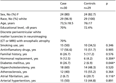
Table 1. Sample outline studied.
Case Controls p
n=30 n=29
Sex, No (%) F 24 (80) 24 (82.7)
Race, No (%) white 29 (96.9) 29 (100)
Age, years 73,5±18.5 76±17
Educational level, <8 years 70% 72.4% Discrete periventricular white
matter lucencies in neuroimaging
(CT or MRI) with encephalic atrophy 70%
Smoking use, yes 15 (50) 10 (34,5) 0.346
Antinflamatory drugs, yes 17 (56.6) 15 (51.7) 0.905 Familial history, yes 8 (26.7) 5 (17.2) 0.576 Hormonal replacement, yes 9 (12.5) 6 (4.2) 0.304* Diabetes mellitus, yes 8 (26.7) 2 (6.9) 0.044* Hypercholesterolemy, yes 18 (60) 14 (48.3) 0.520 Atherosclerosis, yes 12 (40) 15 (55.2) 0.364 Atrial fibrilation, yes 2 (6.7) 6 (20.7) 0.116* Arterial hypertention, yes 15 (50) 13 (44.8) 0.891 The data were presented in numbers (percentage) and p calculated by χ2corrected by Yates test;
We evaluated 29 Caucasian control individuals (24 women and 5 men), without dementia according to neurophychological tests matched for age, sex and edu-cational level with case group.
The sample outline was demonstrated on Table 1. Consent for participation in the study was provided by the subjects themselves or their legal guardians.
Ethics – The research protocol was approved by Scientific and Ethics Committes of the University.
Laboratory methods – DNA from each patient was isolated from peripheral leukocytes of 1 mL venous blood, anticoagulated with ethylenediaminetetraacetic acid (EDTA) by kit GFX Genomic Blood DNA Purification (Amersham Biosciences, USA). Blood samples were stored at 20oC for analysis.
APOE genotyping was performed by amplification of the third exon of the APOE gene by polymerase chain reaction (PCR) with primers described by Wenham et al.23,
followed by an enzymatic clivage with the restriction en-zyme HhaI (RFLP)20. Fragments of 72bp and 48bp are
pro-duced in APOE4, fragments of 91bp and 83bp are produ-ced in APOE2 an 91bp and 48bp are generated in APOE3. (Fig 1).
To detect the mutant allele of MTHFR, the same leu-kocytes DNA samples were examined. The PCR was per-formed with the primers described by Nishiyama et al. using HinfI restriction enzyme to identify the muta-tion20.The mutant allele generated two fragments: 175bp
and 23bp, while the wild-type is not clived and is iden-tified by a 198bp fragment (Fig. 2).
Data analysis – Allele frequences for patients with AD and control subjects were estimated by counting alle-les and calculating sample proportions.
Frequences of APOE and MTHFR genotypes in cases and controls were compared using the chi-square test cor-rected by Fisher test (Stat calc/Epi Info 6).
The APOE genotypes were submitted to chi-square linear tendency test.
The frequences of MTHFR mutant and AD were eval-uated after stratificated analysis adjust by Mantel-Haens-zel method.
RESULTS
The APOE alleles frequencies in our control sam-ple were 83% E3, 10% E4 and 7% E2 and in case sample, 63% E3, 35% E4 and 2% E2. This diference
Fig 1. APOE genotypes: polyacrilamide (15%) gel electrophoresis stained with ethidium bromide. Some representa-tive genotypes and relevant molecular sizes are indicated.
was significant (p=0.002) calculated by Exact Fisher test.
The genotyping distribution and its impact/sig-nificance were analised using the chi-square linear tendency test (Table 2).
The MTHFR alleles frequencies were 65% allele C, 35% allele T in case group while in controls we-re 71% and 29% we-respectively. This difewe-rence was not significant (p=0.508χ2test)
The interaction of APOE4 allele and MTHFR T (mutation) was demonstrated in Table 3. The geno-types distribution in cases was 35% CC (wild gene homozigose), 57% CT and 6% TT (mutation gene homozigose) while controls it was 52% CC, 38% CT and 10% TT. This diference was not statistical sig-nificant (p=0.401Exact Fisher test).
DISCUSSION
The control allele frequencies in our sample we-re 83% E3, 10% E4 and 7% E2. All controls wewe-re Caucasians. In Caucasian populations the E3 allele is the most commonly ocurring: it occurs about 77%. The average frequencies of E4 and E2 are 15% and 8% respectively. The E4 allele frequence varies ac-cording to the race. It reaches up to 30-35% in Afri-can and Asian populations. The north European po-pulation presents larger frequencies (Finland 22.7%, Sweden 20.3%) than southern countries (Italy 9.4%)24. Andrade et al. found in a populational
stu-dy in south Brazilian region 81% E3, 11.5% E4 and 7.5% E2 allele frequencies in caucasians while for Afrobrazilians they found 70% E3, 22.5% E4 and 7.5% E2 allele frequencies25. Schwanke et al. found
in an elderly population in Veranópolis (south Bra-zilian region) 84% E3, 11% E4 and 5% E2 allele fre-quencies26. The control allele frequencies in our
sam-ple were similar to others Caucasian population des-cribed in literature.
The E4 allele frequencies were significantly hig-her in case group than control.The E4 allele frequen-ce was 10% in control group against 35% in cases and similar to values reported by Andrade et al
(11.5% in controls and 39% in cases)25. Almeida
in São Paulo population find 22.1% E4 in cases and 8.9% in controls27.
Our study confirms the E4 allele presence as an AD risk factor for this Caucasians sample accord-ing to literature data7,8,10,12,13,20,28-30. Based on these,
it is obvious that APOE4 has a massive impact on the development of AD.
The E2 allele presence was significantly differ-ent in both groups (p=0.002 Exact Fisher Test). Only 2% of the demented presented E2 alleles whe-reas 7% of normal individuals presented it.
It was demonstrated that AD patients with E2E3 genotype compared with E3E3 presented less β-loid densities in brain cortex moreover reduced ami-loid angiopathy31. Review articles have suggested
a protective effect of E2 allele in AD patients10,12.
Nevertheless, Molero in an aging study performed in Maracaibo (Venezuela), Almeida in São Paulo (Brazil) and Andrade in Porto Alegre (Brazil) did not find significant association between E2 allele in cases and controls25,27,28. In our sample the E2
al-lele presence suggested a protective effect for AD.
Our genotipic distribution showed a strong ten-dency to increase the risk (OR) to develop AD de-pendent on genotype. The E2E3 genotype presen-ted like the most protective genotype for AD (OR=1) The genotypes that brings near the E4 homozigose determined an exponential increase in odds ratio (E3E3 OR=41E3E4 OR=42 ; E4E4 OR=43) (Table 2).
Austin Bradford-Hill describes 8 elements that make evident an effect-cause association. Among
Table 2. APOE genotype distribuition.
Cases Controls
n n=30 n=29 OR* pFisher
E4E4 4 4 (13.3) 0 (0.0) 63.0 0.029 E4E3 17 12 (40.0) 5 (17.2) 15.9 0.049 E4E2 e E3E3 35 14 (46.6) 21 (72.4) 4.7 0.283 E3E2 3 0 (0.0) 3 (10.3) 1.0
χ2linear tendence test, p< 0.001; * Agresti ajusted.
Table 3. Association between MTHFR allele T and AD after adjust to stratifed analysis to allele E4 effect.
Allele status Cases Controls OR (IC95%) E4 Allele (+)
T Allele (MTHFR) n=17 n=6
Positive 10 (58.8) 4 (66.7) 0.71 (0.07 a 6.95) Negative 7 (41.2) 2 (33.3) p=0.999 E4 Allele (-)
T Allele (MTHFR) n=13 n=23
Positive 9 (69.2) 10 (43.3) 2.92 (0.57 a 15.94) Negative 4 (30.8) 13 (56.6) p=0.254
Total 1.80* (0.51 a 6.30)
p=0.473
them, temporality (causes precede the effects), as-sociation force (a high related or absolute risk), a dose-effect relation (high doses are related with effect variations) and consistence (other studies, in different times, in different places, with differ-ent patidiffer-ents come to the same evidence). The E4 allele presence precedes AD manifestation, the 63
odds ratioof E4E4 genotype was remarkable abso-lute risk, the E4 allele quantity increases exponen-tially the AD risk and a lot of studies in different parts of the world comes to the same evidence. So our data confirm a predisposition to develop AD in individuals that present E4 allele and make clear several Bradford-Hill elements32. Sixty-five percent
of AD individuals did not present APOE4 allele in our sample. In this cases other risk factors (genet-ics or environmental) would be acting in the AD development.
Regland et al. related 40% of heterozigose pre-valence in general population (CT), 11% of polymor-phism homozigose (TT) and 49% wild gene homo-zigoses (CC)9. Nishiyama et al. describe 39% CC,
45% CT and 15% TT similar to Japanese and Cana-dian population data20. Our results (38%CT, 10%TT
and 52%CC) resemble genotypic distribution relat-ed by Regland9.
The MTHFR allele frequence did not show signi-ficant difference in control and case groups. Nishi-yama et al found an increased proportion of senile demented individuals in the MTHFR mutation gro-up, particularly in men. This mutation was more associated with AD than with vascular dementia20.
In our work no association with MTHFR mutation and AD was found. It could be probably because of the high polymorphism prevalence in general population associated to a limited group sample (30 cases and 29 controls). It would be need 60 cas-es and 120 controls to exprcas-ess really diferenccas-es.
Regland et al. found an inverse correlation bet-ween MTHFR mutation and APOE4, suggesting that MTHFR mutation is 1.8 times more frequent in the absence of APOE49. The association rate of
MTHFR T allele and AD cases after adjusted for al-lele E4 effect was not significant, despite it suggest-ed a small tendency of allele T individuals to devel-op AD (OR=1.80) (Table 3).
Clark et al. Postiglione et al. Brunelli et al. and Prince et al. did not find a significant relation bet-ween MTHFR mutation and AD either19,33-35. Seripa
et al found no difference in MTHFR polymorphism distribuition between AD cases and elderly controls
in both American cohort and Italian cohort36. Religa
et al. found that plasma total homocysteine is in-creased in AD patients and depended on the MTH-FR T/T genotype (mutation homozigoze) in the presence of low folate levels, however the distribui-tion of MTHFR C677T polymorphism in the Polish po-pulation does not differ in AD and controls37. Our
negative results, in individuals with Probable AD, confirmed the lack of association between AD and C/T polymorphism in the MTHFR gene.
The main conclusions of this study were: APOE4 allele was a risk factor for AD; APOE2 allele was a protective factor for AD; the presence of allele E4 demonstrated a dose-dependent effect, increasing exponentially compared with genotype E2E3; there was a discreet tendency of MTHFR allele T presence in AD patients adjusted to allele E4 efect, but it was not significant; the MTHFR mutation did not de-monstrate significant difference in cases and con-trols.
REFERENCES
1. Prince DL, Sisodia SS, Borchett DR. Alzheimer disease:when or why? Nature Genetics 1998;19:314-316.
2. Goate A, Chartier-Harlin MC, Mullan M, et al. Segregation of a missen-se mutation in the amyloid precursor protein gene with familial Alzhei-mer’s disease. Nature 1991;349:704-706.
3. Sherrington R, Froelich S, Sorbi S, et al. Alzheimer’s disease associat-ed with mutations in presenilin 2 is rare and variably penetrant. Hum Molec Genet 1996;5:985-988.
4. Levy-Lahad E, Wijsman EM, Memens E, et al. A familial Alzheimer’s disease locus on chromosome 1. Science 1995;269:970-973.
5. Tanzi RE, Bertram L. New frontiers in Alzheimer’s disease genitics. Neu-ron 2001;32:181-184.
6. Strittmatter W, Saunders AM, Schmechel D, et al. Apolipoprotein E: high-avidity binding to β-amyloid and increased frequency of type 4 allele in late- onset familial Alzheimer disease. Proc Natl Acad Sci USA 1993; 90:1977-1981.
7. Mayeux R, Stern Y, Ottman R, et al. The apolipoprotein E4 allele in pa-tients with Alzheimer’s disease. Ann Neurol 1993;34:752-754. 8. Saunders AM, Strittmatter WJ, Schmechel D, et al. Association of
apoli-poprotein E allele E4 with late-onset familial and sporadic Alzheimer’s disease. Neurology 1993;43:1467-1472.
9. Regland B, Blennow K,Germgard T, et al. The role of polymorphic ge-nes apolipoprotein E and methylenotetrahidrofolate reductase in the development of dementia of Alzheimer type. Dement Geriatr Cogn Disord 1999;10:245-251
10. Farrer LA, Cupples LA, Haines JL, et al. Effects of age, sex, end ethnici-ty on the association between apolipoprotein E genoethnici-type and Alzheimer disease: a meta-analysis. JAMA 1997;278:1349-1356.
11. Blacker D, Haines JL, Rods L, et al. ApoE4 and age at onset of Alzheimer’s disease: the NIMH.genetics iniciative. Neurology 1997;48:139-147. 12. Lendon CL, Ashall F, Goate AM. Exploring the etiology of Alzheimer
disease using molecular genetics. JAMA 1997;277:825-831.
13. Tol J, Roks G, Slooter AJC, et al. Genetic and environmental factors in Alzheimer’s disease. Rev Neurol (Paris) 1999;155:(Suppl 4):S10-S16. 14. Blacker D, Tanzi RE. The genetics of Alzheimer disease: current status
and future prospects. Arch Neurol 1998;55:294-296.
15. Selhub J, Bagley LC, Miller J, et al. B vitamins, homocysteine, and neurocog-nitive function in the elderly. Am J Clin Nutr 2000;71(Suppl):S614-S620. 16. Snowdon DA, Tully CL, Smith CD, et al. Serum folate and the
severi-ty of atrophy of the neocortex in Alzheimer disease: findings from the Nun Study. Am J Clin Nutr 2000;71:993-998.
18. Nourhashémi F, Gillette-Guyonnet S, Andrieu S, et al. Alzheimer dise-ase: protective factors. Am J Clin Nutr 2000;71(Suppl):S643-S649. 19. Clarke R, Smith AD, Jobst KA, et al. Folate, vitamin B12, and serum total
homocysteine levels in confirmed Alzheimer disease. Arch Neurol 1998;55:1449-1455.
20. Nishiyama M, Kato Y, Hashimoto M, et al. Apolipoprotein E, methyle-netetrahydrofolate reductase (MTHFR) mutation and the risk of senile dementia- an epidemiological study using the polymerase chain reac-tion (PCR) method. Epidemiology 2000;10:163-172.
21. Mc Khann G, Drachman D, Folstein M, et al. Clinical diagnosis of Alzheimer’s disease: report of the NINCDS-ADRDA work group under the auspices of Department of Health and Human Service Task force on Alzheimer’s Disease. Neurology 1984;34:939-944.
22. Cummings Jl, Benson DF.Dementia of the Alzheimer type: an invento-ry of diagnostic clinical features. JAGS 1986;34:12-19.
23. Wenham PR, Price WH, Blundell G. Apolipoprotein E genotyping by one-stage PCR. Lancet 1991;337:1158-1159.
24. Simopoulus AP. Genetic variations and nutrition. Nutrition Reviews 1999;57:S10-S19.
25. Andrade FM, Larrandaburu M, Callegari-Jacques SM, et al. Association of apolipoprotein E polymorphism with plasma lipids and Alzheimer’s disease in a Southern Brazilian population. Braz J Med Biol Res. 2000;33:529-537.
26. Schwanke CHA, Cruz IBM, Leal NF, et al. Analysis of association between apolipoprotein E polymorphism and cardiovascular risk fac-tors in na elderly population with longevity. Arq Bras Cardiol 2002;78:571-579.
27. Almeida OP, Shimokomaki CM. Apolipoprotein E4 and Alzheimer’s disease in São Paulo-Brazil. Arq Neuropsiquiatr 1997;55:1-7. 28. Molero AE, Pino-Ramirez G, Maestre GE. Modulation by age and
gen-der of risk for Alzheimer’s disease and vascular dementia associated with the apolipoprotein E-E4 allele in Latin Americans: findings from Maracaibo Aging Study. Neurosci Letters2001;307:5-8.
29. Myers AM, Goate AM. The genetics of late-onset Alzheimer’s disease. Curr Opin Neurol 2001;14:433-440.
30. Selkoe DJ. Alzheimer’s disease: genes, proteins and therapy. Phisiol Rev 2001;81:741-766
31. Lippa CF, Smith TW, Saunders AM, et al. Apolipoprotein E epsilon 2 and Alzheimer’s disease: genotype influences pathologic phenotype. Neurology 1997;48:515-519.
32. Bradford-Hill A. The environment and disease: association or Causation? J R Soc Med 1965;58:295-300.
33. Postiglione A, Milan G, Ruoceo A, et al. Plasma folate, vitamine B12, and total homocystein and homozygosity for the C677T mutation of the 5,10methylene tetrahydrofolate reductase gene in patients with Alzheimer’s dementia: a case-control study. Gerontology 2001;47:324-329. 34. Brunelli T, Bagnoli S, Giusti B, et al. The C677T methylenetetrahydrofola-te reductase mutation is not associamethylenetetrahydrofola-ted with Alzheimer’s disease. Neu-rosci Letters 2001;315:103-105.
35. Prince JA, Feuk L, Sawyer SL, et al. Lack of replication of association findings in complex disease: an analysis of 15 polymorphisms in pri-or candidate genes fpri-or sppri-oradic Alzheimer’s disease. Eur J Hum Genet 2001 Jun;9:437-444.
36. Seripa D, Forno GD, Matera MG et al. Methylenetetrahydrofolate re-ductase and angiotensin converting enzyme gene polymorphisms in two genetically and diagnostically distinct cohort of Alzheimer patients. Neurobiol Aging, 2003;24:933-939.