RESEARCH ARTICLE
FRPR-4 Is a G-Protein Coupled Neuropeptide
Receptor That Regulates Behavioral
Quiescence and Posture in
Caenorhabditis
elegans
Matthew D. Nelson1,2☯*, Tom Janssen3☯¤, Neil York2, Kun He Lee1, Liliane Schoofs3, David M. Raizen1
1Department of Neurology, Perelman School of Medicine, University of Pennsylvania, Philadelphia, Pennsylvania, United States of America,2Department of Biology, Saint Joseph’s University, Philadelphia, Pennsylvania, United States of America,3Functional Genomics and Proteomics lab, University of Leuven, Leuven, Belgium
☯These authors contributed equally to this work.
¤ Current address: Center for Innovation and Stimulation of Drug Discovery, Bioincubator 2, Gaston Geenslaan 2, Leuven, Belgium
*mnelson@sju.edu
Abstract
Neuropeptides signal through G-protein coupled receptors (GPCRs) to regulate a broad array of animal behaviors and physiological processes. TheCaenorhabditis elegans
genome encodes approximately 100 predicted neuropeptide receptor GPCRs, butin vivo
roles for only a few have been identified. We describe here a role for the GPCR FRPR-4 in the regulation of behavioral quiescence and locomotive posture. FRPR-4 is activated in cell culture by several neuropeptides with an amidated isoleucine-arginine-phenylalanine (IRF) motif or an amidated valine-arginine-phenylalanine (VRF) motif at their carboxy termini, including those encoded by the geneflp-13. Loss offrpr-4function results in a minor feed-ing quiescence defect after heat-induced cellular stress. Overexpression offrpr-4induces quiescence of locomotion and feeding as well as an exaggerated body bend posture. The exaggerated body bend posture requires the geneflp-13. Whilefrpr-4is expressed broadly, selective overexpression offrpr-4in the proprioceptive DVA neurons results in exaggerated body bends that requireflp-13in the ALA neuron. Our results suggest that FLP-13 and other neuropeptides signal through FRPR-4 and other receptors to regulate locomotion pos-ture and behavioral quiescence.
Introduction
Neuropeptides modulate multiple homeostatic aspects of animal physiology, including water balance, sexual drive, appetite, and sleep. Neuropeptides affect behavior via their interaction with membrane bound receptors, most of which have seven transmembrane domains and a11111
OPEN ACCESS
Citation:Nelson MD, Janssen T, York N, Lee KH, Schoofs L, Raizen DM (2015) FRPR-4 Is a G-Protein Coupled Neuropeptide Receptor That Regulates Behavioral Quiescence and Posture in Caenorhabditis elegans. PLoS ONE 10(11): e0142938. doi:10.1371/journal.pone.0142938
Editor:Erik C. Johnson, Wake Forest University, UNITED STATES
Received:August 17, 2015
Accepted:October 28, 2015
Published:November 16, 2015
Copyright:© 2015 Nelson et al. This is an open access article distributed under the terms of the
Creative Commons Attribution License, which permits unrestricted use, distribution, and reproduction in any medium, provided the original author and source are credited.
Data Availability Statement:All relevant data are within the paper and its Supporting Information files.
Funding:This work was supported by the National Institute of Health grants R01NS064030 (DMR), R01NS0884322 (DMR), and T32HL07713 (MDN) (http://nih.gov/).
couple to hetero-trimeric G-proteins, which in turn couple to intracellular effector proteins. Recent work in invertebrate model systems has provided insight into the physiological function
of neuropeptide signaling pathwaysin vivo[1].
Nematodes, like other animals, contain a large number of neuropeptide and neuropeptide
receptors. In the genome of the nematodeCaenorhabditis elegans, there are over 100 genes
pre-dicted to encode neuropeptides, classified as insulin-like, orins, neuropeptide-like, ornlp, and
FMRFamide-like peptides, orflp[2–4], and at least 91 genes predicted to encode neuropeptide
receptors [5–7]. While a number of GPCRs have been shown to interact with specific peptides
in cell culture systems, in only a few cases have cognate peptide / receptor pairs and the physio-logical process they control been identified. For example, learning and reproductive behaviors are modulated by NTC-1 acting on NTR-1, a conserved signaling pathway related to
mamma-lian vasopressin/oxytocin [8,9]. The identification of the NTC-1/NTR-1 signaling pathway in
C.elegansemphasizes the evolutionary conservation of neuropeptide signaling pathways, and
suggests thatin vivoidentification of other ligand-receptor pairs inC.eleganswill provide
insight into other conserved aspects of animal physiology. This study focuses on a previously
unstudied GPCR encoded by the FMRFamide-like peptide receptor-4 genefrpr-4, and its
potentialin vivoligands.
Previously, we have shown that the FLP-13 FMRFamide-like neuropeptides are required for quiescent behavior after environmental exposure to cellular stressors [10], a behavior that
enhances recovery from the stress [11]. FMRFa, aDrosophilapeptide related to peptides
encoded by theC.elegansFLP peptides, signals through its receptor FR to regulate recovery
sleep in response to cellular stress [12,13]. In this study, we provide evidence that FRPR-4,
which is aC.elegansortholog ofDrosophilaFR, can act bothin vitroandin vivoas a receptor
for FLP-13 neuropeptides, and functions specifically in the DVA proprioceptive neuron to reg-ulate body posture.
Results
FRPR-4 is a G-protein coupled receptor related to
D
.
melanogaster
FR
Our initial interest in FRPR-4 stemmed from the motivation to identify the mechanism by which FLP-13 neuropeptides promote quiescence of locomotion and feeding in response tocel-lular stress [10]. InD.melanogaster, FMRFamide peptides similar toC.elegans flp-13-derived
peptides signal through the FR receptor to regulate an analogous recovery sleep, which occurs
in response to heat or infectious stress [12]. Thus, we hypothesized that a homolog of FR inC.
elegansmay be a FLP-13 receptor. Phylogenetic analysis of all predicted neuropeptide receptors fromC.elegansandD.melanogastershowed that FR is related to a group of closely-related
paralogous GPCRs, including one encoded by the genefrpr-4(S1 Fig).frpr-4encodes a
recep-tor of the Rhodopsin class A type, and is predicted to be a neuropeptide receprecep-tor [5].
Using 3’-rapid amplification of cDNA ends (3’RACE) [14], we identified three isoforms of
frpr-4, which differed in their last exon and 3’-untranslated region (UTR) (S2 FigandS1 Table). We named these isoforms FRPR-4A, B and C. Each isoform contained an apparent
1390-bp retrotransposon flanked by 55-bp terminal inverted repeats in the large 3’intron. The
gene model identified by our cDNA analysis differed at both the 5’end and 3’end from the
gene model predicted on WormBase (www.wormbase.org). The 5’end in our
experimentally-verified gene model was shorter, and the 3’end contained two additional exons. The sequence
of one of the new 3’exons identified by our cDNA analysis was highly conserved in the
pre-dictedfrpr-4mRNA from the nematodeCaenorhabditis briggsae(S3 Fig), supporting the
Based on the similarity betweenDrosophilaFR andC.elegansFRPR-4, we hypothesized that FRPR-4 is activated by FMRFamide-like peptides and may regulate quiescent behavior in C.elegans.
FRPR-4 is activated by FLP-13 FMRF-like peptides
in cellulo
To determine if FLP-13 or other peptides can activate FRPR-4, we used anin vitrocell
expres-sion system (in cellulo). We cloned FRPR-4A (S2 FigandS1 Table) into a mammalian
expres-sion plasmid and transiently expressed the protein in Chinese Hamster Ovary (CHO) cells that
also expressed the Ca2+sensitive photoprotein aequorin targeted to the mitochondria and the
promiscuous Galpha16subunit. Galpha16causes Ca2+flux in response to receptor activation
regardless of the type of G-protein that couples to the receptorin vivo[15]. We tested a library
of 262 known and predictedC.elegansneuropeptides at a concentration of 10μM for their
abil-ity to elicit a Ca2+flux in cells expressing FRPR-4A. The peptide library contained peptides of
the FMRFamide-like peptide (FLP) family as well as other neuropeptide-like proteins (NLPs).
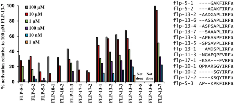
Peptides encoded by the genesflp-5,flp-10,flp-11,flp-13andflp-17were all capable of
activat-ing FRPR-4Ain celluloat 10μM, suggesting that FRPR-4A is activated by FLPsin vivo. Peptides
derived from these genes have similar C-terminal endings consisting of an amidated isoleu-cine-arginine-phenylalanine (IRFa) motif or an amidated valine-arginine-phenylalanine (VRFa) motif, suggesting that the C-terminus of the peptides plays a prominent role in FRPR-4 receptor activation. However, since other peptides containing this motif (e.g. FLP-2, FLP-4) did not activate FRPR-4, other amino acids must confer specificity to the activation. To further explore differences between activating and non-activating peptides, we compared FLP peptides with the (I/V)RF motif that activated FRPR-4A to (I/V)RF peptides that did not activate FRPR-4A (S4 Fig). In general, peptides that activated FRPR-4A were longer (9.4±1.4 amino
acids) than peptides that did not activate FRPR-4A (7.9±1.3 amino acids. p = 0.002, Student’s t
test). However, we did not identify additional consistent differences between the two peptide groups in either specific amino acids or in types of amino acids (charged, polar, or hydropho-bic) outside of the IRF or VRF motif.
The response amplitude elicited by these FLP peptides was variable. Peptides encoded by
the genesflp-5,flp-11, andflp-13elicited FRPR-4 dependent Ca2+flux at concentrations of 100
nM or lower, whereas peptides encodes byflp-10andflp-17elicited a detectable Ca2+signal
only at a concentration of 10μM or greater. In general, neuropeptides are believed to signalin
vivoat concentration in the picomolar to nanomolar range [16,17]. At the lowest peptides
con-centrations,flp-13-derived peptides elicited the largest magnitude FRPR-4-dependent Ca2+
sig-nals (Fig 1), suggesting that they are the most potent activators of FRPR-4A. We then tested a
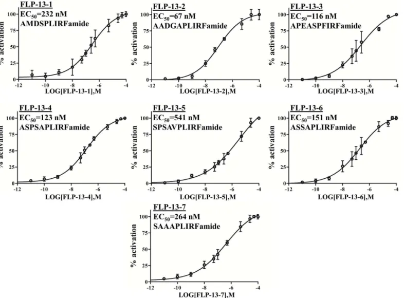
range of concentrations for each of the sevenflp-13derived peptides and found that they
acti-vated FRPR-4 with EC50values ranging from 67 nM to 541 nM (Fig 2). These values are similar
to the those measured for activation of peptide dispersing factor (PDF) receptor by its ligand
PDF, a well characterized ligand receptor pair; the PDF/PDFR EC50values range from 34 nM
to 361 nM [18]. Ourin celluloresults suggest that peptides encoded by the genesflp-5,flp-11,
andflp-13are the best candidates for beingin vivoFRPR-4A ligands. Becauseflp-13was the
strongestin celluloactivator of FRPR-4A and because of our initial interest in FLP-13, we
focused our efforts onin vivointeractions betweenfrpr-4andflp-13.
FRPR-4 regulates behavioral quiescence
We reasoned that if FRPR-4 were anin vivoreceptor for somnogenic FLP-13 neuropeptides,
then manipulatingfrpr-4activity should have similar effects to those observed when we
manip-ulatedflp-13gene activity. That is, overexpression offrpr-4might promote quiescence and
loss-of-function offrpr-4might produce defects in quiescence [10]. The phenomenon of the same phenotype arising from increased activity of the neuropeptide or its receptor has been
previously observed during genetic dissection of theC.elegansegg-laying circuit [19] and social
feeding behaviors [20]. To test whetherfrpr-4can induce quiescence in normally active
ani-mals, we made transgenic animals expressing extra copies of the genomic region that included thefrpr-4coding region as well as 5 kb of DNA upstream of the start site of translation and the
3’UTR we had identified using 3’RACE (see above). Our aim was to increase the copy number
of FRPR-4 in cells where it is normally expressed, and, by doing so, increase FRPR-4 signaling and thus amplify its physiological roles.
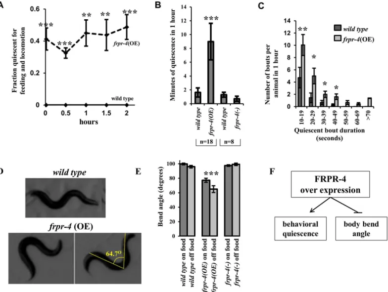
We observed a cohort of adult animals housed on a lawn of bacterial food on an agar surface every 30 minutes. We considered an animal quiescent if we could discern no movements of its body or of its feeding organ, the pharynx, for 15 seconds. Wild-type animals were nearly con-tinually active under these cultivation conditions and thus had only brief pauses of movement
and feeding (S1 Movie,left well). In contrast, animals carrying multiple transgenic copies of
thefrpr-4gene showed spontaneous bouts of behavioral quiescence (Fig 3AandS1 Movie,
right well). At every observation time point, up to 40% of animals carrying an integrated multi-copy transgene of FRPR-4 but 0% of wild-type animals was quiescent (Fig 3A). Four
strains carrying extrachromosomal transgenes with multiplefrpr-4copies showed similar
effects (S5 Fig). Therefore,frpr-4overexpression promotes spontaneous bouts of behavioral
quiescence.
To corroborate our assessments of quiescence, we used a machine vision approach to
mea-sure total quiescence as well as bout frequency and duration [21,22]. We measured quiescence
in pairs of animals, consisting of one animal overexpressingfrpr-4 and one control wild-type
animal.frpr-4over-expressing animals had increased total quiescence (Fig 3B). The increased
quiescence can be explained by an increased number of quiescent bouts (21 ± 4 infrpr-4
over-Fig 1. FLP-13 and other FMRFamide-like peptides activate FRPR-4A in a mammalian cell-culture system.Among 262C.elegansneuropeptides, peptides encoded by the genesflp-5,flp-10,flp-11,flp-13andflp-17activate FRPR-4, withflp-13derived peptides eliciting the largest amplitude responses. The magnitude of aequorin response to the peptides presented at concentrations ranging from 1 nM to 100μM and is shown relative to the magnitude of the response to the FLP-13-7 peptide.
expressing adults; 7 ± 3 in wild-type adults; p<0.001, Wilcoxon Rank Sum Test), as well as an
increased average bout duration (20 ± 3 seconds infrpr-4over-expressing animals; 13 ± 1 in
wild-type animals; p<0.05) (Fig 3C). This analysis suggests thatfrpr-4promotes both the
induction and maintenance of the quiescent behavioral state. Quiescent animals moved when mechanically stimulated (S2 Movie), indicating that they were not paralyzed or injured.
As a complementary approach to studying the effects of over-expressingfrpr-4, we studied
the effects of a loss offrpr-4function on stress-induced quiescence. Theok2376allele contains
a 1540 nucleotide deletion, which removes approximately 300 nucleotides of thefrpr-4
pro-moter as well as the first two exons of thefrpr-4gene (S2 Fig). This deletion is predicted to
make a truncated protein lacking three N-terminal transmembrane domains. However,
because there are potentially alternative start sites for translation,ok2376may retain four
C-terminal transmembrane domains as well as the intracellular C-C-terminal domain.
FLP-13 neuropeptides released from the ALA neuron are partially required for the
sleep-like quiescent behavior that occurs following cellular stress induction [10]. Thus, iffrpr-4were
Fig 2. FRPR-4A is activiated by FLP-13 peptidesin cellulo.Effect of the seven FLP-13 peptides (FLP-13a to g) on the intracellular Ca2+production in
CHO/mtAEQ/Galpha16cells expressing the full-length FRPR-4A. Data are presented as means±s.e.m. percent of maximal activation (N = four trials per
experiment).
doi:10.1371/journal.pone.0142938.g002
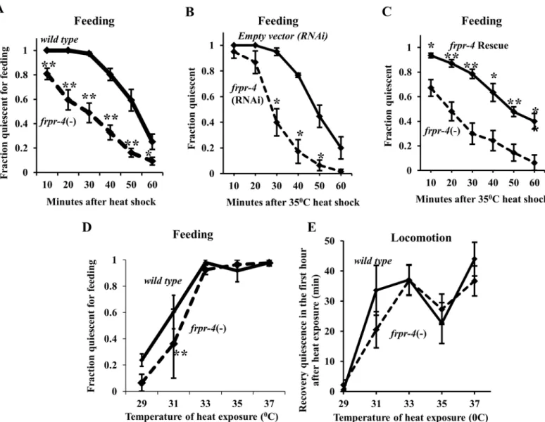
a FLP-13 receptor, thenfrpr-4mutants should also be deficient in this stress-induced quiescent
response. We observed a small but significant reduction in the fraction offrpr-4(ok2376)
ani-mals that were quiescent during recovery from a 30 minute 35°C heat exposure (seemethods–
protocol 1) (Fig 4A). Reduction offrpr-4function via RNA interference (RNAi) had a similar
attenuating effect on heat-induced feeding quiescence under the same conditions (Fig 4B),
sug-gesting thatfrpr-4(ok2376) is a reduction-of-function allele. Transgenic expression of a fosmid
containing thefrpr-4gene restored the heat-induced feeding quiescence offrpr-4(ok2376)
mutant animals to that of wild-type animals (Fig 4C).
Fig 3. Overexpression offrpr-4induces behavioral quiescence and a decreased body bend angle during locomotion.(A) A fraction of animals expressing an integrated multi-copy array of thefrpr-4gene display behavioral quiescence (no feeding or locomotion) when inspected at half-hour intervals over a two-hour period (N69, Fisher’s exact test,***p<.0005). (B) Locomotion quiescence of one day old adult animals measured using machine vision analysis. In each experiment, pairs of worms were observed, one of each of the two genotypes grouped in a bracket. Animals expressing the integrated multi-copy array offrpr-4,qnIs195, are more quiescent than wild-type animals (N = 18, Student’s t-test,***p<.001). There is no significant difference (p>0.05) between wild-type andfrpr-4(ok2376) worms. (C)frpr-4over-expressing animals display more frequent bouts and longer-duration bouts. (N = 18, Wilcoxon rank sum test,**p<.005,*p<.05). (D)frpr-4over-expressing animals display exaggerated body bends in comparison to wild-type animals. A line is drawn from the peak of one bend to the peak of the bend on the opposite side of the animal and the angle formed by the two lines was measured using imageJ software. Animals that over-expressfrpr-4have a significantly reduced bend angle, both on and off food (N>10, Student’s t-test,***p<.001). (E) A summary offrpr-4over-expressing phenotypes.
Previously, we found that worms are highly sensitive to differences in exposure temperature: small differences in heat exposure temperature resulted in large differences in behavioral
quies-cence following the exposures [10]. We therefore assessed the animals’quiescent response to a
range of temperature exposures. To insure that the duration of temperature exposure was the same in all animals, we placed the animals on pre-heated plates to start the 30-minute tempera-ture exposure, and removed them from the heated plates to complete the temperatempera-ture exposure (seemethods–protocol 2) [10]. Similar to our prior observations, the magnitude of the recov-ery quiescent response increased with higher exposure temperatures. Following exposure to 31
degrees Celsius but not following exposures to other temperatures,frpr-4mutants showed a
small defect in feeding quiescence (Fig 4D). In contrast to this defect in feeding quiescence, no
Fig 4. FRPR-4 is partially required for the feeding quiescence response to heat shock.Reducingfrpr-4function by mutation (A) or by RNA interference (B) impairs the feeding quiescence response to a 30-minute 35°C heat shock (Protocol 1 (Seemethods); Student’s t-test, average of 10 trials, N20 worms per trial,*p<.05,**p<.005). (C) A fosmid containing the wild-typefrpr-4gene restores infrpr-4(ok2376)mutants the feeding quiescence response to a 30-minute 35°C heat shock (Protocol 1 (Seemethods)) but not at other temperatures (Student’s t-test, average of 4 trials for each temperature, N20 worms per trial,**p<.005.) (D)frpr-4(ok2376) animals suppress the feeding quiescence in response to a 30-minute 33°C heat shock but not at the other
temperatures tested (Student’s t-test, average of 3 trials, N20 worms per trial,**P<.005). (E)frpr-4(ok2376) worms display normal locomotion quiescence in response to heat stress at all temperatures tested (Average of 2 trials, 12 worms per trial).
doi:10.1371/journal.pone.0142938.g004
exposure temperature resulted in a locomotion quiescence defect (Fig 4E). While the effects of
removingfrpr-4function on stress-induced feeding quiescence was statistically-significant, it
was smaller than the effects of removingflp-13function [10], suggesting that other receptors
are contributing to the quiescence. Unlike the case offlp-13, whose mRNA is induced by heat
shock [10],frpr-4mRNA was not changed by heat shock (S6 Fig).
Because FRPR-4 was activated by FLP-13 peptidesin cellulo,frpr-4overexpression
pro-moted quiescent behaviour, andfrpr-4mutants had a defect in stress-induced quiescence, we
hypothesized that FLP-13 peptides are the activators of the FRPR-4 receptorin vivo. Based on
this hypothesis, we predicted that the elevated quiescence phenotype of animals over express-ingfrpr-4would be attenuated by removing theflp-13gene. We additionally predicted that frpr-4loss-of-function would attenuate the quiescent phenotype observed with overexpression offlp-13[10]. To test these predictions, we over-expressedfrpr-4in theflp-13(tm2427)null
mutant background and over-expressedflp-13in thefrpr-4(ok2376)mutant background.
Con-trary to our predictions, the elevated locomotion quiescence (S7A Fig) and the quiescence bout
frequency (S7B Fig) offrpr-4over-expressing animals were not significantly reduced by the
flp-13(tm2427)mutation. In addition, thefrpr-4(ok2376)mutation did not attenuate the quies-cence induced by FLP-13 overexpression (S7C Fig). Together, these experiments suggest that
(1) FRPR-4 is not the (sole)in vivoreceptor for FLP-13 peptides, and (2) FLP-13 peptides are
not the (sole) ligands for all three FRPR-4 receptor isoforms or FRPR-4 may have ligand-inde-pendent activity.
FRPR-4 affects body posture by acting in the DVA neuron in a
flp-13
dependent fashion
In the course of the experiments in which we closely observed animals for quiescent behaviour,
we noted a body posture phenotype offrpr-4over-expressing animals.C.elegansworms crawl
on an agar surface with a wave of posteriorly directed ventral/dorsal body bends made in the
vector perpendicular to the agar surface. We noted that body bends made byfrpr-4
over-expressing animals were deeper than those made by wild-type animals. The deeper bends were the result of a reduction in the angle produced by the body bends during locomotion (Fig 3D). To quantify this phenotype, we measured the angle produced by the body bend in first day adult worms (Fig 3D). To reduce the chance that the altered body posture might be explained by difference in posture between quiescent and active worms, we measured the body angle
bends only during active bouts.frpr-4over-expressing animals had a significantly reduced
bend angle compared to wild-type animals during movement both in the presence and in the
absence of food (Fig 3E,S3andS4Movies). Thus, FRPR-4 is capable of promoting reduced
body bend angles and thus exaggerated body bends (Fig 3F).
Althoughflp-13was not required for the quiescence-inducing effects offrpr-4
overexpres-sion, we observed that the exaggerated body bend posture induced byfrpr-4overexpression
was absent in theflp-13(tm2427) mutants (Fig 5AandS5 Movie). To quantify this suppression,
we again measured the angle produced by the body bends. Theflp-13(tm2427) mutation
sup-pressed thefrpr-4-overexpression bend phenotype (Fig 5B).
A similar body bend phenotype has been reported in animals that had abnormal activity of the DVA neuron [23]. We therefore hypothesized that FRPR-4 functions in the DVA neuron
to alter its activity. To determine iffrpr-4is expressed in the DVA neuron, we constructed a
fluorescent translational reporter that contained>5kb offrpr-4upstream regulatory DNA and
the entire coding sequence of FRPR-4A, replacing the stop codon with a sequence encoding
green fluorescent protein (S2 Fig). We observed broad expression of thefrpr-4::gfptranslational
pharyngeal muscles, and neurons (S8 Fig). We observed membrane localization of the green fluorescence, as would be expected for a GPCR. We observed spontaneous bouts of behavioral quiescence during the adult stage in two transgenic lines (S5 Fig), suggesting that the
transla-tional reporter protein was functransla-tional. To facilitate identification of cells expressingfrpr-4, we
also generated transcriptional reporters in whichgfpcontaining a nuclear localization signal
was expressed under the control of the 5kb of DNA immediately upstream of thefrpr-4start
site of translation (S8 Fig). As in the case of the translational reporter, we observed broad expression of the transcriptional reporter. Using the transcriptional reporter, we identified frpr-4expression in the paired RIA neurons (S8 Fig), which have been implicated in the regula-tion of locomoregula-tion quiescence [24]; the paired I1 pharyngeal neurons, which regulate pharyn-geal pumping rate [25] and connect the pharynpharyn-geal and somatic nervous systems [26]; the
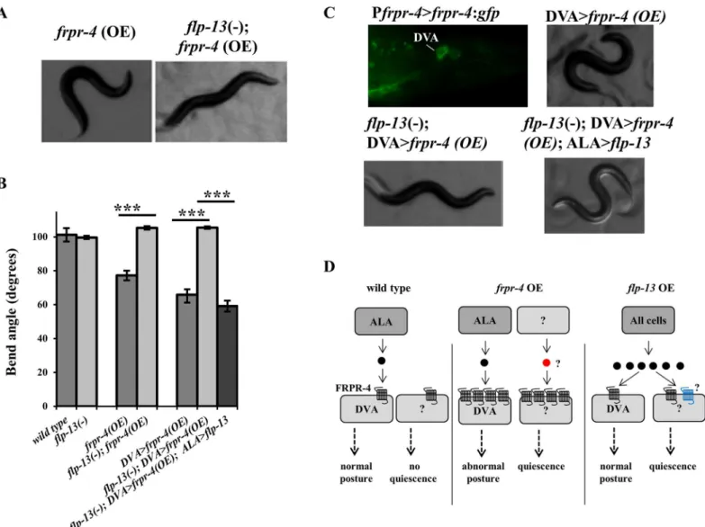
Fig 5. FLP-13 signals through FRPR-4 in DVA to regulate posture.(A-B) Overexpression offrpr-4from its endogenous promoter reduces the animals bend angle. Theflp-13(tm2427) deletion suppresses this phenotype and expression offlp-13in the ALA neuron restores this phenotype (Student’s t-test, N>10,***P<.001). (C) A strain carrying anfrpr-4:gfptranslational reporter shows GFP localization to the membrane of the DVA neuron. Overexpression of frpr-4in the DVA neuron using the promoter from the genetwk-16results in a decrease in bend angle, which is suppressed by theflp-13(tm2427) deletion and then restored by reinstatingflp-13in the ALA neuron. (D) A model of the effects offrpr-4orflp-13overexpression on posture and behavioral quiescence. Black circle denote FLP-13 peptides; red circle denotes an unknown peptide. Black transmembrane-domain receptors denote FRPR-4. Blue seven-transmembrane-domain receptors denote an unknown receptor mediating the quiescent effects of FLP-13 peptides.
doi:10.1371/journal.pone.0142938.g005
AVE neurons (S8 Fig), command interneurons that regulate locomotion [27] and are post-syn-aptic to the quiescence-generating ALA neuron [28], and the PVM neurons (S8 Fig), which are mechanosensory [29]. Additionally, as with our translational reporters, we saw bright expres-sion in the DVA neuron (Fig 5C).
To determine if thefrpr-4-induced body bend phenotype was explained by its activity in the
DVA neuron, we used the DVA-specifictwk-16promoter [30,31] to over-expressfrpr-4.
These animals, like those over-expressingfrpr-4under the control of its endogenous promoter,
showed the exaggerated body bend phenotype (Fig 5B and 5CandS6 Movie), consistent with
the notion thatfrpr-4affects posture via its activity in the DVA neuron. Theflp-13(tm2427)
mutation suppressed the body bend phenotype of animals over-expressingfrpr-4in the DVA
neuron (Fig 5andS7 Movie). These genetic interactions betweenfrpr-4andflp-13together
with ourin celluloanalyses suggest that FRPR-4 is anin vivoFLP-13 receptor.
We previously showed that the ALA neuron releases FLP-13 peptides to induce quiescence in response to cellular stress [10]. We hypothesized that FLP-13 peptides are released from
ALA to regulate body bend amplitude. To test this hypothesis, we restoredflp-13in the ALA
neuron in theflp-13(tm2427); DVA>frpr-4(OE) background. Expression in ALA was
accom-plished by expressing the genomic sequence offlp-13under the control of theida-1promoter
[32]. Restoration offlp-13in ALA did indeed result in a re-emergence of the exaggerated
bend posture phenotype (Fig 5B and 5CandS8 Movie). These data suggest that FLP-13 is
released from ALA to signal through FRPR-4 in the DVA neuron to regulate posture during locomotion.
Discussion
Using a combination of cell culture experiments and genetic analyses, we provide evidence that FRPR-4 is a G-protein coupled receptor that is activated by IRFamide and VRFamide neuro-peptides, and that it can promote both behavioral quiescence and exaggerated body bends. In
addition, we show anin vivogenetic interaction betweenfrpr-4and the P(F/L)IRFamide
encoding geneflp-13.
While overexpression of eitherflp-13orfrpr-4induces quiescence, the quiescent phenotype
caused by either gene is not dependent on the presence of the other gene (S7 Fig). In addition, frpr-4mutants have only a small defect in stress-induced feeding quiescence (Fig 4). Are these
results consistent with FRPR-4 being a receptor for FLP-13in vivoto promote quiescent
behav-ior? One possibility is that FLP-13 neuropeptides act through other receptors in addition to FRPR-4, and conversely, that FRPR-4 is activated by other neuropeptides in addition to the
FLP-13 neuropeptides. Ourin cellulodata are consistent with this notion, since several
neuro-peptides activate FRPR-4. In addition, FRPR-4 has five closely-related paralogs (S1 Fig), which could conceivably act as alternative receptors to FLP-13 peptides. There is precedence for genetic redundancy within neuropeptide signalling pathways. Suppression of the egg-laying defective phenotype caused by increased activity of the EGL-6 GPCR is accomplished by removal of both genes encoding neuropeptide ligands for this receptor; removal of one gene does not fully suppress the phenotype [19]. In addition, we identified three isoforms of FRPR-4
(S2 FigandS1 Table), which are primarily different at their C-termini. Ourin celluloanalyses
tested only the FRPR-4A isoform, but it is possible that the B and C isoforms have different affinities for FLP-13, or may be activated by different ligands.
With respect to the body posture phenotype, we can draw firmer conclusions regarding FLP-13/FRPR-4 interactions. FRPR-4 overexpression specifically in the DVA neuron causes
exaggerated body bends, which requireflp-13expressed in the ALA neuron. This genetic
the model that FLP-13 released from ALA acts directly on FRPR-4 in the DVA neuron to mod-ulate posture.
How do we reconcile this model with our observations that neither aflp-13loss-of-function
mutation nor afrpr-4loss-of-function mutation affects body posture and thatflp-13
overex-pression does not mimic the deeper body bend posture offrpr-4 over expressing animals? We
propose that normally, the amount of expressed FRPR-4 receptor sets a limit on signaling via
this pathway. Withfrpr-4expression under typical cultivation conditions, signaling is too low
to affect body posture (Fig 5D–left panel). Reducing FRPR-4 signaling yet further, by removing
the gene or by removing its ligand FLP-13, would have no effect on body posture, consistent
with our observations. However, increasedfrpr-4receptor expression in DVA causes increased
signaling in DVA to affect body posture. And, whileflp-13is required for the posture effects of
frpr-4overexpression, it is not required for the quiescence phenotype, which must be regulated
by additional unidentified peptides or by ligand-independent activity (Fig 5D–middle panel).
Finally, we propose that because of the limiting effects of wild typefrpr-4expression,flp-13
overexpression does not induce a body posture phenotype, but does induce quiescence in an frpr-4-independent fashion, suggesting that one or more unidentified FLP-13 receptors exist
(Fig 5D–right panel).
The strong phenotype observed withfrpr-4multi-copy overexpression suggests a strategy
for identifyingin vivoroles of other neurotransmitter receptor GPCRs. If a receptor shows low
expression and signaling under typical laboratory cultivation conditions, the phenotypic differ-ence between absdiffer-ence and presdiffer-ence of the gene may be difficult to detect. Consistent with this notion, in a systematic study examining the phenotypic consequences of reduction of function of neurotransmitter receptor GPCRs, only approximately 15 (of over 60 tested) resulted in a discernible phenotype [7]. In contrast, the difference between multiple copy expression of the gene and the wild-type, low expressing, condition, may be far more apparent, and lead to spe-cific hypotheses regarding the normal function of the gene, which can then be tested using fine phenotypic analysis of the loss-of-function mutants.
Materials and Methods
Animal husbandry and strains
Animals were cultivated on NGM agar and fed the OP50E.coliderivative strain DA837 [33].
The following strains were used in this study:N2(Bristol),EG4322ttTi5605 II; unc-119(ed3)
III,TM2427flp-13(tm2427)IV,RB1837frpr-4(ok2376)II,NQ291unc-119(ed3)III;qnEx155
[frpr-4(+), Pmyo-2>mCherry;unc-119(+)],NQ308unc-119(ed3)III;qnEx195[frpr-4(+),
Pmyo-2>mCherry,unc-119(+)],NQ385frpr-4(ok2376)II,NQ408unc-119(ed3)III;
qnEx196[Pfrpr-4>frpr-4:gfp; Pmyo-2>mCherry;unc-119(+)],NQ459unc-119(ed3)III;qnEx233[Pfrpr-4>
frpr-4:gfp;Pglr-3>mCherry,unc-119(+)],NQ460unc-119(ed3)III;qnEx234[Pfrpr-4>frpr-4:gfp:
frpr-43’UTR; Pglr-3>mCherry;unc-119(+)],NQ465unc-119(ed3)III;qnEx248[Pfrpr-4>NLS:
gfp;Pglr-3>mCherry;unc-119(+)],NQ480qnIs195[frpr-4(+); Pmyo-2>mCherry;unc-119(+)],
NQ588flp-13(tm2427)IV;qnIs195[frpr-4(+), Pmyo-2>mCherry,unc-119(+)],NQ601flp-13
(tm2427)IV;qnEx310[Pida-1>flp-13,Prab-3>mCherry],NQ602flp-13(tm2427)IV (Outcrossed
to N2 3 times),NQ648qnEx347[Pfrpr-4:NLS:gfp,Popt-3:mCherry],NQ743frpr-4(ok2376)IV;
qnEx405[frpr-4(+)(FOSMID-WRM0630bE03,Pmyo-2>gfp],NQ744frpr-4(ok2376)IV;
qnEx406[frpr-4(+)(FOSMID-WRM0630bE03,Pmyo-2>gfp],NQ745frpr-4(ok2376)IV;
qnEx407[frpr-4(+)(FOSMID-WRM0630bE03,Pmyo-2>gfp],NQ756qnEx415[Ptwk-16>
frpr-4,Ptwk-16>frpr-4:gfp,myo-2>mCherry],SJU20flp-13(tm2427)IV;qnEx415[Ptwk-16>frpr-4,
Ptwk-16>frpr-4:gfp,myo-2>mCherry],SJU47flp-13(tm2427)IV;qnEx415[Ptwk-16>frpr-4,
Ptwk-16>frpr-4:gfp,myo-2>mCherry];qnEx310[Pida-1>flp-13,Prab-3>mCherry]
The strain RB1837, containingfrpr-4(ok2376), was obtained from the CGC.frpr-4(ok2376)
was outcrossed to N2 three times to create the strain NQ385. The presence of theok2376
dele-tion was detected by PCR.
Molecular biology, transgenics and integrations
We constructed DNA constructs using overlap-extension polymerase chain reaction (PCR), as
previously described [34]. Oligonucleotides used are listed inS2 Table.Constructs were made
by amplifying sequences from genomic DNA, GFP from the Andy Fire vector pPD95.75, NLS: gfp from the Andy Fire vector pPD122.13, and mCherry from pCFJ90 (Addgene).
Transgenic animals were created by microinjection [35] using a Leica DMIRB inverted DIC microscope equipped with an Eppendorf Femtojet microinjection system. Either the wild-type
strain N2 or theunc-119mutant strain EG4322 animals were injected with 2–50 ng/μl of each
construct in combination with one of the following injection markers: 5ng/μl pCFJ90
(Pmyo-2>mCherry), or 5ng/μl pPD118.33 (Pmyo-2>gfp). The DNA mix was adjusted to a final
con-centration of 150 ng/μl by adding 1 kb DNA ladder (New England Biolabs) or the plasmid
pCFJ151 (unc-119(+)). The fosmid WRM0630bE03 was injected intofrpr-4(ok2376) animals at
a concentration of 2 ng/μl. For behavioral experiments using transgenic animals carrying
extra-chromosomal arrays, at least two lines were analyzed. The integrated transgeneqnIs195was
constructed as previously described [36] by UV irradiation of strains carrying the
extrachro-mosomal transgenesqnEx195[36] and then out-crossed to the wild-type strain four times
before analysis.
Microscopy and fluorescence
For GFP and differential interference contrast imaging, animals were mounted on 5% agar pads, immobilized with 15mM levamisole and observed through a 63X or 100X oil-immersion objective lens on a Leica DM5500B microscope. Leica LAS software was used to capture and analyze images.
RNA interference
A 3.6 kb genomic fragment spanning a portion of thefrpr-4gene (C54A12.2) (SeeS2 Fig) was
amplified from genomic DNA using the primers oNQ627 and oNQ628 (S1 Table), which
con-tained T7 5’tails. The PCR product was purified (QIAquick PCR Purification Kit, Qiagen) and
used in anin vitroT7 RNA polymerase transcription kit (New England Biolabs) to produce
double stranded RNA. Double stranded RNA was injected into the intestine or gonad of
wild-type, first-day old adults at a concentration of 200–300 ng/μl. The progeny of the injected
worms were analyzed for their ability to become quiescent in response to heat stress.
Feeding quiescence after heat exposure
On the day prior to the experiments, 15–25 L4 animals were transferred to Petri dishes
con-taining 12 mL of 1.7% NGM agar seeded with DA837 bacteria. Two different experimental approaches were used to heat stress first day adult animals. Protocol 1 (mild heat shock): On the day of the experiment, the plates housing the worms were wrapped in parafilm and sub-merged in a 35°C water bath for 30 minutes. During a single experiment, the various genotypes were staggered with regards to the time that they entered and exited the water bath. After
removing the plates from the water bath, the worms were observed at room temperature (21–
assessing the effect of exposure temperature on feeding quiescence, animals were subjected to precisely 30 minutes of heat at a specific temperature. NGM agar plates seeded with OP50 were pre-heated to the desired temperature for 20 minutes by submerging them in a water bath.
Room temperature (21–23°C) first day adults were transferred to the pre-heated plates and
immediately submerged in a water bath set at the same temperature for 30 minutes. Immedi-ately after the heat exposure, the worms were transferred to room temperature plates seeded with bacteria and then assessed every 10 minutes for 60 minutes for the presence of pharyngeal pumping. In all cases, Statistical comparisons were made between genotypes tested simulta-neously. Experiments were performed by investigators blinded to the genotype of the animals.
Locomotion quiescence after heat exposure
To measure locomotion quiescence, we monitored first day adult worms cultivated on an agar
surface in concave polydimethylsiloxane (PDMS) wells seeded with DA837 bacteria [22,24]
following heat exposure. First, NGM plates were preheated in a water bath for 20 minutes to the desired heat exposure temperature. Animals grown on room temperature plates were trans-ferred to the pre-warmed plates and heat shocked for 30 minutes. Following heat exposure, sin-gle worms were transferred to the agar surface within a PDMS well. The microchip loaded with the worms was placed in a 50 ml petri dish, along with a wet Kimwipe, to prevent desiccation, and the plate was then sealed with Parafilm. Using a USB 2.0 Monochrome Industrial Camera
(The Imaging Source1), dark-field images were taken every ten seconds for 90 minutes. Images
were analyzed with the frame subtraction algorithm [21,37,38] using custom MATLAB
software.
Locomotion Quiescence analysis for
frpr-4
overexpression
We placed one first-day old adult in each of two adjacent concave polydimethylsiloxane (PDMS) wells filled with NGM agar and seeded with DA837 bacteria. In each experiment, we placed one experimental animal into one well and one control animal into an adjacent well. The PDMS was then placed on a Diagnostics Instruments microscope base and illuminated for bright-field microscopy, using white light supplied to the base with a fiber optic cable from a Schott DCR III light source. A camera (659 × 494 pixels, scA640-70fm, Basler Vision Technol-ogies) mounted on a Zeiss Stemi 2000 stereomicroscope captured an image of both wells every 10 seconds with an 8-bit grayscale resolution. At this magnification and camera acquisition
set-ting, the spatial resolution was 12.5 micrometers2per pixel. We monitored animals for one
hour beginning 15 minutes after transfer to the PDMS wells and used a machine vision frame
subtraction principle to identify 10-second epochs of behavioral quiescence [21,37,38].
Quantitative PCR
First-day adult animals were collected prior to heat shock and then at seven additional time intervals (0, 15, 30, 45, 60, 120, and 180 minutes) following a 37°C heat shock for 30 minutes. Total RNA was collected from each group of worms using an RNAeasy mini kit (Qiagen), and cDNA was synthesized using the SuperScript one-step RT-PCR system (Invitrogen). We per-formed three or more biological replicates, each of which was perper-formed using several 100 first day adult animals, during each collection time point, and for each biological replicate we calcu-lated the average of two technical replicates. Real-time PCR was performed using Taqman Gene Expression Mastermix on an Applied Biosystems 7500 platform at the core services within the Penn Center for AIDS Research, an NIH-funded program (P30 AI 045008).
Oligo-nucleotides used are listed inS2 Table. Relative mRNA concentrations were determined by the
delta-delta method [39] by normalization to the expression of the genepmp-3, which has been shown to show little expression variance [40].
Identification of
frpr-4
gene structure
To determine the 3’end offrpr-4and identify potentially different isoforms, we used a 3’RACE
method [14]. We collected wild-type worms of mixed stages from 5–10 plates in which the
bac-teria had recently depleted, isolated total RNA using an RNAeasy mini kit (Qiagen), and gener-ated a cDNA library using SuperScript one-step RT-PCR system (Invitrogen). We used the
primer oNQ549 (Identical to QT, as described by Scotto-Lavino et al [14]) to generate the
cDNA library. We then used the FRPR-4 gene-specific primer oNQ578 together with oNQ550
(Identical to Qo[14]) in an initial round of PCR to amplify a portion of thefrpr-4cDNA. This
PCR product was then used as the template in a PCR reaction with the nested primers oNQ579
and oNQ551(Identical to Qi[14]) to amplify the final cDNA ends. Finally, we cloned the
cDNAs into a pCR2.1-TOPO Vector (Invitrogen) and sequenced the cloned inserts.
Receptor ligand interactions
in cellulo
Total RNA was collected using an RNAeasy mini kit (Qiagen) from wild-type animals har-vested from mixed stage populations grown on five NGM plates. cDNA was synthesized using the SuperScript one-step RT-PCR system (Invitrogen). FRPR-4A cDNA was PCR amplified, directionally cloned into the pcDNA3.1(+) TOPO expression vector (LifeTechnologies), and sequenced to confirm that no errors were introduced during the PCR or cloning steps.
Receptor activation was studied in Chinese hamster ovary cells (CHO) stably expressing
apo-aequorin (mtAEQ) targeted to the mitochondria as well as the human Galpha16subunit.
The CHO/mtAEQ/Galpha16cells were cultured in Ham’s F12 medium (Sigma), containing 10%
fetal bovine serum (FBS), 100 UI/ml of penicillin/streptomycin, 250μg/ml Zeocin and 2.5μg/
ml Fungizone (Amphoterin B). Cell lines were grown at 37°C in a humidified atmosphere of
5% CO2and were diluted fifteen-fold every third day. CHO/mtAEQ/Galpha16cells were
tran-siently transfected with the FRPR-4 cDNA construct or the empty pcDNA3.1(+) vector using
the FuGENE 6 transfection reagent (Promega), according to the manufacturer’s instructions.
Cells expressing the receptor were collected 2 days post-transfection in BSA medium (DMEM/
HAM’s F12 with 15 mM HEPES, without phenol red, supplemented with 0.1% BSA) and
loaded with 5μM coelenterazine h (Invitrogen) for 4 hours to reconstitute the holo-enzyme
aequorin. The cells were plated at a density of 25,000 cells/well and exposed to synthetic
pep-tides at a concentration of 10μM in BSA medium. Aequorin bioluminescence was recorded for
30 seconds on a Mithras LB 940 luminometer (Berthold Technologies) in quadruplicate. For dose-response evaluations, after 30 seconds of ligand-stimulated calcium measurements,
Triton X-100 (0.1%) was added to the well to obtain a measure of the maximum cell Ca2+
response. BSA medium without the peptides was used as a negative control and 1μM ATP was
used to check the functional response of the cells. Cells transfected with the pcDNA3.1 empty
vector were used as a negative control for the effect of the receptor. EC50values were calculated
from dose-response curves, constructed using a computerized nonlinear regression analysis, with a sigmoidal dose-response equation (Sigmaplot 9.0).
Measuring body bend angle
Wild-type,frpr-4(ok2376),flp-13(tm2427),frpr-4(OE),flp-13(tm2427);frpr-4(OE),
DVA>frpr-4(OE),flp-13(-);DVA>frpr-4(OE) andflp-13(-); DVA:frpr-4(OE); ALA:flp-13
camera, ImageSource) mounted on a Leica Microsystems MZ10F stereomicroscope (seeS3–S7 Movies). Videos were analyzed using ImageJ software [41]. Videos of 10 or more individual animals for each genotype were captured. For each individual worm, we drew lines from the
peak of one bend to the peak of the opposite bend (SeeFig 3). We did this for a minimum of 3
body bends for each individual and averaged the bend angles measured for that individual. We then calculated the average and standard error of the mean of all individuals within a given genotype.
Phylogenetic Tree construction
We first alignedC.elegans[5] andDrosophila melanogaster[42] predicted neuropeptide
recep-tors using ClustalW version 2.0 [43]. The alignment was then used as a template to construct a maximum-likelihood tree using the MEGA6 software [44].
Supporting Information
S1 Fig. A phylogenetics tree consisting of allD.melanogasterandC.eleganspredicted neu-ropeptide receptors, as listed in references [5,42].FRPR-4 is most closely related toD. mela-nogasterFR.
(TIF)
S2 Fig. The gene model and isoforms of thefrpr-4gene predicted by wormbase.org (top model) and experimentally determined (bottom three models) by 3’RACE.Arrows denote
inverted repeats flanking intronic DNA, which separates the two parts of the 3’UTR (gray) in
isoform A and separates the last two coding exons in isoforms B and C. Theok2376deletion
removes the first two exons of all three isoforms. It also removes 300 nucleotides of upstream
regulatory DNA. The location ofgfpin the strains NQ408, NQ459 and NQ460 and the location
of the DNA used as template for generating double stranded RNA in the RNAi experiments are marked at the bottom of the figure.
(TIF)
S3 Fig. Alignment ofC.elegansFRPR-4 withC.briggsaeFRPR-4 supports the gene struc-ture determined by cDNA sequencing.Blue denote the most 5’experimentally-determined coding exon. Red denotes the sixth coding exon of FRPR-4A and FRPR-4B, which is absent in
FRPR-4C (not shown) and inC.briggsae. Green denotes the most 3’coding exon, which is
present in FRPR-4A and inC.briggsae. Grey denotes the 3’RACE validated 3’-untranslated
region (3’UTR).C.briggsaedoes not possess the retrotranspon that was observed in the 3’UTR
of theC.elegans frpr-4gene.
(TIF)
S4 Fig. Alignment of (I/V)RF neuropeptides that activate and do not activate FRPR-4A.
Hydrophobic amino acids are yellow, charged amino acids are red, and polar but uncharged amino acids are blue. On average the peptides that do not activate are shorter (mean±SD = 7.9 ±1.3 amino acids) than those that activate (9.4±1.4 amino acids; p = 0.002). No other feature is consistently different between the two groups.
(TIF)
S5 Fig. Transgenic lines that over express eitherfrpr-4orfrpr-4:gfpinduce behavioral qui-escence.A significant fraction of first-day old adult transgenic animals carrying additional
cop-ies of thefrpr-4gene (middle bar) or additional copies offrpr-4:gfptranslational reporters
(right bar) are quiescent. Shown is the average ± s.e.m of three trials using two independent
transgenic lines of each genotype, with each trial containing 20–30 animals of each genotype.
(Students t-test,P<.001). (TIF)
S6 Fig.frpr-4mRNA is not induced by heat stress.
(TIF)
S7 Fig. FLP-13 peptides are not required forfrpr-4-induced quiescence andfrpr-4is not required forflp-13-induced quiescence.Machine vision analysis shows that theflp-13
(tm2427) mutation does not significantly suppress the elevated total quiescence (A), quiescence
bout frequency (B), or quiescence bout duration (B) induced byfrpr-4overexpression. (C)
Direct observation shows that thefrpr-4(ok2376) mutation does not suppress the elevated
qui-escence induced byflp-13overexpression. Shown is the average ± s.e.m fraction of animals
qui-escent for feeding and locomotion two hours after heat-shock promoter induced expression of flp-13. Shown in A and B is the average of>15 trials with 1 animal of each genotype per trial.
Shown in C is the average of 2 trials with>25 animals per trial.
(TIF)
S8 Fig.frpr-4has a broad expression pattern.(A) Transgenic animals carrying anfrpr-4:gfp translational reporter show GFP localization to the membrane of multiple neurons, including
the RIA neurons (identified using thePglr-3>mCherrymarker) and PVM neuron (identified
based on location and morphology), as well as body muscle. (B) Transgenic animals expressing
a Pfrpr-4>NLS:gfptranscriptional reporter shows additional expression in the AVE neuron
(which co-expresses the geneopt-3, marked in red in the left panel in B), the I1 pharyngeal
neu-ron (identified based on location and morphology) and other head neuneu-rons. In the left panel in
A and in the right panel in B, the RIA neurons co-express the geneglr-3, which is marked in
red. (TIF)
S1 Movie. Several first-day old adult wild-type animals (on the left) and first-day old adult transgenic animals expressing an integrated multi-copy array of the genefrpr-4(strain NQ480, on the right).Whereas the wild-type animals are continuously active,frpr-4 over-expressing animals have spontaneous bouts of locomotion quiescence. The movie is played at 16 times the real speed.
(MP4)
S2 Movie. Two first-day old adult animals over-expressingfrpr-4(strain NQ480).The left worm is foraging whereas the right worm is quiescent. In response to two dish taps, both worms move, one forward and the other backwards. The movie is played at real speed. (MP4)
S3 Movie. One first day adult N2 (wild-type) animal moving on an unseeded agar surface.
The movie is played at 16 times the real speed. (MP4)
S4 Movie. One first day adult NQ480 (frpr-4(OE)) animal moving on an unseeded agar sur-face.Body bends are deeper than those of N2 animals (reflecting smaller bend angles of body bends by the transgenic animals). The movie is played at 16 times the real speed.
(MP4)
S5 Movie. A first day adult NQ588 (flp-13(-);frpr-4(OE)) animal moving on an unseeded plate.Body bends appear similar to those of N2 animals. The movie is played at 16 times the real speed.
S6 Movie. A first day adult NQ756 (DVA>frpr-4(OE)) animal moving on an unseeded
plate.Body bends are deeper than those of N2 animals. The move is played at 8 times the real speed.
(MP4)
S7 Movie. A first day adult NQ756 (flp-13(-); DVA>frpr-4(OE)) animal moving on an
unseeded plate.Body bends appear similar to those of N2 animals. The movie is played at 8 times the real speed.
(MP4)
S8 Movie. A first day adult SJU47 (flp-13(-); DVA>frpr-4(OE); ALA>flp-13) animal
mov-ing on an unseeded plate.Body bends are deeper than those of N2 animals. The movie is played at 8 times the real speed.
(MP4)
S1 Table. Sequences of the cDNA of the experimentally verified FRPR-4 isoforms and the presumed transposable element located in the most 3’intron offrpr-4.
(DOCX)
S2 Table. Oligonucleotides used in this study.Most DNA construct was made using
overlap-extension PCR, as previously described.£The FRPR-4A cDNA was amplified from aC.elegans
cDNA library (SeeMaterials and Methods).§The PCR product used to make dsRNA (See
Materials and Methods) was amplified from genomic DNA with PCR-engineered tails contain-ing T7 promoters.
(DOCX)
Acknowledgments
This manuscript is dedicated to the memory of N.Y. Strains were provided by the CGC, which
is funded by NIH Office of Research Infrastructure Programs (P40 OD010440). The strain
flp-13(tm2427) was provided by the National BioResource Project (PI, Shohei Mitani). We thank N. Trojanowski and R. McCloskey for comments on this manuscript.
Author Contributions
Conceived and designed the experiments: MDN DMR TJ LS NY. Performed the experiments: MDN TJ NY KHL. Analyzed the data: MDN TJ NY LS DMR. Contributed reagents/materials/ analysis tools: MDN DMR LS. Wrote the paper: MDN DMR.
References
1. Taghert PH, Nitabach MN. Peptide neuromodulation in invertebrate model systems. Neuron. 2012; 76 (1):82–97. doi:10.1016/j.neuron.2012.08.035PMID:23040808; PubMed Central PMCID:
PMC3466441.
2. Li C, Nelson LS, Kim K, Nathoo A, Hart AC. Neuropeptide gene families in the nematode Caenorhabdi-tis elegans. Ann N Y Acad Sci. 1999; 897:239–52. PMID:10676452.
3. Peymen K, Watteyne J, Frooninckx L, Schoofs L, Beets I. The FMRFamide-Like Peptide Family in Nematodes. Front Endocrinol (Lausanne). 2014; 5:90. doi:10.3389/fendo.2014.00090PMID: 24982652; PubMed Central PMCID: PMC4058706.
4. Li C, Kim K. Neuropeptide gene families in Caenorhabditis elegans. Adv Exp Med Biol. 2010; 692:98–
137. PMID:21189676.
5. Janssen T, Lindemans M, Meelkop E, Temmerman L, Schoofs L. Coevolution of neuropeptidergic sig-naling systems: from worm to man. Ann N Y Acad Sci. 2010; 1200:1–14. doi:10.1111/j.1749-6632. 2010.05506.xPMID:20633129.
6. Hobert O. The neuronal genome of Caenorhabditis elegans. WormBook: the online review of C elegans biology. 2013:1–106. doi:10.1895/wormbook.1.161.1PMID:24081909.
7. Keating CD, Kriek N, Daniels M, Ashcroft NR, Hopper NA, Siney EJ, et al. Whole-genome analysis of 60 G protein-coupled receptors in Caenorhabditis elegans by gene knockout with RNAi. Current biol-ogy: CB. 2003; 13(19):1715–20. PMID:14521838.
8. Beets I, Janssen T, Meelkop E, Temmerman L, Suetens N, Rademakers S, et al. Vasopressin/oxyto-cin-related signaling regulates gustatory associative learning in C. elegans. Science. 2012; 338 (6106):543–5. doi:10.1126/science.1226860PMID:23112336.
9. Garrison JL, Macosko EZ, Bernstein S, Pokala N, Albrecht DR, Bargmann CI. Oxytocin/vasopressin-related peptides have an ancient role in reproductive behavior. Science. 2012; 338(6106):540–3. doi: 10.1126/science.1226201PMID:23112335; PubMed Central PMCID: PMC3597094.
10. Nelson MD, Lee KH, Churgin MA, Hill AJ, Van Buskirk C, Fang-Yen C, et al. FMRFamide-like FLP-13 Neuropeptides Promote Quiescence following Heat Stress in Caenorhabditis elegans. Current biology: CB. 2014. doi:10.1016/j.cub.2014.08.037PMID:25264253.
11. Hill AJ, Mansfield R, Lopez JM, Raizen DM, Van Buskirk C. Cellular stress induces a protective sleep-like state in C. elegans. Current biology: CB. 2014; 24(20):2399–405. doi:10.1016/j.cub.2014.08.040 PMID:25264259; PubMed Central PMCID: PMC4254280.
12. Lenz O, Xiong J, Nelson MD, Raizen DM, Williams JA. FMRFamide signaling promotes stress-induced sleep in Drosophila. Brain Behav Immun. 2015. doi:10.1016/j.bbi.2014.12.028PMID:25668617. 13. Meeusen T, Mertens I, Clynen E, Baggerman G, Nichols R, Nachman RJ, et al. Identification in
Dro-sophila melanogaster of the invertebrate G protein-coupled FMRFamide receptor. Proceedings of the National Academy of Sciences of the United States of America. 2002; 99(24):15363–8. doi:10.1073/ pnas.252339599PMID:12438685; PubMed Central PMCID: PMC137722.
14. Scotto-Lavino E, Du G, Frohman MA. 3' end cDNA amplification using classic RACE. Nat Protoc. 2006; 1(6):2742–5. doi:10.1038/nprot.2006.481PMID:17406530.
15. Beets I, Lindemans M, Janssen T, Verleyen P. Deorphanizing g protein-coupled receptors by a calcium mobilization assay. Methods in molecular biology. 2011; 789:377–91. doi: 10.1007/978-1-61779-310-3_25PMID:21922422.
16. Scimemi A, Beato M. Determining the neurotransmitter concentration profile at active synapses. Mol Neurobiol. 2009; 40(3):289–306. doi:10.1007/s12035-009-8087-7PMID:19844813; PubMed Central PMCID: PMCPMC2777263.
17. van den Pol AN. Neuropeptide transmission in brain circuits. Neuron. 2012; 76(1):98–115. doi:10. 1016/j.neuron.2012.09.014PMID:23040809; PubMed Central PMCID: PMCPMC3918222.
18. Janssen T, Husson SJ, Lindemans M, Mertens I, Rademakers S, Ver Donck K, et al. Functional charac-terization of three G protein-coupled receptors for pigment dispersing factors in Caenorhabditis ele-gans. J Biol Chem. 2008; 283(22):15241–9. doi:10.1074/jbc.M709060200PMID:18390545; PubMed Central PMCID: PMC3258896.
19. Ringstad N, Horvitz HR. FMRFamide neuropeptides and acetylcholine synergistically inhibit egg-laying by C. elegans. Nat Neurosci. 2008; 11(10):1168–76. doi:10.1038/nn.2186PMID:18806786; PubMed Central PMCID: PMC2963311.
20. Rogers C, Reale V, Kim K, Chatwin H, Li C, Evans P, et al. Inhibition of Caenorhabditis elegans social feeding by FMRFamide-related peptide activation of NPR-1. Nature neuroscience. 2003; 6(11):1178–
85. doi:10.1038/nn1140PMID:14555955.
21. Raizen DM, Zimmerman JE, Maycock MH, Ta UD, You YJ, Sundaram MV, et al. Lethargus is a Caenor-habditis elegans sleep-like state. Nature. 2008; 451(7178):569–72. doi:10.1038/nature06535PMID: 18185515.
22. Yu CC, Raizen DM, Fang-Yen C. Multi-well imaging of development and behavior in Caenorhabditis elegans. Journal of neuroscience methods. 2014; 223:35–9. doi:10.1016/j.jneumeth.2013.11.026 PMID:24321627; PubMed Central PMCID: PMC3972622.
23. Li W, Feng Z, Sternberg PW, Xu XZ. A C. elegans stretch receptor neuron revealed by a mechanosen-sitive TRP channel homologue. Nature. 2006; 440(7084):684–7. doi:10.1038/nature04538PMID: 16572173; PubMed Central PMCID: PMC2865900.
24. Nelson MD, Trojanowski NF, George-Raizen JB, Smith CJ, Yu CC, Fang-Yen C, et al. The neuropep-tide NLP-22 regulates a sleep-like state in Caenorhabditis elegans. Nature communications. 2013; 4:2846. doi:10.1038/ncomms3846PMID:24301180; PubMed Central PMCID: PMC3867200. 25. Trojanowski NF, Padovan-Merhar O, Raizen DM, Fang-Yen C. Neural and genetic degeneracy
26. Albertson DG, Thomson JN. The pharynx of Caenorhabditis elegans. Philosophical transactions of the Royal Society of London Series B, Biological sciences. 1976; 275(938):299–325.
27. Chalfie M, Sulston JE, White JG, Southgate E, Thomson JN, Brenner S. The neural circuit for touch sensitivity in Caenorhabditis elegans. The Journal of neuroscience: the official journal of the Society for Neuroscience. 1985; 5(4):956–64. PMID:3981252.
28. White JG, Southgate E, Thomson JN, Brenner S. The structure of the nervous system of the nematode Caenorhabditis elegans. Philosophical transactions of the Royal Society of London Series B, Biological sciences. 1986; 314(1165):1–340. PMID:22462104.
29. Chalfie M, Sulston J. Developmental genetics of the mechanosensory neurons of Caenorhabditis ele-gans. Developmental biology. 1981; 82(2):358–70. PMID:7227647.
30. Puckett Robinson C, Schwarz EM, Sternberg PW. Identification of DVA interneuron regulatory sequences in Caenorhabditis elegans. PLOS one. 2013; 8(1):e54971. doi:10.1371/journal.pone. 0054971PMID:23383017; PubMed Central PMCID: PMC3557239.
31. Salkoff L, Butler A, Fawcett G, Kunkel M, McArdle C, Paz-y-Mino G, et al. Evolution tunes the excitabil-ity of individual neurons. Neuroscience. 2001; 103(4):853–9. PMID:11301195.
32. Cai T, Fukushige T, Notkins AL, Krause M. Insulinoma-Associated Protein IA-2, a Vesicle Transmem-brane Protein, Genetically Interacts with UNC-31/CAPS and Affects Neurosecretion in Caenorhabditis elegans. The Journal of neuroscience: the official journal of the Society for Neuroscience. 2004; 24 (12):3115–24. doi:10.1523/JNEUROSCI.0101-04.2004PMID:15044551.
33. Davis MW, Somerville D, Lee RY, Lockery S, Avery L, Fambrough DM. Mutations in the Caenorhabditis elegans Na,K-ATPase alpha-subunit gene, eat-6, disrupt excitable cell function. The Journal of neuro-science: the official journal of the Society for Neuroscience. 1995; 15(12):8408–18. PMID:8613772. 34. Nelson MD, Fitch DH. Overlap extension PCR: an efficient method for transgene construction. Methods
Mol Biol. 2011; 772:459–70. doi:10.1007/978-1-61779-228-1_27PMID:22065455.
35. Stinchcomb DT, Shaw JE, Carr SH, Hirsh D. Extrachromosomal DNA transformation of Caenorhabditis elegans. Mol Cell Biol. 1985; 5(12):3484–96. PMID:3837845; PubMed Central PMCID:
PMCPMC369179.
36. Mello C, Fire A. DNA transformation. Methods Cell Biol. 1995; 48:451–82. PMID:8531738. 37. Belfer SJ, Chuang HS, Freedman BL, Yuan J, Norton M, Bau HH, et al. Caenorhabditis-in-drop array
for monitoring C. elegans quiescent behavior. Sleep. 2013; 36(5):689–98G. doi:10.5665/sleep.2628 PMID:23633751; PubMed Central PMCID: PMC3624823.
38. Zimmerman JE, Raizen DM, Maycock MH, Maislin G, Pack AI. A video method to study Drosophila sleep. Sleep. 2008; 31(11):1587–98. PMID:19014079; PubMed Central PMCID: PMC2579987. 39. Livak KJ, Schmittgen TD. Analysis of relative gene expression data using real-time quantitative PCR
and the 2(-Delta Delta C(T)) Method. Methods. 2001; 25(4):402–8. PMID:11846609.
40. Hoogewijs D, Houthoofd K, Matthijssens F, Vandesompele J, Vanfleteren JR. Selection and validation of a set of reliable reference genes for quantitative sod gene expression analysis in C. elegans. BMC Mol Biol. 2008; 9:9. PMID:18211699. doi:10.1186/1471-2199-9-9
41. Schneider CA, Rasband WS, Eliceiri KW. NIH Image to ImageJ: 25 years of image analysis. Nature methods. 2012; 9(7):671–5. PMID:22930834.
42. Hewes RS, Taghert PH. Neuropeptides and neuropeptide receptors in the Drosophila melanogaster genome. Genome Res. 2001; 11(6):1126–42. doi:10.1101/gr.169901PMID:11381038; PubMed Cen-tral PMCID: PMC311076.
43. Thompson JD, Gibson TJ, Higgins DG. Multiple sequence alignment using ClustalW and ClustalX. Curr Protoc Bioinformatics. 2002; Chapter 2:Unit 2 3. doi:10.1002/0471250953.bi0203s00PMID: 18792934.
44. Tamura K, Stecher G, Peterson D, Filipski A, Kumar S. MEGA6: Molecular Evolutionary Genetics Anal-ysis version 6.0. Molecular biology and evolution. 2013; 30(12):2725–9. doi:10.1093/molbev/mst197 PMID:24132122; PubMed Central PMCID: PMC3840312.