The Design of Simple Bacterial Microarrays:
Development towards Immobilizing Single
Living Bacteria on Predefined Micro-Sized
Spots on Patterned Surfaces
Nina Bjørk Arnfinnsdottir1, Vegar Ottesen1, Rahmi Lale2, Marit Sletmoen1*
1Biophysics and Medical Technology, Department of Physics, Norwegian University of Science and Technology, NO-7491 Trondheim, Norway,2Department of Biotechnology, Norwegian University of Science and Technology, NO-7491 Trondheim, Norway
Abstract
In this paper we demonstrate a procedure for preparing bacterial arrays that is fast, easy, and applicable in a standard molecular biology laboratory. Microcontact printing is used to deposit chemicals promoting bacterial adherence in predefined positions on glass surfaces coated with polymers known for their resistance to bacterial adhesion. Highly ordered arrays of immobilized bacteria were obtained using microcontact printed islands of polydopamine (PD) on glass surfaces coated with the antiadhesive polymer polyethylene glycol (PEG). On such PEG-coated glass surfaces, bacteria were attached to 97 to 100% of the PD islands, 21 to 62% of which were occupied by a single bacterium. A viability test revealed that 99% of the bacteria were alive following immobilization onto patterned surfaces. Time series im-aging of bacteria on such arrays revealed that the attached bacteria both divided and ex-pressed green fluorescent protein, both of which indicates that this method of patterning of bacteria is a suitable method for single-cell analysis.
Introduction
The awareness of the challenges connected to population averages, i.e. their inherent masking of the behavior of minority subpopulations, explains why single-cell analysis is increasingly used in multiparametric analysis of microbial cells [1,2]. Single molecule studies have revealed that a major strength of studying processes at the level of individual cells lies in the direct mea-surement of distributions of properties, rather than their ensemble averages [3,4]. This aware-ness is in the biological research community accompanied by a growing demand for sensitivity and throughput in single-cell studies. For many purposes, the possibility to correlate the behav-ior of an individual cell prbehav-ior to, during and after changing its environmental conditions is also required. High resolution temporal imaging of bacterial microarrays allows a high number of a11111
OPEN ACCESS
Citation:Arnfinnsdottir NB, Ottesen V, Lale R, Sletmoen M (2015) The Design of Simple Bacterial Microarrays: Development towards Immobilizing Single Living Bacteria on Predefined Micro-Sized Spots on Patterned Surfaces. PLoS ONE 10(6): e0128162. doi:10.1371/journal.pone.0128162
Academic Editor:Etienne Dague, LAAS-CNRS, FRANCE
Received:January 27, 2015
Accepted:April 22, 2015
Published:June 3, 2015
Copyright:© 2015 Arnfinnsdottir et al. This is an open access article distributed under the terms of the Creative Commons Attribution License, which permits unrestricted use, distribution, and reproduction in any medium, provided the original author and source are credited.
Data Availability Statement:All relevant data are within the paper and its Supporting Information files.
Funding:The Research Council of Norway is acknowledged for the support to the Norwegian Micro- and Nano-Fabrication Facility, NorFab (197411/V30).
individual bacterial cells to be followed over time [5]. This approach thus allows for insight into overall population behavior as a function of time.
A bacterial microarray can be defined as a supporting material onto which bacteria are at-tached in a regular and well defined pattern. Different strategies have been proposed for the preparation of bacterial microarrays. They can be divided into two main categories. The first category includes strategies where the bacteria are deposited directly onto the substrate in a predefined pattern. The second category is characterized by the use of surface patterning tech-niques allowing the surface to be patterned in such a way that bacteria only attach to specific areas of the pattern.
Many of the studies belonging to the first category rely on deposition of droplets containing the bacteria [6–10]. They are therefore limited in resolution by the size of the droplets that can be deposited, and only dip-pen nanolithography (DPN) has been used to deposit single bacte-ria [8]. DPN based deposition of single bacteria does however require the bacteria to be sus-pended in a glycerol or tricine containing solution since the deposited droplet size is viscosity dependent. Another limitation of this approach is connected to the requirements for dedicated instrumentation to make each array, complicating the possibility for mass production. Alterna-tively, bacteria can be directly deposited using microcontact printing (μCP) [9,10].μCP is a
simple, fast and reproducible way of patterning large areas (up to cm2) on a substrate with few restrictions on the substrates available for patterning [11–13]. However, usingμCP to deposit
bacteria entails a risk of harming the bacteria due to exposure to altered environmental condi-tions during the stamping process.
The second category of approaches, i.e. allowing bacteria to attach to predefined spots on a patterned surface, minimizes the direct handling of bacteria and the risk of exposing them to air. Surface patterning involves either chemical or topographic micro scale patterns on a cho-sen substrate. Surfaces with pillars in the same size range as a single bacterium have been shown to produce regular patterns of bacteria [14]. SingleE.colicells have been successfully im-mobilized in holed arrays on a silicon substrate [15]. The production of topographical patterns does however require the use of time consuming lithographic techniques and access to clean-room facilities. Chemical patterning is commonly obtained byμCP which has successfully
been used for patterning of surfaces for selective adhesion of bacteria. Single bacterial arrays have been achieved by using both gold coated silicon oxide wafers [16] and glass substrates [17, 18]. When aiming at optimizing the bacterial microarray technology for use in biologically ori-ented laboratories, the possibility of preparation on transparent microscopy slides is an advan-tage, and this requirement conflicts with the use of gold coated silicon oxide wafers.
Furthermore, the need for modification of the bacteria to be immobilized, in order to introduce reactive surface groups to be used for the immobilization [17] restricts the applicability of the technique. This restriction has been overcome by altering the chemicals used for the bacterial adhering areas of the patterned glass surfaces [18]. The chemicals used in producing these ar-rays are, however, classified as hazardous. In addition, the glass surfaces must be activated by oxygen plasma before patterning, which requires equipment that is not standard in an ordinary biology lab. Further optimization of the experimental approach for production of bacterial mi-croarrays is therefore needed.
(dopamine) (PD). Dopamine and its analogues are an essential part of the adhesive proteins that mussels use to attach to a variety of surfaces under wet conditions [21]. PD has been shown to produce a thin film which has also proven itself to be very useful for binding of mole-cules [22], giving rise to the interest in using PD for immobilization of both eukaryotic and bacterial cells [23,24]. Bacteria can also be immobilized by patterning antibodies for the specif-ic bacteria [25], or through streptavidin—biotin interactions provided that the cell-surface pro-teins of bacteria are chemically biotinylated [17].
To avoid unspecific adhesion of bacteria to areas that are not functionalized with bacterial adhering chemicals, the substrate is often coated with a passivating chemical. Polyethylene gly-col (PEG), bovine serum albumin (BSA) and poly(vinyl) alcohol (PVA) are known to prevent protein adsorption when coated on surfaces, and are therefore used to inhibit bacterial adhe-sion. A lattice of BSA printed on glass cover slips has been shown to inhibitE. coliadhesion when the lattice features where smaller than the bacteria [26]. PEG is commonly used in order to avoid bioadhesion [27–29], and has also been useed in combination with PD to patternE. colion polystyrene surfaces [23]. PVA hydrogels have been shown to resist protein adsorption [30] and have been used in studies aimed at making patterns of eukaryotic cells [24,31].
In this paper we propose an approach for the preparation of bacterial microarrays usingμCP of bioadhesive chemicals to glass substrates coated with antiadhesive chemicals in
order to selectively immobilize bacteria onto predefined spots on the substrate (Fig 1a). In this studyPsaudomonas putidaKT2440 was used, which is a non-pathogenic bacterial strain that has a GRAS (generally regarded as safe) status. They are suitable bacterial bio-platforms due to their metabolic and stress-endurance properties [32]. The design features of the elastomer stamps are evaluated to optimize the probability of capturing single bacteria on the adhesive spots of the array.
Materials and Methods
Stamp production
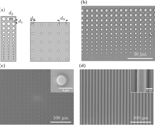
The master mold for stamp production was produced by photolithography. A 4@silicon wafer (Siltronix) was spincoated with the positive photoresist Microposit S1818 (Microresist Tech-nology) before exposure to UV light through a quartz mask (Computographics) for the desired pattern. The photoresist thickness was 2.3μm, resulting in stamp features of that hight. Three
different patterns where used (Fig 1). The first pattern consists of slits of width 5μm
inter-spaced by 5μm opaque lines (Fig 1d). The second pattern consists of 13 circular holes of
diam-eter increasing from 0.8μm to 4.4μm on an opaque background (Fig 1a, left side). This pattern
was produced in four versions, each characterized by a vertical separation distanced1of 3, 4, 6
or 8μm between the circular holes and a fixed horizontal distance,d2, between the center of
each hole of 7.4, 8.4, 10.4 or 12.4μm. The third pattern consists of circular holes with a
diame-ter,dh, of 3.5μm with a separation distance between the circular holes equal to either 10 or
15μm (Fig 1a, right side). After development, the wafer was covered by PDMS (Sylgard 184
Patterning of glass surfaces using
μ
CP and PDMS stamps
The surface patterning techniqueμCP was used to introduce circular spots or lines coated with
chosen chemicals introducing the surface properties needed in order to obtain bacterial arrays. The PDMS stamp was incubated with a drop of the selected chemical (10 to 30 minutes) fol-lowed by blow drying with nitrogen and placed pattern side down on the substrate to be pat-terned. A pressure was applied onto the PDMS stamp throughout the stamping period by placing a weight of 100 grams ontop of the stamps, in order to obtain good contact between the features of the stamp and the substrate.
The reproducibility of theμCP process was investigated by stamping cleaned glass cover
slips (borosilicate glass, VWR international) employing PDMS stamps incubated in a solution containing qdot 655 ITK amino (PEG) quantum dots (Life Technologies) diluted in MilliQ Fig 1. (a): The patterns on the photolithography masks used to produce PDMS stamps.The first pattern (left) consisted of 13 circular holes of diameter increasing from 0.8μm to 4.4μm on an opaque background. The mask contained four quadrants, each characterized by a vertical separation distanced1of 3, 4, 6 or 8μm between the circular holes and a fixed horizontal distanced2between the center of each hole of 7.4, 8.4, 10.4 or 12.4μm. The pattern on the
second photolithography mask (right) consisted of circular holes with a diameterdhof 3.5μm with a separation distanced3between the circular holes equal to either 10 or 15μm. (b), (c) and (d): SEM micrographs of gold coated PDMS stamps intended for patterning of glass surfaces byμCP. The stamps shown in
(b) and (c) are produced using the photolithography masks schematically illustrated in 1(a). The stamp depicted in (d) was obtained using a photolithography mask with slits of width 5μm interspaced by 5μm.
water to a concentration of 10 nM. The patterns where imaged with a Zeiss 510 Meta micro-scope with a 20x objective (NA = 0.5, liquid). The size of the introduced quantum dot coated areas was determined using the analyze particles function in ImageJ software, and the diameter was calculated based on these results.
For patterning of surfaces intendedused for preparation of bacterial microarrays, a Willco-dish kit (Willco Wells) was used. The Willco-dish facilitates covering the microarrays in liquid during investigation, and this kit allows for patterning of the glass bottom of the Willco-dish before assemblement of the dish. Prior to being patterned, the glass surfaces were cleaned by immer-sion in a 1:1 V/V solution of puriss grade hydrochloric acid (Sigma-Aldrich) and methanol (Sigma-Aldrich) for 20 minutes before rinsing in MilliQ water and drying by a stream of nitro-gen gas. To avoid bacterial adhesion the glass surfaces were passivated through coating with the chemicals BSA, PVA or PEG prior to patterning usingμCP. The coatings were introduced
using the following procedures: BSA (Sigma) was dissolved in phosphate buffered saline (PBS, Sigma) to a concentration of 1 mg/mL and added to the glass surface for incubation for 20 min-utes. After incubation the glass surface was rinsed in MilliQ water and dried by a stream of ni-trogen. Coating with PVA was obtained by dissolving 22 kDa poly(vinyl) alcohol (PVA) from BDH Chemicals to 1 wt % in MilliQ water and spincoating this onto on the glass surface before curing on a hotplate at 130°C for 30 minutes. PEGylation of the surfaces was acheived by im-mersion for 60 minutes in a solution containing poly-L-lysine (20 kDa) grafted with PEG(2 kDa) (in the further referred to as PLL-g- PEG) from Susos was dissolved in MilliQ water to a concentration of 1mg/mL. After incubation the excess liquid was removed and the glass was rinsed in PBS before rinsing in MilliQ water and dried by a stream of nitrogen gas.
In order to promote bacterial adhesion onto defined spots on the surface, the passivated sur-faces were patterned usingμCP with one of three chemicals, PD, PLL or PEI, all characterized
by their expected ability to promote bacterial adhesion. The chemicals were patterned using the following procedures: Dopamine hydrochloride (Sigma-Aldrich) was dissolved in TRIS buffer (Sigma-Aldrich, pH = 8.5) (final concentration equal to 1 mg/mL) in order to initiate the polymerisation into polydopamine. A drop of this solution was transferred to a PDMS stamp for incubation for 30 minutes. PLL: Poly-L-lysine (Mw 15.000–30.000, FITC Labeled, Sigma-Aldrich) was dissolved in MilliQ water to a consentration of 1mg/mL and incubated on a PDMS stamp for 10 minutes. PEI: poly(ethyleneimine) (Mw 750,000 by LS, 50 wt % in H2O,
Sigma-Aldrich) was dilluted in MilliQ water to a 1% wt solution before incubation on a PDMS stamp for 10 minutes. After incubation the stamps were dried with a stream of nitrogen and the stamps were placed pattern side down on the glass bottomslides of Willco-dishes. After pat-terning of the glass bottom slides, the Willco-dishes where assembled following the
manufacturers instructions.
Patterned surfaces with PD islands on PEGylated surfaces were imaged using Multimode V AFM (Digital Instruments/VEECO) equipped with J scanner operated in tapping mode under ambient conditions. Silicon nitride cantilevers PPP-NCH (Nanosensors, nominal resonant fre-quency 204–497 kHz and nominal spring constant 10–130 N/m) were used. Overlapping of trace and retrace signal was used as a prerequisite for adequate and high-quality
image acquisition.
Bacterial strain, plasmid, growth media, and DNA transformation
In this study thePseudomonas putidaKT2440 (TOL plasmid cured derivative [33]) was uti-lized.P. putidaKT2440 was grown in LB (10g/L tryptone; 5g/L yeast extract; 5g/L NaCl) sup-plemented with 50μg/mL kanamycin at 30°C over-night in shake flasks. The plasmid
Pm promoter. This plasmid harbors the positively regulated XylS/Pm positive regulator/pro-moter system which can be induced by the passively diffusing 3-methylbenzoic acid (MB) (Sigma-Aldrich), a mini-RK2 replicon for vegetative replication, and a kanamycin gene as anti-biotic resistance marker. Plasmid pSB-M1g was transferred intoP. putidaKT2440by electropo-ration [35].
Immobilization of bacteria onto
μ
CP patterned glass surfaces
In order to obtain bacterial microarrays, the chemically patterned glass bottomed Willco-dishes obtained as described above, were incubated for 5 minutes with the over night grownP. putidaKT2440 culture in LB medium. Once rinsed in distilled water in order to remove any unattached bacteria, LB was added to the dish to minimize the stress induced in the
attached bacteria.
The viability of attached bacteria was investigated using a live/dead assay (LIVE/DEAD Bac-Light bacterial viability kit from Life Technologies AS). The live/dead assay was added to bacte-rial microarrays in Willco-dishes immediately after bactebacte-rial attachment to the arrays. When using the live/dead assay, bacteria with intact cell membranes are expected to emit green fluo-rescent light when illuminated with the appropriate excitation light, and these bacteria were considered alive. Bacteria with damaged cell membrane emit read fluorescent light, as a nucleic acid stain can reach the bacterial DNA, and these bacteria were considered dead.
As a proof of concept, the immobilizedP. putidaKT2440 harbouring the plasmid pSB-M1g, while on a microscope, was induced with MB. This was achieved by changing the liquid cover-ing the bacteria from LB to LB containcover-ing 0.3 mM MB. The presence of the inducer initiates the expression of the GFP from the positively regulated XylS/Pm positive regulator/promoter system [34]. Upon induction the expression of GFP in the bacteria was followed using time laps imaging.
The bacterial arrays were inspected using a Leica SP5 confocal microscope.
Results and Discussion
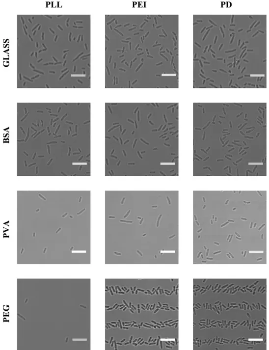
When aiming at controlling bacterial adhesion, optimization of surface chemistry is essential. In the present study, in addition to clean glass, three different anti-adhesion coatings where in-vestigated: BSA, PVA and PEG. These were investigated in combination with three chemicals commonly used to promote bacterial adhesion: PD, PLL and PEI. The twelve resulting combi-nations were all evaluated in order to identify the optimal combination for selective bacterial adhesion onto predefined surface areas. For these investigations PDMS stamps with lines of 5μm width where used (Fig 1d). After incubation with bacterial suspensions containing the
Fig 2. Images of glass surfaces and glass surfaces precoated with chemicals reducing bacterial adhesion, patterned with chemicals promoting bacterial adhesion, immersed in a solution containing bacteria and finally rinsed and covered with LB.Results obtained for the three chemicals potentially reducing bacterial adhesion (BSA, PVA or PEG) are shown. The substrates are patterned with one of three chemicals promoting bacterial adhesion (PLL, PEI or PD) usingμCP with a PDMS stamp with 5μm lines (Fig
1d) and immersed in a solution containing bacteria. All scalebars are 10μm. The combination of chemicals
investigated in each experiment is indicated on the figure. The surfaces were rinsed in MilliQ water after the incubation with bacteria (P. putidaKT2440) in order to remove weakly adhering bacteria. During imaging the surfaces were covered with LB in order to minimize stress induced in the attached bacteria. The images are obtained by using transmission light microscopy, and were captured on a Leica TCS SP5 with a
40 × objective (water, N.A. = 1.2).
used as an anti-adhesion coating in this study. PEG, on the other hand, efficiently prevent bac-terial adhesion to areas in between the patterned bacbac-terial adhering chemicals PEI and PD (Fig 2).
Of the three bacterial adhesion promoting chemicals tested, only PEI and PD produce well defined patterns of adhered bacteria on PEG films (Fig 2). The lack of patterns of adhered bac-teria on surfaces patterned with PLL is thought to be the result of the PLL dissolving in the liq-uid covering the patterned surfaces as the patterns of FITC-labeled PLL could not be observed using a fluorescence microscope. The bacterial arrays are intended to be used for study of immobilised bacteria covered in LB to minimize stress, and PLL was thus not included in the further studies. No observed difference in suitability between PEI and PD was observed on striped patterns. However, for patterns with smaller feature sizes, deposition of PD resulted in an improved tendency for immobilisation of bacteria relative to PEI (data not shown). Patterns of PD on PEGylated surfaces were therefore chosen for the further investigations.
Immobilization of single bacteria onto adhesive spots on a patterned surface does not only require optimization of the surface chemistry, pattern features like spot size and inter-spot dis-tance must also be optimized. To this end two different photolithography masks were designed and used to obtain PDMS stamps presenting pillars of varying diameter and separated by vary-ing inter pillar spacvary-ing (Fig 1). The design presented inFig 1aon the left side was inspired by a previously published design used for immobilizingE. coli[16] and consisted of 13 circular holes of increasing diameter on an opaque background. 11 out of the 13 circular features in the designed pattern on the first mask were successfully reproduced in the PDMS stamps (Fig 1b). The results obtained based on this mask, guided the design of a second mask. The pattern on the second mask consisted of circular holes with a diameter of 3.5μm with a separation
dis-tanced3between the circular holes equal to either 10 or 15μm (Fig 1a, right side). This pattern
was successfully reproduced in the PDMS stamps (Fig 1c).
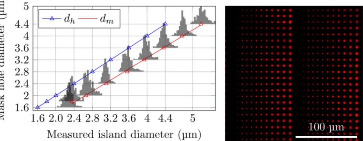
The PDMS stamps were used to deposit chemicals on glass surfaces. In order to evaluate the the successfulness of the deposition over relatively large areas (up to 9 mm2), surfaces patterned using PDMS stamps coated with quantum dots were used (Fig 3, right). The patterns obtained
Fig 3. Right: Fluorescence micrograph of quantum dots deposited on a cleaned glass coverslip usingμCP with PDMS stamps.Such images were used to study the reproducibility of the obtained patterns. Left: Distributions of observed diameters of the nine largest stamped islands compared to the mask hole diameters (blue triangles and corresponding blue linear regression line). Island diameters calculated from the area of each island as determined based on the ImageJ software and fluorescence micrographs of quantum dots. The red triangle indicate the most probable island diameterdmand the red line is the linear regression obtained based ondmobtained for the eight largest stamped islands.
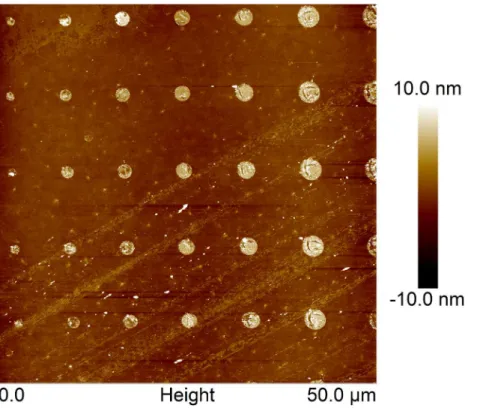
were reproducible over large areas (data not shown). Image analysis revealed a narrow distribu-tion of island sizes (Fig 3, left). The sizes and size distributions of the islands of deposited quan-tum dots were found to be independent of the precise area of the stamp used to produce the printed features. The variation observed between different stamps produced using the same photolithography mask and identical parameter settings during stamp production was also in-significant. Furthermore, the size of the islands were compared with the size of the holes in the photolithography mask used when preparing the PDMS stamp. The most probable measured diameter (Fig 3, red triangles), defined as the peaks of the histograms presented inFig 3were compared to the designed diameter on the photolithography mask (Fig 3, blue triangles). The deposited islands were found to be larger than the holes in the photolithography mask (Fig 3, left). This is a systematic effect caused by the photolithography process and it can be tuned by adjusting the exposure dose. The patterns of stamped PD on PEGylated glass matches both the stamp features and the patterns of deposited quantum dots, as confirmed by AFM imaging of an array of PD on PEGylated glass (Fig 4).
Having identified PD and PEG as an effective combination of bacterial promoting and pre-venting chemicals, PD wereμCP onto PEG coated surfaces using PDMS stamps with pilars of
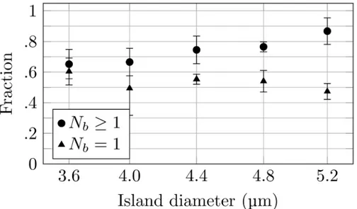
increasing diameter (Fig 1b). The obtained patterned surfaces gave bacterial arrays which suc-cessfully reproduced the pattern on the stamp. The preparation of single bacterial arrays re-quires that the bacterial adhering spots have a size that is sufficiently large to allow stable attachment of one bacterium, yet sufficiently small to minimize the probability for adherence of multiple bacteria. The number of bacteria immobilized on each spot of the arrays was deter-mined by manual inspection of dry arrays for increased contrast in the images and revealed a correlation between the spot size and the number of bacteria adhering to the spot.Fig 5
Fig 4. Tapping mode AFM height topographs of PD printed on PEGylated glass.
displays this analysis for deposited spots with a measured size in the range 3.6 to 5.2μm.
Guid-ed by these observations and the documentGuid-ed relationship between mask hole diameter and measured island size (Fig 3) a photolithography mask with holes of a size equal to 3.5μm was
chosen for the further studies. This is a compromise between a high probability of obtaining full coverage of the array, which is obtained for spot sizes large enough to capture several bacte-ria, and obtaining single bacterial arrays, which requires a spot size so small that a relatively large fraction of the spots remains unoccupied after incubation. The 3.5μm spot size should
give a large degree of coverage, while still keeping the average number of bacteria on each spot small enough for data analysis to recognize single bacteria. In addition to the spot sizes, the inter-spot distances were also evaluated. The arrays obtained revealed that even for separation distances equal to 8μm, i.e. the largest distance included in the photolithography mask (Fig
1a), a fraction of the spots were bridged by the bacteria. This was especially apparent for the larger spots. Based on these findings, the pattern for a second photolithography mask was de-signed. The pattern had the following characteristics: holes of 3.5μm diameter separated by
ei-ther 10 or 15μm (Fig 1c).
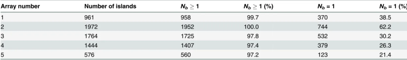
PD coated PDMS stamps prepared using the second photolithography mask allowed prepa-ration of regular bacterial arrays on PEGylated glass surfaces. The number of bacteria immobi-lized on each adhesive island on the arrays were determined (Table 1). The inspection of five parallelμCP PD arrays on PEG coated surfaces revealed that the fraction of spots occupied by
one or more bacteria was between 97 and 100% whereas the fraction of spots with single bacte-rial occupancy varied from 21.4 to 62.2% (Table 1). The amount of bacterial adhesion to the PEG coated areas was insignificant, as was the fraction of spots bridged by bacteria. The pro-posed method for the making of bacterial microarrays has several advantages compared to pre-viously proposed methods, in the sense that it does not require modification of the bacteria and the surface modification procedure is fast and does not involve harmful chemicals. The size of the islands obtained in the current study, being approximately 10μm2, is also significantly
Fig 5. Quantitative analysis of the number ofP. putidaKT2440 adhering onto each of the PD islands on the obtained microarrays.For each PD island size, both the fraction of the spots having one or more bacteria attached (Nb1) as well as the fraction of the spots with only one bacterium attached (Nb= 1), were determined.
smaller than the sizes used in other recently published procedures [23]. This small island size explains the low number of bacteria attached to each functionalized surface spot.
The observed variation in the fraction of spots displaying single attached bacteria (Table 1) might be due to variations in the feature sizes in the PDMS stamps. A small increase in the di-ameter of the PD islands will lead to an increased probability of adherence of multiple bacteria to each spot. The exposure dose during the photolithography process for making the mold, the amount of PDMS shrinkage during curing and the pressure applied during PDMS surface stamping are parameters that might influence the feature size of the stamped pattern and fur-ther optimization of these steps are fur-therefore likely to furfur-ther increase the probability for single bacterial occupancy. In addition, relatively large separation distance between the pillars of the PDMS stamp used may complicate the reproducibility of the stamping process.
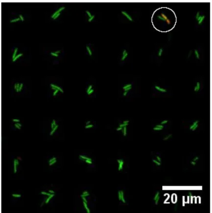
The viability of bacteria attached to PD patterns on PEGylated surfaces was investigated using a live/dead viability kit. Bacteria with intact cell membranes are stained green and consid-ered alive, whereas bacteria with damaged cell membrane are stained read as a nucleic acid stain can reach the bacteria DNA and are thereby considered dead. The live/dead assays re-vealed that the majority of the bacteria were viable while being immobilized onto patterned substrates (Fig 6). Out of a total of 3101 attached bacteria, 99.1% where stained green.
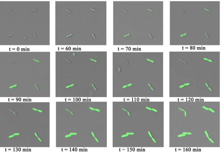
Arrays ofP. putidaKT2440 were exposed to MB, leading to expression of GFP from the positively regulated XylS/Pm system. Upon induction the expression was followed by micros-copy. The introduction of MB was achieved by exchanging the medium covering the bacteria with LB containing 0.3 mM of MB. Bright field and fluorescent images of the bacteria were cap-tured every ten minutes after adding the inducer (Fig 7). A green fluorescent signal was ob-served from the bacteria within an hour after adding the inducer. The obob-served fluorescence intensity increased over time. This time laps imaging also revealed that the bacteria were divid-ing while bedivid-ing immobilized on the array (Fig 7). These observations, along with the live/dead assay, show that the bacteria not only survive the immobilization process, but also that any stress induced by the immobilization does not significantly affect their growth. A variation in fluorescence intensity was observed between individual bacteria, and time from introduction of the inducer to the expression of GFP also varied between bacteria. This is an example of ob-served stochastic gene expression that leads to population heterogeneity. Such heterogeneity is masked in studies performed using methods that provide insight into average properties of bacterial populations.
Table 1. Quantitative analysis of the number of bacteria immobilized onto each adhesive PD island of bacterial microarrays prepared on glass sur-faces coated with PEG.
Array number Number of islands Nb1 Nb1 (%) Nb= 1 Nb= 1 (%)
1 961 958 99.7 370 38.5
2 1972 1952 100.0 744 62.2
3 1764 1725 97.8 532 30.2
4 1444 1407 97.4 379 26.3
5 576 560 97.2 123 21.4
The arrays were prepared usingμCP with PDMS stamps obtained using a photolithography mask withdhequal to 3.5μm (Fig 1d).Nb1: one or more bacteria per island.Nb= 1: one bacterium per island.
Conclusion
Several conditions must be satisfied for a bacterial array to be an effective tool to study bacterial populations. The time and cost of making the array should be minimized, and the techniques used should preferably be applicable in a standard molecular biology laboratory. The chosen substrate should be transparent to allow for simple detection using optical imaging techniques while the bacteria are covered with liquid medium. In addition, the patterning technique cho-sen should not adversely affect the immobilized bacteria and the immobilization method should not require surface modification of the bacteria.
The present paper proposes a procedure for preparing microarrays of live bacteria that meets such criteria. In the proposed procedure, the substrates are patterned usingμCP.
Differ-ent combinations of chemicals for surface functionalization were evaluated. More precisely, the commonly used passivating chemicals PEG, PVA and BSA were tested in combination with the bacterial adhering chemicals PEI, PLL and PD. PEG-coated glass slides with printed PD patterns were shown to be effective at selectively immobilizing bacteria onto predefined areas on the surface. The design features of the PDMS stamps, including the diameter of each pillar on the stamp and the distance separating them, allowed the preparation of arrays of the bacte-riaP. putidaKT2440 displaying high regularity, as reflected by the fraction of spots occupied by one or a few bacteria ranging from 97.2 to 100%. The proposed method for the preparation Fig 6. Fluorescence image reflecting the viability ofP. putidaKT2440 immobilized on arrays of PD islands on a PEGylated glass surface.Live bacteria are stained green, dead bacteria are stained red and the image is an overlay of both green and red fluorescent images. A single dead (red) bacteria is observed (white circle). The image is obtained for arrays covered in liquid using a Leica SP5 with a 10 × objective (N.A. = 0.4).
of bacterial arrays can be applied to any microorganisms for which a surface coating is identi-fied that gives a high probability for attachment of the microorganism to the islands as well as a surface coating that gives a low probability for attachment to areas outside the islands. Howev-er, in the current study optimalisation of the surface coating for bacteria other thanP. putida KT2440 was not performed. Furthermore, a live/dead assay revealed that 99.1% of the bacteria were alive after immobilization onto the array and bacteria attached to these arrays both divide and express GFP upon induction. The presently developed microarray with a large selectivity for single bacterial adherence to polydopamineμCP domains, and maintaining bacterial
viabil-ity can be expected to support studies addressing bacterial heterogeneviabil-ity.
Acknowledgments
The Research Council of Norway is acknowledged for the support to the Norwegian Micro-and Nano-Fabrication Facility, NorFab (197411/V30). We are grateful to Katarzyna Maria Psonka-Antonczyk for assistance with AFM imaging.
Fig 7. Time laps images ofP. putidaKT2440 immobilized on PD islands printed on a PEGylated glass surface.The images are obtained for surfaces covered with medium between 0 and 160 minutes after changing the medium from LB to LB containing the inducer MB. MB induces the expression of GFP in the bacteria. The images are overlays of transmission light images and fluorescence images—both obtained using a Leica SP5 with a 20 × objective (N.A. =
0.7).
Author Contributions
Conceived and designed the experiments: NBA VO MS. Performed the experiments: NBA VO. Analyzed the data: NBA VO. Contributed reagents/materials/analysis tools: RL. Wrote the paper: NBA MS.
References
1. de Souza N (2011) Single-cell methods. Nature Methods 9: 35–35. doi:10.1038/nmeth.1819
2. Lidstrom ME, Konopka MC (2010) The role of physiological heterogeneity in microbial population be-havior. Nature chemical biology 6: 705–12. doi:10.1038/nchembio.436PMID:20852608
3. van Oijen AM (2008) Cutting the forest to see a single tree? Nature chemical biology 4: 440–3. doi:10.
1038/nchembio0808-440PMID:18641617
4. van Oijen AM (2011) Single-molecule approaches to characterizing kinetics of biomolecular interac-tions. Current opinion in biotechnology 22: 75–80. doi:10.1016/j.copbio.2010.10.002PMID:21036593
5. Locke JCW, Elowitz MB (2009) Using movies to analyse gene circuit dynamics in single cells. Nature reviews Microbiology 7: 383–92. doi:10.1038/nrmicro2056PMID:19369953
6. Mossoba MM, Al-Khaldi SF, Kirkwood J, Fry FS, Sedman J, Ismail AA (2005) Printing microarrays of bacteria for identification by infrared microspectroscopy. Vibrational Spectroscopy 38: 229–235. doi:
10.1016/j.vibspec.2005.04.006
7. Kim JH, Lee DY, Hwang J, Jung HI (2009) Direct pattern formation of bacterial cells using micro-drop-lets generated by electrohydrodynamic forces. Microfluidics and Nanofluidics 7: 829–839. doi:10.
1007/s10404-009-0441-6
8. Kim J, Shin YH, Yun SH, Choi DS, Nam JH, Ryong S, et al. (2012) Direct-write patterning of bacterial cells by dip-pen nanolithography. Journal of the American Chemical Society 134: 16500–3. doi:10.
1021/ja3073808PMID:22992015
9. Xu L, Robert L, Ouyang Q, Taddei F, Chen Y, Lindner AB, et al. (2007) Microcontact printing of living bacteria arrays with cellular resolution. Nano letters 7: 2068–72. doi:10.1021/nl070983zPMID:
17585831
10. Weibel DB, Lee A, Mayer M, Brady SF, Bruzewicz D, Yang J, et al. (2005) Bacterial printing press that regenerates its ink: contact-printing bacteria using hydrogel stamps. Langmuir 21: 6436–42. doi:10.
1021/la047173cPMID:15982051
11. Kumar A, Biebuyck HA, Whitesides GM (1994) Patterning Self-Assembled Monolayers: Applications in Materials Science. Langmuir 10: 1498–1511. doi:10.1021/la00017a030
12. Xia Y, Whitesides GM (1998) SOFT LITHOGRAPHY. Annual Review of Materials Science 28: 153–
184. doi:10.1146/annurev.matsci.28.1.153
13. Whitesides GM, Ostuni E, Takayama S, Jiang X, Ingber DE (2001) Soft lithography in biology and bio-chemistry. Annual review of biomedical engineering 3: 335–73. doi:10.1146/annurev.bioeng.3.1.335
PMID:11447067
14. Hochbaum AI, Aizenberg J (2010) Bacteria pattern spontaneously on periodic nanostructure arrays. Nano letters 10: 3717–21. doi:10.1021/nl102290kPMID:20687595
15. Rozhok S, Fan Z, Nyamjav D, Liu C, Mirkin CA, Holz RC (2006) Attachment of motile bacterial cells to prealigned holed microarrays. Langmuir 22: 11251–4. doi:10.1021/la0609726PMID:17154612
16. Rozhok S, Shen CKF, Littler PLH, Fan Z, Liu C, Mirkin CA, et al. (2005) Methods for fabricating microar-rays of motile bacteria. Small 1: 445–51. doi:10.1002/smll.200400072PMID:17193470
17. Cerf A, Cau JC, Vieu C (2008) Controlled assembly of bacteria on chemical patterns using soft lithogra-phy. Colloids and Surfaces B: Biointerfaces 65: 285–291. doi:10.1016/j.colsurfb.2008.04.016PMID:
18556179
18. Cerf A, Cau JC, Vieu C, Dague E (2009)Nanomechanical properties of dead or alive single-patterned bacteria. Langmuir 10: 5731–6. doi:10.1021/la9004642
19. Wong I, Ding X, Wu C, Ho CM (2012) Accurate and Effective Live Bacteria Microarray Patterning on Thick Polycationic Polymer Layer Co-Patterned with HMDS. RSC advances 2: 7673–7676. doi:10.
1039/C2RA20938APMID:23418622
20. Colville K, Tompkins N, Rutenberg AD, Jericho MH (2010) Effects of poly(L-lysine) substrates on at-tached Escherichia coli bacteria. Langmuir 26: 2639–44. doi:10.1021/la902826nPMID:19761262
22. Lee H, Dellatore SM, Miller WM, Messersmith PB (2007) Mussel-inspired surface chemistry for multi-functional coatings. Science 318: 426–30. doi:10.1126/science.1147241PMID:17947576
23. Sun K, Xie Y, Ye D, Zhao Y, Cui Y, Long F, et al. (2012) Mussel-inspired anchoring for patterning cells using polydopamine. Langmuir 28: 2131–6. doi:10.1021/la2041967PMID:22085048
24. Beckwith KM, Sikorski P (2013) Patterned cell arrays and patterned co-cultures on polydopamine-mod-ified poly(vinyl alcohol) hydrogels. Biofabrication 5: 045009. doi:10.1088/1758-5082/5/4/045009
PMID:24280598
25. Howell SW, Inerowicz HD, Regnier FE, Reifenberger R (2003) Patterned Protein Microarrays for Bacte-rial Detection. Langmuir 19: 436–439. doi:10.1021/la026365+
26. Oh YJ, Jo W, Lim J, Park S, Kim YS, Kim Y (2008) Micropatterning of bacteria on two-dimensional lat-tice protein surface observed by atomic force microscopy. Ultramicroscopy 108: 1124–1127. doi:10.
1016/j.ultramic.2008.04.055PMID:18571856
27. Kingshott P, Griesser HJ (1999) Surfaces that resist bioadhesion. Current Opinion in Solid State and Materials Science 4: 403–412. doi:10.1016/S1359-0286(99)00018-2
28. Caro A, Humblot V, Méthivier C, Minier M, Salmain M, Pradier CM (2009) Grafting of lysozyme and/or poly(ethylene glycol) to prevent biofilm growth on stainless steel surfaces. The journal of physical chemistry B 113: 2101–9. doi:10.1021/jp805284sPMID:19166331
29. Kingshott P, Wei J, Bagge-Ravn D, Gadegaard N, Gram L (2003) Covalent Attachment of Poly(ethyl-ene glycol) to Surfaces, Critical for Reducing Bacterial Adhesion. Langmuir 19: 6912–6921. doi:10.
1021/la034032m
30. Barrett DA, Hartshorne MS, Hussain MA, Shaw PN, Davies MC (2001) Resistance to Nonspecific Pro-tein Adsorption by Poly(vinyl alcohol) Thin Films Adsorbed to a Poly(styrene) Support Matrix Studied Using Surface Plasmon Resonance. Analytical Chemistry 73: 5232–5239. doi:10.1021/ac010368u
PMID:11721924
31. Peterbauer T, Heitz J, Olbrich M, Hering S (2006) Simple and versatile methods for the fabrication of ar-rays of live mammalian cells. Lab on a chip 6: 857–63. doi:10.1039/b601803cPMID:16804589
32. Lieder S, Nikel PI, de Lorenzo V, Takors R (2015) Genome reduction boosts heterologous gene ex-pression in Pseudomonas putida. Microbial cell factories 14: 23 doi:10.1186/s12934-015-0207-7
PMID:25890048
33. Ramos-González MI, Campos MJ, Ramos JL (2005) Analysis of Pseudomonas putida KT2440 gene expression in the maize rhizosphere: in vivo [corrected] expression technology capture and identifica-tion of root-activated promoters. Journal of bacteriology 187: 4033–41. doi:
10.1128/JB.187.12.4033-4041.2005PMID:15937166
34. Balzer S, Kucharova V, Megerle J, Lale R, Brautaset T, Valla S (2013) A comparative analysis of the properties of regulated promoter systems commonly used for recombinant gene expression in Escheri-chia coli. Microbial cell factories 12: 26 doi:10.1186/1475-2859-12-26PMID:23506076
35. Choi KH, Kumar A, Schweizer HP (2006) A 10-min method for preparation of highly electrocompetent Pseudomonas aeruginosa cells: Application for DNA fragment transfer between chromosomes and plasmid transformation. Journal of Microbiological Methods 64: 391–397 doi:10.1016/j.mimet.2005.