Vol.55, n. 4: pp.569-576, July-August 2012
ISSN 1516-8913 Printed in Brazil BRAZILIAN ARCHIVES OF
BIOLOGY AND TECHNOLOGY
A N I N T E R N A T I O N A L J O U R N A L
Influence of Growth Media and Temperature on Bacterial
Adhesion to Polystyrene Surfaces
Ana Eliza Zeraik*and Marcia Nitschke
Instituto de Química de São Carlos; Universidade de São Paulo; Av. Trabalhador São Carlense, 400; C.P.: 780; 13560-970; São Carlos - SP - Brasil
ABSTRACT
Bacterial adhesion to inert surfaces is a complex process influenced by environmental conditions. In this work, the influence of growth medium and temperature on the adhesion of Pseudomonas aeruginosa, Serratia marcescens, Staphylococcus aureus, Micrococcus luteus and Listeria monocytogenesto polystyrene surfaces was studied. Most bacteria demonstrated the highest adhesion when cultured in TSYEA, except S. marcescens, which showed to be positively influenced by the pigment production, favored in poor nutrient media (lactose and peptone agar). P. aeruginosa adhesion to polystyrene increased at low temperatures whatever the medium used. The culture medium influenced the surface properties of the bacteria as assessed by the MATS test.
Key words: Bacterial adhesion, biofilms, hydrophobicity, polystyrene.
INTRODUCTION
The high biofilm resistance to antibiotics and disinfectants and consequently the problems they
cause in food processing and medical
environments, turns biofilms the focus of many studies worldwide (Bower et al. 1999). Biofilm formation begins with the microbial adhesion to a substratum. Thus, control of the adhesive process is one of the main goals to combat the biofilm formation. To inhibit the bacterial adhesion, it is important to understand the facts that influence and the forces involved in this process (Goulter et al. 2009).
Studies have shown that the bacteria can adhere and form the biofilm on different solid materials, such as metal, glass, rubber and plastic (Hood and Zottola 1997). Plastic materials are widely used in the food industry (cutting surfaces, packages,
tanks, pipes) and medical devices (prosthesis, catheters) where biofilms are undesirable.
Bacterial attachment to inert surfaces is influenced by the properties of both, substratum and bacterial cell, such as charge, hydrophobicity, surface roughness, the presence of fimbriae, flagella and production of exopolysaccharides (EPS) (Donlan 2002). The properties of the bacterial cells are
affected by the environmental conditions
(temperature, pH or composition of the culture medium); hence, alterations in these conditions can affect the bacterial adhesion (Faille et al. 2002; Bonaventura et al. 2008).
The adhesion process of bacteria to the surfaces include interactions, such as van der Waals, Lewis
acid-base, hydrophobic and electrostatic
Carballo 2000) and that the hydrophobic effect may be the primary driving force for the adhesion of most pathogens (Duncan-Hewitt 1990). However, a great diversity of results is found in the literature; in some cases, there is a correlation between the hydrophobicity and adhesion (Zita and Hermansson 1997; Marin et al. 1997) and in others, this correlation is not found (Chae et al. 2006; Li and McLandsborough 1999). Therefore, a study involving the bacterial adhesion in a variety of growth conditions and temperatures on a hydrophobic substratum is relevant to the actual context.
The aim of this work was to evaluate the influence of different culture media on the hydrophobicity
and adhesion of Listeria monocytogenes,
Staphylococcus aureus, Pseudomonas aeruginosa,
Micrococcus luteus and Serratia marcescens on
the polystyrene surfaces, as well as the influence of temperature shifts (25 ºC and 4 ºC) on bacterial adhesion.
MATERIALS AND METHODS
Bacterial strains and growth conditions
Listeria monocytogenes ATCC 19112,
Staphylococcus aureus ATCC 25923,
Pseudomonas aeruginosa ATCC 27853,
Micrococcus luteus ATCC 4698 and Serratia
marcescens ATCC 8100 obtained from Fundação
Oswaldo Cruz Culture Collection were stored at
-20ºC on tryptic soy broth supplemented with 20% glycerol (v/v). The cells were grown on TSYEA (trypticase soy agar 40 g/L and yeast extract 6 g/L), lactose agar (lactose 5 g/L, peptone 5 g/L, beef extract 3 g/L and agar 20 g/L), and peptone agar (peptone 10 g/L, NaCl 5 g/L, Na2HPO4 9 g/L, KH2PO4 1.5 g/L and agar 20 g/L).
The strains were cultured in each medium in a slant at 35 ºC for 24 h and then transferred to agar plates and incubated again at 35 ºC for 24 h. The cells were scraped from agar and suspended in
NaCl 0.15 mol L-1 solution to reach a
concentration of approximately 1x10⁹-1x10¹⁰ CFU mL-1 (cell number was determined by the drop method developed by Miles and Misra 1938). This bacterial cell suspension, after growing on different media, was utilized for further adhesion assays.
Bacterial adhesion assay
The adhesion tests were performed by dispensing 200 µL of bacterial suspensions, prepared as previously described, in 96 well polystyrene microtiter plate (02623-Kartell, Italy). The time of contact for the adhesion of cells to polystyrene was 4 h, and each test was conducted under different temperatures: 35 ºC (no temperature shift relating to growth temperature in agar medium), 25 ºC and 4 ºC during the 4 h of adhesion assay. The quantification of bacterial adhesion was performed by the crystal violet staining technique according to Rodrigues et al. (2006). The unattached cells were removed by washing the wells three times with water. The adherent microorganisms were fixed with 200 µL of methanol for 15 min. The wells were then stained for 15 min with 200 µL crystal violet (1% w/v aqueous solution), rinsed under the running tap water and left to dry. The bound dye was re-solubilized with 200 µL of glacial acetic acid (33%, v/v) and the optical density of each well was measured by an automated plate reader (Thermoplate) at 630 nm.
Physicochemical characterization of cell
surfaces
The MATS test (microbial adhesion to solvents), developed by Bellon-Fontaine et al. (1996), was performed to evaluate the Lewis acid-base properties and the hydrophilic/ hydrophobic nature of bacterial surfaces under different nutritional conditions (growth media). The pairs of solvents used were: Chloroform (an acidic solvent) and hexadecane (apolar), diethyl ether (a basic solvent) and hexane (apolar).
Experimentally, 2.4 mL of a bacterial suspension (in NaCl 0.15 mol L-1) containing approximately 108 CFU mL-1 and 0.4 mL of the solvent under investigation was shaken vigorously in vortex for 2 min, forming an emulsion.
The mixture was allowed to stand for 15 min to ensure the complete separation of the two phases and the absorbance of the aqueous phase was measured at 400 nm. The percentage of bound cells to each solvent was calculated by the equation: % Adh = (1 – A/A0) x 100, where A0 was the absorbance of the bacterial suspension before mixing and A was the absorbance after
Statistical analysis
To evaluate if the changes in the adhesive behavior of the bacteria were significant, the analysis of variance (ANOVA) was performed, with a confident level of 95% using Origin Software 7.5 (Origin Lab Coorporation). The results presented were calculated using data from at least three independent experiments and eight replicates each.
RESULTS
A preliminary adhesive assay was performed to evaluate if the bacterial strains were able to adhere to the polystyrene surface, as well as the time necessary to promote the adhesion. The test was performed using TSYEA medium (rich in nutrients). Figure 1 displays the adhesive behavior, showing that all the strains were able to adhere to
the polystyrene surface under the tested
conditions. From this experiment, the time of 4 h was selected for the futures experiments, since at this period, all the selected bacteria showed a good adherence to the plastic surface.
Adhesion assay under different growth media and temperature shifts
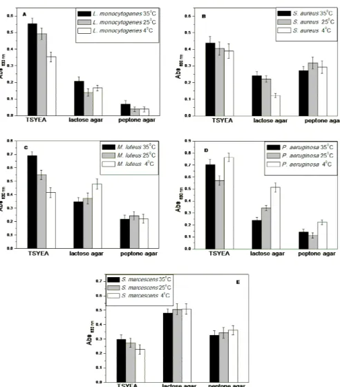
The adhesion assays of the five bacteria under different conditions are displayed in Figure 2. L.
monocytogenes showed the highest adherence when cultured in TSYEA. When peptone agar was the culture medium, L. monocytogenes presented the lowest adhesion values; these results were in agreement with the data of Dubravka et al. (2007) and Hood and Zotolla (1997) that showed that L. monocytogenes cells were better biofilm producers in rich nutrient media, whereas the decrease in concentration of nutritive compounds reduced their growth.
For S. aureus, M. luteus and P. aeruginosa, the
adhesion was also higher in TSYEA, the richest medium studied. The growth medium that resulted
the lowest adhesion of M. luteus and P.
aeruginosa was peptone agar while for S. aureus it
was the lactose agar. On the contrary, S.
marcescens presented lower attachment to polystyrene surface when grown in TSYEA and higher in lactose agar.
Regarding the effect of temperature shifts, a pattern behavior was observed only in TSYEA, where the adhesion decreased with the decrease of temperature; adhesion was higher at 35 ºC and lower at 4 ºC for most of the bacterial strains,
except for P. aeruginosa, that presented an
opposite behavior, showing higher adhesion at 4 ºC in all media studied. This temperature shifts could induce a stress in the strains that could affect the adhesion.
Figure 2 - Adhesion assays of bacterial suspensions of A) Listeria monocytogenes B)
Staphylococcus aureus C) Pseudomonas aeruginosa D) Micrococcus luteus and E)
Serratia marcescens after cultivation on different growth media and adhesion temperatures.
Physicochemical characterization of cell
surfaces
The affinity of the bacteria studied for the solvents was performed to evaluate the surface properties
of each strain when grown in the different nutritive media. The data are shown in Tables 1, 2 and 3 for the bacteria cultured in TSYEA, lactose agar and peptone agar, respectively.
Table 1 - MATS test to bacterial cells cultured in TSYEA.
% Adhesion
Bacteria Chloroform Hexadecane Diethyl ether Hexane
L. monocytogenes 83.75 ± 1.67a 76.21 ± 4.82b 19.25 ± 1.50c 67.26 ± 2.78d
S. aureus 96.98 ± 0.87a 94.94 ± 1.65b 44.46 ± 6.30c 94.58 ± 3.53b
M. luteus 91.87 ± 0.79a 89.69 ± 2.00b 47.79 ± 6.83c 74.20 ± 3.2d
P. aeruginosa 16.54 ± 3.37a 4.12 ± 1.48b 27.44 ± 1.89c 2.92 ± 1.08d
Table 2 - MATS test to bacterial cells cultured in lactose agar.
% Adhesion
Bacteria Chloroform Hexadecane Diethyl ether Hexane
L. monocytogenes 88.18 ± 1.73a,c 81.57 ± 5.13a 56.02 ± 4.07b 75.06 ± 5.75c
S.aureus 99.41 ± 0.16a 97.77 ± 0.79a 23.98 ± 3.86b 93.33 ± 2.26c
M. luteus 92.00 ± 1.40a 88.28 ± 2.63a 63.42 ± 2.19b 81.73 ± 4.41c
P. aeruginosa 41.08 ± 4.52a 18.52 ± 3.24b 39.04 ± 3.90a 15.00 ± 1.31b
S. marcescens 69.54 ± 4.58a 76.05 ± 3.95b 96.42 ± 0.98c 56.91 ± 4.65d (a) - (d) Within each line, values with the same letters are not significantly different (P < 0.05).
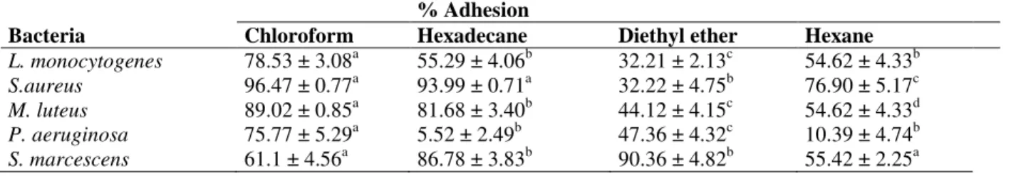
Table 3 - MATS test to bacterial cells cultured in peptone agar
% Adhesion
Bacteria Chloroform Hexadecane Diethyl ether Hexane
L. monocytogenes 78.53 ± 3.08a 55.29 ± 4.06b 32.21 ± 2.13c 54.62 ± 4.33b
S.aureus 96.47 ± 0.77a 93.99 ± 0.71a 32.22 ± 4.75b 76.90 ± 5.17c
M. luteus 89.02 ± 0.85a 81.68 ± 3.40b 44.12 ± 4.15c 54.62 ± 4.33d
P. aeruginosa 75.77 ± 5.29a 5.52 ± 2.49b 47.36 ± 4.32c 10.39 ± 4.74b
S. marcescens 61.1 ± 4.56a 86.78 ± 3.83b 90.36 ± 4.82b 55.42 ± 2.25a (a) - (d) Within each line, values with the same letters are not significantly different (P < 0.05).
The higher affinity to chloroform when compared to hexadecane was an indicative of the predominance of basic properties on the cell surface, while higher adhesion to the basic solvent (diethyl ether) compared to hexane indicated that the cell surface presented more acidic properties. The percentage of bound cells to hexadecane was used to evaluate the hydrophobicity. According to Chae et al. (2006), cells could be strongly hydrophobic, when the bound cells to hexadecane were higher than 55%, moderately hydrophobic (30-54%), moderately hydrophilic (10-29%) and strongly hydrophilic (<10%).
L. monocytogenes showed the predominance of basic properties in all the growth media. When the bacterium was cultivated in peptone medium, the difference between the cells bound to chloroform and hexadecane were not statistically different.
This was probably due to the strong
hydrophobicity of this strain. However, it did not meant a prevalence of acidic properties, as the adhesion in diethyl ether was lower when compared to hexane. Although considered strongly hydrophobic in all the conditions, in peptone agar, the affinity for hexadecane was lower, and when
grown in this medium, L. monocytogenes also
showed the lowest adhesion to polystyrene surfaces (Fig. 2 A).
S. aureus presented similar characteristics under different nutritive media, prevalence of basic properties, with a percentage of bound cells to
hydrophobic character once the adhesion to hexadecane was higher than 93%.
M. luteus also had a similar behavior in all the growth media studied, a high affinity for the chloroform, suggesting the basic character of the cell surfaces, and a strongly hydrophobicity, which was slightly lower in peptone agar medium that provided the lowest adhesion to polystyrene. S. marcescens cultured in TSYEA showed similar basic and acid properties and a moderately hydrophobic nature. In lactose and peptone agar, the cell surfaces were predominantly acidic and strongly hydrophobic which was in agreement with the higher adhesion on polystyrene observed in these media.
P. aeruginosa adhered preferentially to chloroform and diethyl ether when compared to the apolar solvents, indicating the predominance of basic and acidic properties in all the media tested. However, a higher basic character was observed in peptone agar (75% affinity to chloroform). Although the cell surface was considered strongly hydrophilic in TSYEA and peptone agar and moderately hydrophilic in lactose agar, P. aeruginosa was able to adhere to the hydrophobic polystyrene surface.
DISCUSSION
psychrotrophic bacterium (L. monocytogenes), a Gram positive and thermotoletant bacterium (M. luteus), a Gram positive and mesophilic bacterium (S. aureus) and two Gram negative and mesophilic strains (P. aeruginosa and S. marcescens). All the microorganisms studied were in the stationary phase of growth, thus it was possible to compare the behavior of these strains and gain some insights into the adaptive response under the changes caused by the temperature and growth media.
In order to find the correlation between the availability of nutrients in the growth medium and the adhesive capacity of the bacterial strains, several studies have been made. According to Stepanovic et al. (2004), the nutrient composition of the medium influenced the quantity of produced biofilm in different ways; for Salmonella spp. the biofilm production was enhanced in poor nutrient
medium while L. monocytogenes produced more
biofilm in rich nutrient medium. Hood and Zotolla (1997) studied five bacterial strains and concluded that the medium which produced the highest level of adherent cells was different for each microorganism. It was also shown that the starvation could enhance the adhesion of some microorganisms, while others presented high adhesion rates under optimal growth conditions (van Loosdrecht et al. 1987).
In this study, most bacteria had the adhesion enhanced when cultured in TSYEA, the most
nutritive medium, with the exception of
S. marcescens. The higher hydrophobicity of this strain in lactose and peptone agar (as seen in MATS test) could be associated to the production of a red pigment, the prodigiosin.
The prodigiosin production by S. marcescens cells was shown to be influenced by the growth media. Song et al. (2006) reported that the pigment production could be associated with the activity of casein hydrolases in the cell, once prodigiosin was formed in casein-enriched medium, while when casein was replaced with casitone (pancreatic digest of casein – present in TSYEA), the pigment was hardly produced.
In this study, a significant increase in cells
pigmentation was observed when S. marcescens
was cultured in lactose and peptone agar. Similar results were observed by Solé et al. (1997), which showed that when S. marcescens was cultured in trypticase soy agar no pigment was formed. The present results demonstrated that the pigment production increased the cell hydrophobicity.
Hydrophobic interactions between the cell and the surface could be the reason for increase in the adhesion to polystyrene observed when the bacteria was cultivated in peptone and lactose agar.
Regarding the influence of temperature, only few studies evaluated the changes in bacterial cells caused by the temperature shifts, most studies focus the growth temperature (Hemery et al. 2007; Dubravka et al. 2007). Since differences in the adhesive behavior of bacterial strains submitted to temperature changes were observed, we concluded that the period of 4 h was sufficient to induce alterations in the bacterial surface properties.
Phan-Thanh and Gormon (1995) exposed L.
monocytogenes to heat and cold shock between 49 and 4 ºC during 15 min (for high temperature) to 2-3 h at 4 ºC and showed that around half the number of proteins synthesized changed upon temperature stress. Meylheuc et al. (2001) also stated that the 2 h period that L. monocytogenes was subjected to change in temperature from 37 ºC (growth temperature) to 20 ºC (adhesion temperature) was sufficient to modify the surface characteristics that might alter its bioadhesive behavior.
The highest adhesion values were obtained at 35 ºC and the lowest at 4 ºC when most bacteria were cultured in TSYEA. In the other media, the influence of temperature varied. In peptone agar, the adhesion was not significantly affected by the temperature. After growth in lactose agar,
L. monocytogenes and S. marcescens adhesion
were not significantly affected by the temperature. S. aureus showed the same behavior as in TSYEA while M. luteus showed the opposite behavior, an increased attachment with the decrease of temperature, just as observed for P. aeruginosa in
all the media. The behavior observed for P.
aeruginosa that showed increased attachment at 4 ºC, demonstrated the adaptive response of this strain to low temperatures.
The MATS test showed similar properties among the Gram-positive bacteria (L. monocytogenes, S.
aureus and M. luteus), i.e., the predominance of
the adhesion in some conditions, but the influence of other factors, such as surface charge, EPS production and the presence of fimbriae and flagella should also be considered. It turns difficult to make generalizations concerning the complex adhesion process based only in some surface properties of the bacterial strains.
In summary, this study demonstrated that the growth media influenced the surface properties of the bacterial strains studied and affected their adhesive behavior. Rich nutrient media increased the adhesion to polystyrene. The 4 h contact time at different temperatures showed to be adequate to promote the changes in the adhesion of the strains and these changes were variable according the bacterial strain and growth medium.
ACKNOWLEDGEMENTS
This study was supported by Conselho Nacional de Desenvolvimento Científico e Tecnológico (CNPq) and Coordenação de Aperfeiçoamento de Pessoal de Nível Superior (CAPES).
REFERENCES
Bellon-Fontaine MN, Rault J, van Oss CJ. Microbial adhesion to solvents: a novel method to determine the electron-donor/electron-acceptor or Lewis acid-base properties of microbial cells. Colloids Surf B.1996; 7: 47-53.
Bonaventura G, Piccolomini R, Paludi D, D’orio V, Vergara A, Conter M, et al. Influence of temperature on biofilm formation by Listeria monocytogenes on various food-contact surfaces: relationship with motility and cell surface hydrophobicity. J Appl Microbiol. 2008; 104: 1552-1561.
Bower CK, Daeschel MA. Resistance responses of microorganisms in food environments. Int J Food Microbiol. 1999; 50: 33-44.
Chae MS, Schraft H, Hansen LT, Mackereth R. Effects of physicochemical surface characteristics of Listeria monocytogenes strains on attachment to glass. Food Microbiol. 2006; 23: 250-259.
Donlan RM. Biofilms: microbial life on surfaces.
Emerg Infec Dis. 2002;8: 881-890.
Dubravka M, Ruzica A, Misic D, Branka V, Ratajac R. Investigation of biofilm formation in vitro ability of
Listeria monocytogenes strains isolated from animals.
Acta Vet-Beograd. 2007; 57: 429-440.
Duncan-Hewitt WC. Nature of the hydrophobic effect. In: Doyle RJ, Rosenberg M, editors. Microbial Cell Surface Hydrophobicity. Washington DC: ASM Publications; 1990. p. 39-73.
Faille C, Jullien C, Bellon-Fontaine MN, Slomianny C, Benezech T. Adhesion of Bacillus spores and
Escherichia coli cells to inert surfaces: role of surface hydrophobicity. Can J Microbiol.2002; 48:728-738. Goulter RM, Gentle IR, Dykes, GA. Issues in determining factors influencing bacterial attachment: a review using the attachment of Escherichia coli to abiotic surfaces as an example. Lett Appl Microbiol. 2009; 49: 1-7.
Hamadi F, Latrache H. Comparison of contact angle measurement and microbial adhesion to solvents for assaying electron donor-electron acceptor (acid-base) properties of bacterial surface. Colloids Surf B.2008; 65: 134-139.
Hemery G, Chevalier S, Bellon-Fontaine MN, Haras D, Orange N. Growth temperature and OprF porin affect cell surface physicochemical properties and adhesive capacities of Pseudomonas Fluorescens MF37. J Ind Microbiol Biot. 2007;34: 49-54.
Hood SK, Zottola EA. Biofilms in food processing.
Food Control. 1995;6: 9-18.
Hood SK, Zottola EA. Adherence to stainless steel by foodborne microorganisms during growth in model food systems. Int J Food Microbiol. 1997; 37: 145-153.
Li J, McLandsborough LA. The effects of the surface charge and hydrophobicity of Escherichia coli on its adhesion to beef muscle. Int J Food Microbiol. 1999; 53: 185-193.
Marin ML, Benito Y, Pin C, Fernandez MF, Garcia ML, Selgas MD, et al. Lactic acid bacteria: hydrophobicity and strength of attachment to meat surfaces. Lett Appl Microbiol. 1997; 24: 14-28. Meylheuc T, van Oss CJ, Bellon-Fontaine MN.
Adsorption of biosurfactant on solid surfaces and consequences regarding the bioadhesion of Listeria monocytogenes LO28. J Appl Microbiol. 2001; 91: 822-832.
Miles AA, Misra SS. The estimation of the bactericidal power of the blood. J Hyg. 1938; 38: 732-749. Phan-Thanh L, Gormon T. Analysis of heat and cold
shock proteins in Listeria by two-dimensional electrophoresis. Electrophoresis. 1995;16: 444-450. Rodrigues LR, Banat MI, van Der Mei HC, Teixeira
JA, Oliveira R. Interference in adhesion of bacteria and yeasts isolated from explanted voice prostheses to silicone rubber by rhamnolipid biosurfactants. J Appl Microbiol. 2006; 100: 470-480.
Sinde E, Carballo J. Attachment of Salmonella spp. and
Solé M, Francia A, Rius N, Lorén JG. The role of pH in the ‘glucose effect’ on prodigiosin production by non-proliferating cells of Serratia marcescens. Lett Appl Microbiol.1997; 25: 81–84.
Song MJ, Bae J, Lee DS, Kim CH, Kim JS, Kim SW, et al. Purification and characterization of prodigiosin produced by integrated bioreactor from Serratia sp. KH-95. J Biosci Bioeng. 2006;101: 157-161. Stepanovic S, Cirkovic I, Ranin L, Svabic-Vlahovic M.
Biofilm formation by Salmonella spp. and Listeria monocytogenes on plastic surface. Lett Appl Microbiol. 2004; 38: 428-432.
van Loosdrecht MC, Lyklema J, Norde W, Schraa G, Zehnder AJ. Electrophoretic mobility and hydrophobicity as a measured to predict the initial steps of bacterial adhesion. Appl Environ Microbiol. 1987; 53: 1898-1901.
Zita A, Hermansson M. Effects of bacterial cell surface structures and hydrophobicity on attachment to activated sludge flocs. Appl Environ Microbiol. 1997; 63: 1168–1170.