MUSEU DE ZOOLOGIA DA UNIVERSIDADE DE SÃO PAULO
ISSN 0031-1049 PAPÉIS AVULSOSDE ZOOL., S. PAULO 42(8):183-192 14.VI.2002
1 Departamento de Biologia, Universidade Federal de Sergipe e Instituto Nacional de Pesquisas da
Amazônia. E-mail: cmorato@bol.com.br.
Trabalho recebido para publicação em 16.III.2000 e aceito em 16.VII.2001.
DESCRIÇÃO DE UMA NOVA ESPÉCIE DE MICRURUS DO ESTADO DE RORAIMA, BRASIL (SERPENTES, ELAPIDAE)
CELSO MORATODE CARVALHO1
ABSTRACT
Micrurus pacaraimae, sp. n., from Vila Pacaraima, Roraima, Brasil/ Venezuela border (04°31’N, 61°09’W), is characterized by: 201 ventrals; 43 subcaudals; no supra-anal tubercles; anal divided; black cephalic cap in contact with the nuchal black ring on the posterior margin of the parietals; throat white; black and red single rings on body and tail.
KEYWORDS: Micrurus, Brasil.
INTRODUÇÃO
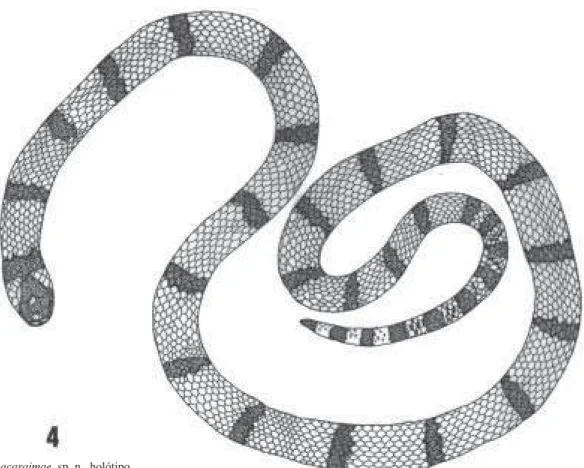
Micrurus pacaraimae, sp. n. (Figs 1-4)
Holótipo: MZUSP 8565, macho, 313 + 42 mm. Brasil: Vila Pacaraima: Rodovia BR – 174 na fronteira com a Venezuela (04°31’N, 61°09’W), 10.i.85, coletada pelo Pelotão Especial de Fronteira para C.M.Carvalho.
Etimologia. Do nome da localidade, Vila Pacaraima.
Diagnose. Colorido dorsal e ventral vermelho e negro no corpo e cauda, garganta branca. Capuz cefálico negro em contato com o anel nucal negro. Anéis negros 23, simples; anéis vermelhos cerca de quatro vezes mais largos que os negros. Ventrais 201, subcaudais 43, anal dividida, tubérculos supra-anais ausentes.
Descrição. Rostral mais larga que alta, pouco visível de cima. Nasal grande, a narina localizada na depressão mediana, voltada para trás. Internasal poligonal, com aproximadamente a metade do tamanho da prefrontal. Frontal pouco mais alta que larga, maior que a sua distância à ponta do focinho e pouco menor que a parietal. Uma preocular grande e visível de cima, sua margem posterior perpendicular à porção mediana do olho. Duas post-oculares pequenas, irregulares, oblíquas. Temporais 1 + 1, a anterior oblíqua, estreita e alongada, a posterior menor, poligonal. Supralabiais 7, 3ª e 4ª em contato com o olho, a 3ª mais alta, em contato com a preocular e com o quadrante antero-inferior; 5ª supralabial separada do olho pela post-ocular inferior. Infralabiais 7, as três anteriores e a borda da quarta em contato com as post-mentais anteriores, que são menores que as posteriores.
Dorsais 15-15-15, lisas sem fossetas apicais. Ventrais 201, subcaudais 43. Anal dividida. A escama ventral que antecede a anal também dividida. Tubérculos supra-anais ausentes.
Cabeça com capuz negro cobrindo o terço superior das primeiras supralabiais, parte das post-oculares e das parietais, unindo-se ao anel nucal negro na primeira escama dorsal mediana. Rostral e nasal marmoreadas. Cinco pequenas manchas claras no topo da cabeça: uma em cada internasal, preenchendo quase toda a escama; uma pequena na borda de cada prefrontal; uma irregular na metade posterior da frontal. O anel nucal negro estende-se ventralmente um pouco para a frente, tocando a margem posterior do segundo par de post-mentais; demais partes ventrais da cabeça brancas, as infralabiais anteriores salpicadas de preto.
manchas negras apicais que ocupam até 1/4 de escama. Margens brancas ausentes nos anéis negros.
Cauda com 9 anéis negros e vermelhos alternados, os últimos levemente salpicados de branco; a escama da extremidade é negra.
DISCUSSÃO
A nova espécie pode ser reconhecida pelo padrão bicolor dos anéis corporais, os negros sem formarem tríades e cerca de quatro vezes mais estreitos que os vermelhos, pela ausência de tubérculos supra-anais e pela disposição do capuz negro cefálico, unido ao anel nucal preto atrás das parietais. Entre as Micrurus de ocorrência na região, com padrão de anéis negros e vermelhos simples e escamas supra-anais lisas, são relevantes no presente contexto as espécies geograficamente próximas M. circinalis e M. psyches, da Venezuela e
regiões vizinhas; M. remotus, M. langsdorffi e M. ornatissimus, do oeste da
Amazônia; e M. paraensis, do Pará e Maranhão (Vanzolini, 1986; Peters & Orejas-Miranda, 1986).
Micrurus circinalis Duméril, Bibron & Duméril, 1854, (MZUSP 8651-52, Trinidad), ocorre em Trinidad, noroeste da Venezuela e norte da Guyana (Roze, 1996: 149). Os caracteres merísticos de M. circinalis são semelhantes aos da espécie nova; as principais diferenças estão no padrão de colorido, aliás regra geral para a maioria das espécies do gênero. O capuz negro de M. circinalis cobre todo o focinho e as parietais; uma faixa branca nucal está presente; a garganta é negra, com estreita faixa branca cobrindo as infralabiais intermediárias e parte das post-mentais posteriores. O capuz negro de M. pacaraimae cobre somente parte da cabeça; faixa branca nucal ausente e a garganta é branca. Os anéis negros de M. circinalis (22-31), com 2-3 escamas de largura, são delimitados por anéis brancos ou amarelos no corpo, seguidos por anel negro estreito; cauda com anéis brancos e vermelhos (somente anéis negros e vermelhos em M. pacaraimae).
Micrurus psyches (Daudin, 1803), ocorre no sudeste da Venezuela, Guianas e Suriname (Roze, 1996:210). A cabeça é toda negra, com um anel branco na porção posterior das parietais. A coloração do corpo consiste de anéis vermelhos e negros, ambos estreitos, aproximadamente da mesma largura, os vermelhos tão melânicos que parecem roxos. Os anéis negros, 27 a 41 em M. psyches (23 em M. pacaraimae), são delimitados por anéis brancos ou amarelos; a cauda tem anéis negros, vermelhos irregulares e brancos.
remotus (Roze, 1987) cobre todo o focinho e as parietais (apenas parcialmente a cabeça em M. pacaraimae); as manchas cefálicas são azuis; a garganta é negra com faixa branca irregular que se estende às infralabiais. Os anéis negros em remotus (25-40), 2-3 escamas dorsais de largura, são delimitados por anéis brancos; os anéis vermelhos têm 4-5 escamas dorsais de largura (7-8 dorsais em M. pacaraimae); 6-11 anéis negros na cauda, separados por anéis vermelhos e pontuações brancas.
Micrurus langsdorffi Wagler, 1868 (MZUSP 8331, Ecuador) (revisão em Cunha & Nascimento, 1982; Vanzolini, 1986; Roze, 1996:185) tem focinho marrom, negro ou amarelo; temporais e parte das supralabiais vermelhas; infralabiais geralmente negras. Cabeça ventralmente negra, marrom ou amarela, geralmente vermelha na porção posterior. Os anéis negros (36-91) são em maior número que em M. pacaraimae (23). Em M. langsdorffi os anéis são negros, amarelos ou marrons, separados por anéis vermelhos; os anéis negros são delimitados por anéis brancos interrompidos ventralmente; faixas brancas ventrais presentes; a cauda é negra com anéis brancos.
Micrurus ornatissimus (Jan, 1858) (revisão em Cunha & Nascimento, 1982; Vanzolini, 1986; Roze, 1996:206) tem a cabeça negra com pontuações claras e garganta negra com manchas brancas. Os anéis negros (38-67), 2-3 escamas de largura, são delimitados por anéis brancos; os anéis vermelhos são aproximadamente da mesma largura que os negros (cerca de quatro vezes mais largos que os negros em M. pacaraimae); a cauda é intensamente melânica.
Finalmente, Micrurus paraensis Cunha & Nascimento, 1973 (MZUSP
8047, Pratinha, Pará – parátipo) (revisão e comentários em Cunha & Nascimento, 1973, 1982; Hoge & Romano-Hoge, 1978; Vanzolini, 1986; Roze 1996:207) tem a cabeça negra com pequena faixa branca interrompida nas parietais, estendendo-se nas temporais, supralabiais posteriores, infralabiais intermediárias e parte das post-mentais posteriores. Os anéis vermelhos de M. paraensis são cerca de seis vezes maiores que os negros (mais largos que os de M. pacaraimae) e separados deles por anéis brancos; a cauda é negra com anéis brancos.
Cinco outras espécies de Micrurus ocorrem na área geral de Roraima, com diversos padrões de colorido: M. hemprichii (Jan, 1858), M. surinamensis (Cuvier, 1817) e M. lemniscatus (Lineu, 1758) apresentam os anéis negros dispostos em tríades; M. karlschmidti Romano, 1972 (Roze, 1996:135) tem
colorido dorsal marrom-escuro uniforme; M. averyi Schmidt, 1939
DISTRIBUIÇÃO
A localidade-tipo de M. pacaraimae, Vila Pacaraima, está no marco de fronteira número oito entre o Brasil e a Venezuela (BV-8). É uma região de formações montanhosas (altitude 920 metros) composta por rochas sedimentares do Grupo Roraima, que se estende de sudeste para noroeste até a Guyana e corresponde à unidade morfoclimática do platô interfluvial Amazonas-Orinoco (Radambrasil, 1975:155). A mais conhecida unidade da Serra Pacaraima é o Monte Roraima, um complexo sedimentar (altitude 2850 metros) com o relevo tabular característico dos tepuis, situado no extremo nordeste daquela Serra, na fronteira entre Brasil, Venezuela e Guyana (Tate, 1932). Aproximadamente 20 quilômetros ao norte da Vila Pacaraima situam-se os tepuis da Gran Sabana venezuelana. Cerca de 50 quilômetros para o sul, descendo a serra através da BR-174, encontra-se a unidade morfoclimática brasileira conhecida como pediplano dos rios Branco e Negro, formada em grande parte pelo lavrado da bacia de Boa Vista (descrição da região em Vanzolini & Carvalho, 1991).
A vegetação da região inclui áreas florestadas, com raras árvores emergentes, sub-bosque bem estruturado, com muitas palmeiras no primeiro estrato; as capoeiras têm as costumeiras características estruturais e fisionômicas desorganizadas, com plantas pioneiras (as embaúbas, diversas espécies de Cecropia são as mais comuns). Há áreas abertas recobertas por gramíneas e ciperáceas (várias espécies de Bulbostylis), arbustos esparsos ou agrupados (o mais freqüente é o caimbé, Curatella americana) e rochas expostas de diversas maneiras e fragmentações. Alguns rios que recortam o lavrado de Roraima têm suas origens na Serra Pacaraima; correm para o sul e deságuam nos rios Surumu, Miang, Tacutu e Uraricoera; os dois últimos formam o rio Branco.
Micrurus pacaraimae foi coletada nas margens de um pequeno igarapé que percorre um vale de 30-40 metros, em área de mata, com muita rocha exposta. Este pequeno igarapé é um dos formadores do rio Miang (Heyer, 1994), o principal formador do rio Surumu, que deságua no rio Tacutu.
O clima da região (Radambrasil, 1975) é caracterizado por chuvas de maio a setembro. A precipitação anual varia em torno de 1500-1800 mm; o mês mais seco é janeiro (aproximadamente 30 mm). A temperatura anual oscila entre 22°C a 30°C; junho e julho são os meses com temperaturas mais baixas.
AGRADECIMENTOS
Papéis
A
vulsos de Zoologia
18
9
REFERÊNCIAS
Cunha, O. R & F. P. Nascimento, 1973. Ofídios da Amazônia. IV. As cobras corais (gênero Micrurus) da região leste do Pará (Ophidia:Elapidae). Nota preliminar. Publ. Avuls. Mus. Para. Emílio Goeldi, Belém, 20:273-286.
Cunha, O. R. & F. P. Nascimento, 1982. Ofídios da Amazônia. XVII. Revalidação de M.langsdorffi
(Wagler, 1824) e distribuição geográfica das duas espécies (Ophidia:Elapidae). Bol. Mus. Para. Emílio Goeldi (Nova Série, Zoologia), Belém, 116:1-17.
Cunha, O. R. & F. P. Nascimento, 1982. Ofídios da Amazônia. XIV. As espécies de Micrurus, Bothrops, Lachesis e Crotalus do sul do Pará e oeste do Maranhão, incluindo áreas de cerrado desse Estado (Ophidia: Elapidae e Viperidae). Bol. Mus. Para. Emílio Goeldi (Nova Série, Zoologia), Belém, 112:1-58.
Cunha, O. R. & F. P. Nascimento, 1993. Ofídios da Amazônia. As cobras da região leste do Pará. Bol. Mus. Para. Emílio Goeldi (Série Zoologia), Belém, 9(1):1-191.
Heyer, W. R. 1994. Hyla benitzi (Amphibia, Anura:Hylidae): first record for Brazil and its biogeographical significance. J. Herp. 28(4):497-499.
Hoge, A. R. & S. A. R. W. L. Romano-Hoge, 1981. Sinopse das serpentes peçonhentas do Brasil (2ª ed.). Mem. Inst. Butantan 42/43 (1978/1979):373-349.
Peters, J.A. & B. Orejas-Miranda, 1986. Catalogue of the Neotropical Squamata: Part I, Snakes. Revised edition (originally published 1970), addenda and corrigenda by P.E.Vanzolini. Washington, D.C.: Smithsonian Institution. 347 p.
Radambrasil, 1975. Folha NA. 20 Boa Vista e parte de NA. 21 Tumucumaque, NB. 20 Roraima e NB. 21; geologia, geomorfologia, pedologia, vegetação e uso potencial da terra. Rio de Janeiro: Departamento Nacional de Produção Mineral (Levantamentos de Recursos Naturais 8): 428 p., 6 mapas. Apêndice: Análise estatística de dados (Vegetação). 260 p. Roze, J. A., 1987. Summary of coral snakes (Elapidae) from Cerro da La Neblina, Venezuela, with
description of a new subspecies. Rev. Franç. Aquariol. 14(3):109-112.
Roze, J. A., 1996. Coral snakes of the Americas: Biology, identification and venoms. Malabar, Florida. Krieger Publishing Company. xii + 328 p.
Tate, G. H. H., 1932. Life zones at Mount Roraima. Ecology 13(3):225-257.
Vanzolini, P. E., 1985. Micrurus averyi Scmidt, 1939, in Central Amazonia (Serpentes, Elapidae).
Papéis Avuls. Zool., São Paulo, 36(8):77-85.
Vanzolini, P. E., 1986. Addenda and corrigenda to Part I Snakes, pp. 1-26, In: Peters, J.A. & B.Orejas-Miranda, Catalogue of the Neotropical Squamata. Part I, Snakes. Washington, D.C. : Smithsonian Institution. 347 p.
Vanzolini, P. E. & C. M.Carvalho, 1991. Two sibling and sympatric species of Gymnophthalmus in
Roraima, Brasil (Sauria:Teiidae). Papéis Avuls. Zool., S. Paulo, 37(12):173-226.
CREDENCIAMENTO E APOIO FINANCEIRO DO
EDITORIAL COMMITTEE
Editor-in-Chief. Hussam Zaher, Serviço de Vertebrados, Museu de Zoologia, Universidade de São Paulo, Caixa Postal 42594, 04299-970, São Paulo, SP, Brazil. E-mail: hzaher@ib.usp.br
Associated Editors. Mario C. C. de Pinna (Universidade de São Paulo, Brazil), Antonio C. Marques (Universidade de São Paulo, Brazil), Sergio A. Vanin (Universidade de São Paulo, Brazil).
Editorial Board. Adriano Kury (Museu Nacional do Rio de Janeiro, Brasil), Aziz N. Ab-Saber (Universidade de São Paulo, Brazil), Carlos Roberto F. Brandão, (Universidade de São Paulo, Brazil), Christian de Muizon (Museum National d’Histoire Naturelle, France), Darrel Frost (American Museum of Natural History, U.S.A.), Gerardo Lamas (Museu Javier Prado de Lima, Peru), H. R. Heyer (National Museum of Natural History, U.S.A.), James Carpenter (American Museum of Natural History, U.S.A.), James Patton (University of Berkeley, U.S.A.), John Maisey (American Museum of Natural History, U.S.A.), Marcos Raposo (Museu Nacional do Rio de Janeiro, Brazil), Marcos Tavares (Universidade Santa Ursula, Brazil), Mario de Vivo (Universidade de São Paulo, Brazil), Miguel T. U. Rodrigues (Universidade de São Paulo, Brazil), Naércio A. Menezes (Universidade de São Paulo, Brazil), Nelson Papavero (Museu Paraense Emilio Goeldi, Brazil), Olivier Rieppel (Field Museum of Natural History, U.S.A), Paulo E. Vanzolini (Universidade de São Paulo, Brazil), Paulo Young (Museu Nacional do Rio de JAneiro, Brazil), Ralf Holzentahl (University of Minesotta, U.S.A.), Randahl Schuh (American Museum of Natural History, U.S.A.), Ricardo Macedo Correa e Castro (Universidade de São Paulo, Brasil), Richard Prum (University of Kansas, U.S.A.), Richard Vari (National Museum of Natural History, U.S.A.), Rudiger Bieler (Field Museum of Natural History, U.S.A.), Ubirajara Martins (Universidade de São Paulo, Brasil), Walter A. P. Boeger (Universidade Federal do Paraná, Brasil).
INSTRUCTIONS FOR AUTHORS
General information. Papéis Avulsos de Zoologia covers primarily the fields of Zoology, publishing original contributions in systematics, paleontology, evolutionary biology, ecology, taxonomy, anatomy, behavior, functional morphology, molecular biology, ontogeny, faunistic studies, and biogeography. PAZ also encourages submission of theoretical and empirical studies that explore principles and methods of systematics.
All contributions must follow the International Code of Zoological Nomenclature and relevant specimens should be properly curated and deposited in a recognized public or private, non-profit institution. Tissue samples should be referred to their voucher specimens and all nucleotide sequence data (aligned as well as unaligned) should be submitted to Genbank (http:// www.ncbi.nlm.nih.gov/Genebank/) or EMBL (http://www.ebi.ac.uk/).
Peer Review. All submissions to Papéis Avulsos de Zoologia are subject to review by at least two referees and the Editor-in-Chief. Three legible copies (including photocopies of original illustrations) and original illustrations must be submitted; all authors will be notified of submission date. Authors may suggest potential reviewers. Communications regarding acceptance or rejection of manuscripts are made through correspondence with the first or corresponding author only. Once a manuscript is accepted providing changes suggested by the reviewers, the author is requested to return a revised version incorporating those changes (or a detailed explanation of why reviewer’s suggestions were not followed) in four weeks upon receiving the communication by the editor. Revised manuscripts must be submitted as both hard copy and disk file (3.5” disk with text in Microsoft Word format).
Proofs. Page-proofs with the revised version will be sent to the first or corresponding author. Page-proofs must be returned to the editor in two weeks, preferably within 48 hours. Failure to return the proof promptly may be interpreted as approval with no changes and/or may delay publication. Only necessary corrections in proof will be permitted. Once page proof is sent to the author, further alterations and/or significant additions of text are permitted only at the author’s expense or in the form of a brief appendix (“note added in proof”).
Submission of manuscripts. Manuscripts should be sent to the Editor-in-Chief (H. Zaher, Museu de Zoologia da USP, Caixa Postal 42694, CEP 04299-970, São Paulo, SP, Brasil). Manuscripts are considered on the understanding that they have not been published or will not appear elsewhere in substantially the same or abbreviated form. The criteria for acceptance of articles are quality and relevance of research, clarity of text, and compliance with the guidelines for manuscript preparation.
Manuscripts should be written preferentially in English, but texts in Portuguese or Spanish will also be considered. Studies with a broad coverage are encouraged to be submitted in English. All manuscripts should include an abstract in English regardless of the original language.
Manuscript form. Manuscripts should not exceed 100 pages of double-spaced typescript on 21 by 29.7 cm (A4 format) or 21.5 by 28 cm (Letter format) paper, with wide margins. The pages of the manuscript should be numbered consecutively.
The text of articles should be arranged in the following order: title page, abstract, body of text, literature cited, tables, appendices, and figure captions. Each of these sections should begin on a new page. All typescript pages must be double-spaced.
(1) Title page. This should include the title, author(s) name(s), institutions, and Key words in English as well as in the language of the manuscript, and a short running title in English. The title should be concise and, where appropriate, should include mention of families and/or higher taxa. Names of new taxa should not be included in titles.
(2) Abstract: All papers should have an abstract in English, regardless of the original language. The abstract is of great importance as it may be reproduced elsewhere. It should be in a form intelligible if published alone in conjunction with the title and should summarize the main facts, ideas, and conclusions of the article. Indicative abstracts are strongly discouraged. Include all new taxonomic names for referencing purposes. Abbreviations should be avoided. It should not include references. Abstracts should not exceed 350 words.
(3) Body of text: The main body of the text should include the following sections: Introduction, Materials and Methods, Results, Discussion, and Acknowledgments at end. Primary headings in the text should be in capital letters and centered; the following text should begin on the next line, indented. Secondary headings should be in capital and lowercase letters and flush left; the following text should begin on the next line, indented. Tertiary headings should be in capital and lower case letters, in italics and indented; the following text should be on the same line and separated from the heading by a hyphen.
(4) Literature cited. Citations in the text should be given as: Silva (1998)…, Silva (1998: 14-20)…, Silva (1998: figs. 1, 2)…, Silva (1998a, b)…, Silva and Oliveira (1998)…, (Silva, 1998)…, (Rangel, 1890; Silva and Oliveira, 1998a, b; Adams, 2000)…, (Silva, pers. comm.)…, (Silva et al., 1998), the latter when the paper has three or more authors. The reference need not be cited when author and date are given only as authority for a taxonomic name. The literature section should be arranged strictly alphabetically and given in the following format:
Journal Article – Silva, H. R., H. Oliveira & S. Rangel. Year. Article title. Journal name, 00:000-000. Names of journals must be spelled out in full.
Books – Silva, H. R. Year. Book title. Publisher, Place, 000 pp.
Articles in Books – Silva, H. R. Year. Article title; pp. 000-000. In: H. Oliveira & S. Rangel (Eds.), Book Title. Publisher, Place.
Articles in Larger Works – Silva, H. R. Year. Article title; pp. 000-000. In: H. Oliveira & S. Rangel (Eds.), Title of Larger Work. Serial Publication 00. Publisher, Place.
Dissertations and Theses – Silva, H. R. Year. Dissertation title. Ph.D. dissertation, University, Place, 000 pp.
Tables. All tables must be numbered in the same sequence in which they appear in the text. Authors are encouraged to indicate where the tables should be placed in the text. They should be comprehensible without reference to the text. Tables should be formatted with horizontal, not vertical, rules. In the text, tables should be referred as Table 1, Tables 2 and 3, Tables 2-6. Use “TABLE” in the table heading.
Illustrations. Figures should be numbered consecutively, in the same sequence they appear in the text. Separate illustrations of a composite figure should be identified by capital letters and referred in the text as so (fig. 1A). Where possible, letters should be placed in the lower right corner of each illustration of a composite figure. Hand-written lettering on illustrations are unacceptable. Illustrations should be mounted on stout, white cardboard. Figures should be mounted in order to minimize blank areas between separate illustrations. High quality color or black and white photographs, and computer generated figures are preferable. Authors are encouraged to indicate where the figures should be placed in the text. Use “(Fig(s).)” and “Figure(s)” for referring to figures in the text, but “FIGURE(S)” in the figure captions and “ (fig(s).)” when referring to figures in another paper.