Isothermal Amplification, Fluorescence Smear
Microscopy and Culture for the Diagnosis of Tuberculosis
Geojith George1, Prem Mony2, John Kenneth1*
1Division of Infectious Disease, St. John’s Research Institute, Bangalore, India,2Division of Epidemiology and Biostatistics, St. John’s Research Institute, Bangalore, India
Abstract
Background:Despite the advent of novel diagnostic techniques, smear microscopy remains as the most practical test available in resource-limited settings for tuberculosis (TB) diagnosis. Due to the low sensitivity of microscopy and the long time required for culture, feasible and accessible rapid diagnostic methods are urgently needed. Loop-mediated Isothermal Amplification (LAMP) is a promising nucleic-acid amplification assay, which could be accessible, cost-effective and more suited for use with unpurified samples.
Methodology/Principal Findings: In the current study, the objective was to assess the efficacy of a LAMP assay for tuberculosis compared with fluorescence smear microscopy as well as Lo¨wenstein-Jensen (LJ) and Mycobacteria Growth Indicator Tube (MGIT) cultures for the diagnosis of pulmonary tuberculosis using sputum samples. Smear microscopy and culture were performed for decontaminated and concentrated sputum from TB suspects and the LAMP was also performed on these specimens. The LAMP and smear microscopy were compared, in series and in parallel, to culture. LAMP and smear microscopy showed sensitivities of 79.5% and 82.1% respectively and specificities of 93.8% and 96.9% respectively, compared to culture. LAMP and smear in series had sensitivity and specificity of 79.5% and 100.0% respectively. LAMP and smear in parallel had sensitivity and specificity of 82.1% and 90.6% respectively.
Conclusions/Significance:The overall efficacies of LAMP and fluorescence smear microscopy in the current study were high and broadly similar. LAMP and smear in series had high specificity (100.0%) and can be used as a rule-in test combination. However, the performance of LAMP in smear negative samples was found to be insufficient.
Citation:George G, Mony P, Kenneth J (2011) Comparison of the Efficacies of Loop-Mediated Isothermal Amplification, Fluorescence Smear Microscopy and Culture for the Diagnosis of Tuberculosis. PLoS ONE 6(6): e21007. doi:10.1371/journal.pone.0021007
Editor:Shuang-yong Xu, New England Biolabs, Inc., United States of America ReceivedMarch 12, 2011;AcceptedMay 16, 2011;PublishedJune 17, 2011
Copyright:ß2011 George et al. This is an open-access article distributed under the terms of the Creative Commons Attribution License, which permits
unrestricted use, distribution, and reproduction in any medium, provided the original author and source are credited.
Funding:The authors acknowledge the salary support for Geojith G., which was provided by a research grant from the Research Council of Norway (179342: TB Trials). The initial funding for this project was also obtained from Aeras Global TB Vaccine Foundation. The funders were involved in this project with regard to sample collection, preliminary processing for smear examination/culture and storage (which has been used in this study to compare the LAMP technique). This was primarily for quality control purposes to determine laboratory functionality and efficacy. The funders had no other role in the study design, data collection and analysis, decision to publish, or preparation of the manuscript. The study received no other external funding.
Competing Interests:The authors have declared that no competing interests exist. * E-mail: [email protected]
Introduction
Tuberculosis (TB) is one of the oldest diseases that still afflict mankind. The dual specters of TB and AIDS have drawn recent attention to the lack of a suitable diagnostics for TB [1,2,3,4]. TB case detection is the first hurdle towards tackling the TB epidemic [5]. However, the culture which is considered as the ‘gold stan-dard’ of TB diagnosis takes 3–6 weeks, leaving the less sensitive smear microscopy as the only feasible rapid test presently. Even the automated liquid culture systems like BACTEC or Mycobac-teria Growth Indicator Tube (MGIT) take 1–6 weeks for growth detection. The utility of microscopy decreases radically in pauci-bacillary and HIV positive TB suspects. Smear negative carriers, even if considered less infectious, can still spread TB [6]. The long delays in diagnosis result in patients dropping out or continuing to spread TB till they are correctly diagnosed, found and treated [7,8]. While treating all suspected cases adds significantly to the cost of TB control programs, it also exposes subjects to unnecessary
drugs and worsens the emergence of drug resistance. A highly sensitive rule-in test can significantly improve the case detection whereas a highly specific rule-out test can reduce the turnaround time and the duration of respiratory isolation as well as avoid unnecessary administration of potentially toxic drugs [9,10,11].
specificity, especially compared to serological tests [17,18,19]. Nucleic acid assays are also more amenable to miniaturization and microfabrication, opening new vistas for cost reduction and auto-mation [20]. Loop-mediated Isothermal Amplification (LAMP) was shown to be a promising nucleic-acid amplification assay, which could be accessible and cost-effective [21]. It could also be more robust than other nucleic acid amplification tests, retaining the specificity across wider pH and temperature gradients and showing lesser inhibition in unpurified samples [22]. Suitability of LAMP as a point of care test for the diagnosis of pulmonary tuberculosis is beginning to be evaluated with clinical samples [23,24].
The LAMP assay was found to be suitable for the laboratory identification of M. tuberculosis (MTB) in culture isolates by the authors previously [25]. In the current study, the objective was to assess the efficacy of a LAMP assay for tuberculosis, alone and in combination with fluorescence smear microscopy as well as Lo¨wenstein-Jensen (LJ) and Mycobacteria Growth Indicator Tube (MGIT) cultures for the detection ofM. tuberculosisfrom archived sputum samples.
Materials and Methods
Ethics Statement
This study was reviewed and cleared by the St. John’s Medical College Hospital ethics review board. Written informed consent was obtained from all the participants. The data were analyzed anonymously.
Participants
To compare ‘LAMP’ with the fluorescence smear microscopy and LJ culture, we used 78 sputum samples obtained from as many TB suspects from the Palamaner region in the state of Andhra Pradesh in southern India.
Procedures
Samples were collected over a period of six months starting from January 2007. These were decontaminated using NALC-NaOH method and stored at220uC. Sputum sample digestion-decontamination, LJ culture [26] and auramine O fluorescence microscopy were performed as per standard literature [27]. MGIT cultures were performed as per the manufacturer’s instructions. Decontaminated sputum was processed (January to February 2010) using the ‘AMPLICOR Respiratory Specimen Preparation Kit’ (Roche Diagnostics GmbH, Mannheim), according to manu-facturer’s instructions and the resulting lysate was stored at220uC. LJ and MGIT cultures were used as the ‘gold standard’ against which other tests could be assessed. The tests were executed and read by experienced personnel, who were blinded to the results of the other tests.
LAMP reaction
TheM. tuberculosis specific LAMP reaction was carried out as published previously [28], but was modified to suit local conditions [25]. Briefly, the primers, along with 5ml of the sample lysate were heat denatured for 3 minutes and then annealed prior to adding the enzyme-dNTP mix. The LAMP reaction was carried out in a final volume of 25ml in a 120 minute format and was terminated by heating at 80uC for 2 minutes. This assay is specific for the rimMsequence ofM. tuberculosisandM. bovis. DNA extracted from M. tuberculosisATCC strain H37Rv was used as the positive control and PCR grade water as the negative control. TheBstpolymerase (Large fragment) was purchased from New England Biolabs Inc. The primers and SYBR Green I were acquired from Sigma-Aldrich Corporation, India. The final reaction volume was 25ml
(including sample volume 5ml).The results were visualized by adding 2ml of 10-fold diluted original SYBR Green I after amplification, and confirmed by agarose gel electrophoresis [29].
Statistical methods
Data was analyzed using SPSS software - version 17 (ßIBM
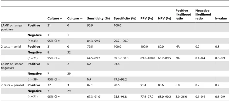
corporation 2010). Sensitivity, specificity [30] as well as positive and negative predictive values (PPV & NPV) [31] were calculated for the LAMP and smear microscopy in comparison with culture as the gold standard. LAMP and smear microscopy testing in series and parallel were also compared against the culture [32]. For this purpose, LAMP and smear microscopy were compared in series (LAMP performed only if smear positive and considered TB positive if both tests are positive or TB negative if either test is negative) and in parallel (LAMP performed for all samples and considered TB positive if either smear or LAMP is positive and TB negative if both are negative). Serial testing improves the specificity but lowers the sensitivity. It also reduces the cost of the second test as it is performed for only those samples positive by the first test. Parallel testing improves the sensitivity, while decreasing the specificity. Cohen’s kappa was calculated as a measure of agree-ment between the tests [33].
Results and Discussion
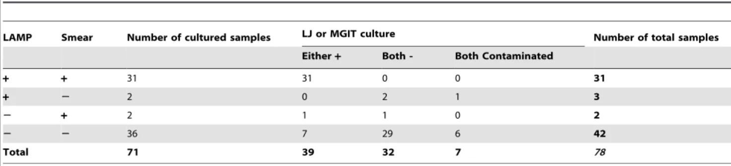
The results of the study are outlined in table 1. Samples were considered to be culture negative if no growth was detected in both LJ and MGIT cultures. Samples were considered to be ‘culture positive’ if growth was detected in either LJ or MGIT cultures. Of the 78 samples tested, 7 showed contamination for both LJ and MGIT cultures and were omitted from analysis. 34 samples were LAMP positive, of which one was detected to be contaminated on both LJ and MGIT cultures. Of the 44 LAMP negative samples, 6 were detected to be contaminated on both LJ and MGIT cultures. 33 samples were smear positive. Of the 45 smear negative samples, 7 were detected to be contaminated on both LJ and MGIT cul-tures. Non-contaminated culture results were available for 33 positive and 38 negative samples each for the LAMP and smear. The performance of LAMP and smear as compared to the LJ or MGIT culture is given in table 2. Efficacy of the LAMP and smear microscopy in series and in parallel is depicted in table 3.
LAMP detects the presence/absence of the genetic material of M. tuberculosis. Smear microscopy detects the morphology and culture methods differentiate based on the physiology of the viable organism. These different approaches could be complementary. In this study, the LAMP was observed to have high sensitivity and specificity for samples with concordant culture and smear results. The overall efficacy of LAMP and fluorescence smear microscopy in the current study was high and broadly similar. However, the performance of LAMP in smear negative samples was found to be suboptimal.
performed serially, the overall sensitivity and specificity observed were 79.5% and 100.0% respectively (n = 71). When both the LAMP and smear microscopy were performed in parallel (n = 71), the sensitivity and specificity observed were 82.1% and 90.6% respectively (Table 3). Commercial NAATs had shown pooled sensitivity and specificity of 96.0% and 85.0% respectively among smear positive samples and 66.0% and 98.0% respectively among smear negative samples[34]. In-house PCR assays were reported to show pooled sensitivity and specificity of 96.0% and 81.0% respectively for smear positive samples [35], indicating that a standardized LAMP can perform potentially better than or equivalent to PCR-based methods.
LAMP does not appear to pick up any additional true positives or pick up any that the smear missed, in smear negative samples. Hence the LAMP assay in the current format may be useful only in diagnosis of smear positive samples or as a rule-in test for smear
negative samples (specificity of 93.6%). The utility of LAMP as a tool to resolve differences between the culture and smear results is questionable, especially when smear negative samples are involved. However, this has to be validated with larger sample sets. Discordant results between the LAMP and smear or culture can arise if non-tuberculous Mycobacteria (NTM) are present [36,37] or if cultures are contaminated [38]. NTM have been reported at high frequencies from southern India [39,40]. The development of a LAMP assay capable of identifying both MTBC and NTM can improve the sensitivity and specificity significantly. In this study, the one sample that was LAMP negative but smear and LJ positive was later identified as MTB by the ‘GenoType MTBC’ assay (HAIN lifesciences GmbH, Germany) as well as by LAMP for culture lysates. These rule out NTM or sequence variation as reasons for amplification failure with this sample and indicate the lower concentration of nucleic acid present or the presence of Table 1.Results of LAMP, smear and culture.
LAMP Smear Number of cultured samples LJ or MGIT culture Number of total samples
Either+ Both - Both Contaminated
+ + 31 31 0 0 31
+ 2 2 0 2 1 3
2 + 2 1 1 0 2
2 2 36 7 29 6 42
Total 71 39 32 7 78
*Positive results marked as ‘+’ and Negative results as ‘2‘. doi:10.1371/journal.pone.0021007.t001
Table 2.Three-way comparison of LAMP, smear and culture.
Culture+ Culture2 Sensitivity (%) Specificity (%) PPV (%) NPV (%)
Positive likelihood ratio
Negative
likelihood ratio k-value
LAMP+ 31 2 79.5 93.8 93.9 79.0 12.7 0.2 0.7
LAMP2 8 30
n = 71 95% CI = 64.5–89.2 79.9–98.3 80.4–98.3 63.7–88.9 3.3–49.1 0.1–0.4 0.6–0.9
Culture+ Culture2
Smear+ 32 1 82.1 96.9 97.0 81.6 26.3 0.2 0.8
Smear2 7 31
n = 71 95% CI = 67.3–91.0 84.3–99.5 84.7–99.5 66.6–90.8 3.8–181.7 0.1–0.4 0.6–0.9
Smear+ Smear2
LAMP+ 31 2 93.9 94.7 93.9 94.7 17.9 0.1 0.9
LAMP2 2 36
n = 71 95% CI = 80.4–98.3 82.7–98.5 80.4–98.3 82.7–98.5 4.6–69.0 0.0–0.2 0.8–1.0
Culture+
Smear+
Culture2
Smear2
LAMP+ 29 0 96.7 100.0 100.0 96.7 NA 0.0 1.0
LAMP2 1 29
n = 59 95% CI = 83.3–99.4 88.3–100.0 88.3–100.0 83.3–99.4 NA 0.0–0.2 0.9–1.0
Culture+
Smear2
Culture2
Smear+
LAMP+ 2 2 22.2 33.3 50.0 12.5 0.3 2.3 NA
LAMP - 7 1
n = 12 95% CI = 6.3–54.7 6.2–79.2 15.0–85.0 2.2–47.1 0.1–1.4 0.5–12.0 NA
inhibitors. All samples negative by LAMP were spiked with M. tuberculosisDNA and re-amplified. As all these samples amplified, presence of inhibitors can be ruled out.
The high sensitivity observed for smear microscopy in this study may be due to the nature of sampling, better lab practices or the high prevalence of TB as observed elsewhere [41,42]. One of the factors reducing the sensitivity of the LAMP may be the lysis of specimens upon storage [43]. Subsequent supernatant removal and NaOH treatment of lysed specimens may reduce the amount of amplifiable DNA available. Washing and NaOH-free methods for sputum processing will be of considerable value to NAATs.
Conclusions
LAMP appears to be suitable for smear positive specimens with concordant culture results, but was ineffective in smear negative samples. LAMP and smear in series had high specificity (100.0%) and can be used as a rule-in test combination. An improved LAMP alone or together with smear microscopy can offer same-day diagnosis and has the potential to reduce the drop-out rate substantially. However, these findings need to be substantiated further with larger sample sizes and in different geographical settings. Inter-reader reproducibility and the performance with smear negative samples also need further evaluation. Suitably modified, LAMP can potentially improve the diagnosis of TB in resource limited settings.
Limitations
In the current study, sample size under certain subtypes was too limited to arrive at statistically significant estimates of efficacy. The sampling was not continuous due to limited availability of stored samples. Speciation of all the cultured samples were not performed and as such it is not possible to determine how many of the culture or smear positives were NTM, which the LAMP will not detect using current set of primers. The HIV status was not ascertained for the study samples. However, it is unlikely that HIV had a significant impact on the findings, as other studies in the same region indicate,1% prevalence for HIV.
Acknowledgments
We acknowledge the TB Trials Group and part funding by the Research council of Norway (TB Trials 179342). We would also like to thank Bijo Thomas and the SETRI lab, Palamaner, Andhra Pradesh.
Author Contributions
Conceived and designed the experiments: JK. Performed the experiments: GG. Analyzed the data: GG JK PM. Contributed reagents/materials/ analysis tools: JK PM. Wrote the paper: GG JK.
References
1. Perkins MD, Cunningham J (2007) Facing the crisis: improving the diagnosis of tuberculosis in the HIV era. Journal of Infectious Diseases 196: 15. 2. WHO (2010) Global tuberculosis control: WHO report 2010.: World Health
Organization. WHO/HTM/TB/2010.7 WHO/HTM/TB/ 2010.7. 3. Pai M, Minion J, Steingart K, Ramsay A (2010) New and improved tuberculosis
diagnostics: evidence, policy, practice, and impact. Current opinion in pulmo-nary medicine 16: 271.
4. Davies P, Pai M (2008) The diagnosis and misdiagnosis of tuberculosis [State of the art series. Tuberculosis. Edited by ID Rusen. Number 1 in the series]. The International Journal of Tuberculosis and Lung Disease 12: 1226–1234. 5. Keeler E, Perkins M, Small P, Hanson C, Reed S, et al. (2006) Reducing the global
burden of tuberculosis: the contribution of improved diagnostics. Nature 444: 49–57. 6. Behr M, Warren S, Salamon H, Hopewell P, de Leon AP, et al. (1999) Transmission ofMycobacterium tuberculosisfrom patients smear-negative for acid-fast bacilli. The Lancet 353: 444–449.
7. Millen S, Uys P, Hargrove J, Van Helden P, Williams B (2008) The effect of diagnostic delays on the drop-out rate and the total delay to diagnosis of tuberculosis. PloS One 3: 1933.
8. Sreeramareddy CT, Panduru KV, Menten J, Van den Ende J (2009) Time delays in diagnosis of pulmonary tuberculosis: a systematic review of literature. BMC Infectious Diseases 9: 91.
9. Moore DF, Guzman JA, Mikhail LT (2005) Reduction in turnaround time for laboratory diagnosis of pulmonary tuberculosis by routine use of a nucleic acid amplification test. Diagnostic microbiology and infectious disease 52: 247–254. 10. Campos M, Quartin A, Mendes E, Abreu A, Gurevich S, et al. (2008) Feasibility of shortening respiratory isolation with a single sputum nucleic acid ampli-fication test. American journal of respiratory and critical care medicine 178: 300. 11. Bailey S, Roper M, Huayta M, Trejos N, Alarcon L, et al. (2011) Missed opportunities for tuberculosis diagnosis. The International Journal of Tubercu-losis and Lung Disease 15: 205–210.
Table 3.Comparison of LAMP and smear in series and parallel.
Culture+ Culture2 Sensitivity (%) Specificity (%) PPV (%) NPV (%)
Positive likelihood ratio
Negative likelihood ratio k-value LAMP on smear
positives
Positive 31 0 96.9 100.0
Negative 1 1
(n = 33) 95% CI = 84.3–99.5 20.7–100.0
2 tests – serial Positive 31 0 79.5 100.0 100.0 80.0 NA 0.2 0.8
Negative 8 32
(n = 71) 95% CI = 64.5–89.2 89.3–100.0 89.0–100.0 65.2–89.5 NA 0.1–0.4 0.6–0.9
LAMP on smear negatives
Positive 0 2 NA 93.6
Negative 7 29
(n = 38) 95% CI = NA 79.3–98.2
2 tests – parallel Positive 32 3 82.1 90.6 91.4 80.6 8.8 0.2 0.7
Negative 7 29
(n = 71) 95% CI = 67.3–91.0 75.8–96.8 77.6–97.0 65.0–90.2 3.0–26.0 0.1–0.4 0.6–0.9
12. Nyendak M, Lewinsohn D, Lewinsohn D (2009) New diagnostic methods for tuberculosis. Current Opinion in Infectious Diseases 22: 174.
13. Lemaire J, Casenghi M (2010) New diagnostics for tuberculosis: fulfilling patient needs first. Journal of the International AIDS Society 13: 40.
14. Lange C, Mori T (2010) Advances in the diagnosis of tuberculosis. Respirology 15: 220–240.
15. Pai M, Minion J, Sohn H, Zwerling A, Perkins MD (2009) Novel and improved technologies for tuberculosis diagnosis: progress and challenges. Clinics in Chest Medicine 30: 701-716, viii.
16. Dinnes J, Deeks J, Kunst H, Gibson A, Cummins E, et al. (2007) A systematic review of rapid diagnostic tests for the detection of tuberculosis infection. Health Technology Assessment 11: 1–196.
17. Morris K (2011) WHO recommends against inaccurate tuberculosis tests. The Lancet 377: 113–114.
18. CDC (2009) Updated Guidelines for the Use of Nucleic Acid Amplification Tests in the Diagnosis of Tuberculosis. Morbidity and Mortality Weekly Report (MMWR) 58: 7–10.
19. Neonakis IK, Gitti Z, Krambovitis E, Spandidos DA (2008) Molecular diagnostic tools in mycobacteriology. Journal of Microbiological Methods 75: 1–11.
20. Kiechle FL, Holland CA (2009) Point-of-care testing and molecular diagnostics: miniaturization required. Clinics in laboratory medicine 29: 555–560. 21. Dowdy D, OBrien M, Bishai D (2008) Cost-effectiveness of novel diagnostic
tools for the diagnosis of tuberculosis. The International Journal of Tuberculosis and Lung Disease 12: 1021–1029.
22. Francois P, Bento M, Hibbs J, Bonetti E, Boehme C, et al. (2011) Robustness of loop–mediated isothermal amplification reaction for diagnostic applications. FEMS Immunology & Medical Microbiology.
23. Neonakis I, Spandidos D, Petinaki E (2011) Use of loop-mediated isothermal amplification of DNA for the rapid detection ofMycobacterium tuberculosis in clinical specimens. European Journal of Clinical Microbiology & Infectious Diseases;DOI 10.1007/s10096-10011-11195-10090.
24. Boehme C, Nabeta P, Henostroza G, Raqib R, Rahim Z, et al. (2007) Operational feasibility of using loop-mediated isothermal amplification for diagnosis of pulmonary tuberculosis in microscopy centers of developing countries. Journal of clinical microbiology 45: 1936.
25. Geojith G, Dhanasekaran S, Chandran S, Kenneth J (2011) Efficacy of Loop Mediated Isothermal Amplification (LAMP) assay for the laboratory identifica-tion ofMycobacterium tuberculosisisolates in a resource limited setting. Journal of Microbiological Methods 84: 71–73.
26. Levin W, Brandon GR, Mc MS (1950) The culture method of laboratory diagnosis of tuberculosis. American Journal of Public Health and The Nation’s Health 40: 1305–1310.
27. Kent P, Kubica G, (US) CfDC (1985) Public health mycobacteriology: a guide for the level III laboratory: US Dept. of Health and Human Services, Public Health Service, Centers for Disease Control .
28. Zhu R-Y, Zhang K-X, Zhao M-Q, Liu Y-H, Xu Y-Y, et al. (2009) Use of visual loop-mediated isothermal amplification of rimM sequence for rapid detection of
Mycobacterium tuberculosis and Mycobacterium bovis. Journal of Microbiological Methods 78: 339–343.
29. Iwamoto T, Sonobe T, Hayashi K (2003) Loop-mediated isothermal amplification for direct detection ofMycobacterium tuberculosiscomplex,M. avium, andM. intracellularein sputum samples. Journal of clinical microbiology 41: 2616–2622.
30. Altman DG, Bland JM (1994) Statistics Notes: Diagnostic tests 1: sensitivity and specificity. BMJ 308: 1552.
31. Altman DG, Bland JM (1994) Statistics Notes: Diagnostic tests 2: predictive values. BMJ 309: 102.
32. Fletcher RH, Fletcher SW (2005) Clinical epidemiology: the essentials: Lippincott Williams & Wilkins.
33. Cohen J (1960) A coefficient of agreement for nominal scales. Educational and psychological measurement 20: 37–46.
34. Greco S, Girardi E, Navarra A, Saltini C (2006) Current evidence on diagnostic accuracy of commercially based nucleic acid amplification tests for the diagnosis of pulmonary tuberculosis. Thorax 61: 783.
35. Greco S, Rulli M, Girardi E, Piersimoni C, Saltini C (2009) Diagnostic accuracy of in-house PCR for pulmonary tuberculosis in smear-positive patients: meta-analysis and metaregression. Journal of clinical microbiology 47: 569. 36. Lee JS, Kim EC, Joo SI, Lee SM, Yoo CG, et al. (2008) The incidence and
clinical implication of sputum with positive acid-fast bacilli smear but negative in mycobacterial culture in a tertiary referral hospital in South Korea. Journal of Korean medical science 23: 767.
37. Vidal R, Juan A, Miravitlles M, Martı´n-Casabona N, Falgueras T (1996) Incidence and significance of acid-fast bacilli in sputum smears at the end of antituberculous treatment. Chest 109: 1562.
38. Lai CC, Tan CK, Lin SH, Liao CH, Chou CH, et al. (2010) Molecular evidence of false-positive cultures forMycobacterium tuberculosisin a Taiwanese hospital with a high incidence of TB. Chest 137: 1065–1070.
39. Chauhan M (1993) Non-tuberculous mycobacteria isolated from an epidemi-ological survey in rural population on Banglore district. Indian Journal of Tuberculosis 40: 195–197.
40. Paramasivan C, Govindan D, Prabhakar R, Somasundaram P, Subbammal S, et al. (1985) Species level identification of non-tuberculous mycobacteria from South Indian BCG trial area during 1981. Tubercle 66: 9–15.
41. Shea Y, Davis J, Huang L, Kovacs J, Masur H, et al. (2009) High sensitivity and specificity of acid-fast microscopy for diagnosis of pulmonary tuberculosis in an African population with a high prevalence of human immunodeficiency virus. Journal of clinical microbiology 47: 1553.
42. Pandey BD, Poudel A, Yoda T, Tamaru A, Oda N, et al. (2008) Development of an in-house loop-mediated isothermal amplification (LAMP) assay for detection of Mycobacterium tuberculosis and evaluation in sputum samples of Nepalese patients. Journal of Medical Microbiology 57: 439–443.