ISSN: 2067-533X
INTERNATIONAL JOURNAL
OFCONSERVATION SCIENCE
Volume 2, Issue 3, July-September 2011: 145-154 www.ijcs.uaic.ro
CHARACTERIZATION OF ANCIENT EGYPTIAN
WALL PAINTINGS, THE EXCAVATIONS
OF CAIRO UNIVERSITY AT SAQQARA
Hussein MAREY MAHMOUD1*, Nikolaos KANTIRANIS2,
Mona ALI1, John STRATIS3
1)
Department of Conservation, Faculty of Archaeology, Cairo University, Giza, Egypt.
2) Department of Mineralogy-Petrology-Economic Geology, Aristotle University, Thessaloniki, Greece. 3) Laboratory of Analytical Chemistry, Aristotle University, Thessaloniki, Greece.
Abstract
The present study aims at characterizing some Egyptian wall paintings discovered during the excavations of Cairo University (since 1988 and recently in 2005) at Saqqara area in the south of Cairo. There, a number of tombs dating back to the 19th dynasty (c.1293–1185 BC) were discovered. The walls of these tombs are carved with bass and raised reliefs and painted with different colours. The characterization of the wall paintings was done by means of optical microscopy (OM), scanning electron microscopy (backscattered electron mode, BSE) equipped with an energy dispersive X-ray detector (EDS), micro XRF spectrometry (µ-XRF), and X-ray diffraction analysis (XRD). The analysis of the examined samples indicated that the blue pigment is Egyptian blue (Cuprorivaite), the green pigment is Egyptian green, the red pigment is red ochre, and the yellow pigment is a blended layer of yellow ochre and orpiment (As2S3). The results will help in providing an image concerning some painting materials used
during the new Kingdom in ancient Egypt.
Keywords: Wall paintings; Excavations, Saqqara; Cross-sections; µ-XRF; BSE−EDS.
Introduction
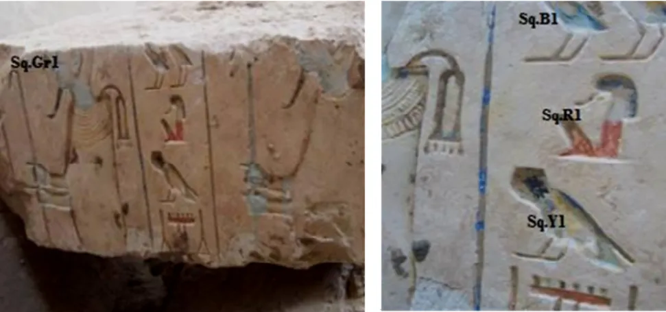
Saqqara is a one section of the great necropolis of Memphis, the Old Kingdom capital and the Kings of the 1st dynasty as well as that of the 2nd dynasty are mostly buried in this section of the Memphis necropolis. Saqqara is located 30 km south of Cairo (29.871264° N 31.216381° E). The excavations of the Faculty of Archaeology have begun since 1984, in south of the way of pyramid of Unas; there, burials from the Roman and Coptic times were discovered. During the years (1984−1988), many tombs from the 19th dynasty have been discovered. In the mission worked in the year 2005 at the area, painted limestone blocks have been discovered in the wheel of tomb of hwi nfr (Fig. 1). The preparation of multi-layered paint-samples and their structural analysis is standard practice for the examination of painted surfaces in art conservation. Mounted cross-sections have been commonly used to study the
*
morphology, stratigraphy and materials of the polychrome of artefacts. They are a valuable source of information for a painting’s layered structure, its pigments and painting technique or for providing indications of over-painting or other restoration procedures. Taking even a tiny sample for a cross-section means removing and destroying a part of the artefact’s original structure.
Fig. 1. The painted limestone block discovered at the excavations of Cairo University at Saqqara area. The codes refer to locations of the collected samples.
Scanning electron microscopy has been extensively used for the material characterization of objects of artistic and archaeological importance, especially in combination with energy dispersive X-ray microanalysis [1]. As a host for this technique, scanning electron microscopy offers powerful means of characterizing materials by utilizing the wide range of signals resulting from the interaction of solid samples with a scanning beam of energetic electrons. X-ray fluorescence (XRF) spectrometry has been used for the investigation of archaeological and historical materials for some fifty years; though, a great variety of spectrometers has been successfully employed, it is only by portable systems that non-destructivity, multi-elemental, fast, and cost-effective; thanks to the possibility of working in situ, portable spectrometers have virtually extended the range of use of XRF to any type of object [2, 3]. Micro X-ray fluorescence (µ-XRF) is one of the latest branches of X-ray fluorescence spectrometry; here “micro” stands for the fact that localized regions of a sample are investigated with spatial resolutions on the micrometer scale [4, 5]. X-ray diffraction analysis of a pure substance is like a fingerprint of the substance, XRD is considered the most famous method used in the identification of crystalline compounds by their diffraction pattern. It plays an important role in the study of works of art and museum objects; also it helps in answering questions related to degradation processes [6].
Materials and methods
Sampling
During the visits of the excavations, digital colour photographs were taken for site observation of the wall paintings to record the state of conservation and to determine the suitable areas for sampling process. Tiny pieces of the wall paintings were carefully chosen from areas that had no aesthetic value or seriously damaged areas. Appropriate representative samples including different visible colours were collected to provide stratigraphic information of paint layers and substrate layers. In areas with good state of conservation, few powder of the colouring materials were removed carefully using a micro-scalpel, beneath the pictorial layers and then stored in different tubes.
Optical microscopy (OM)
In order to study the stratigraphic structure of the painted layers, some samples were embedded in an Epoxy resin (EpoFix), cross-sectioned using variable speed silicon carbide papers and DP-lubricant blue for fine and cool polishing, and mounted on glass slides. The polished cross sections have been investigated under optical microscopy (OM) using a Zeiss (stemi DV4) stereomicroscope with Sony camera (DSC-S85) and under reflected light using a Leitz orthoplan-bionculor (polarized microscope) equipped with camera Nikon Cooplix 990.
Scanning electron microscopy in the backscattered electron mode (BSE)
Scanning electron microscopy (SEM) provides information about mineral morphology, crystal features, and chemical composition. In this study a JEOL JSM-840A scanning electron microscope. The microanalysis was carried out using an energy dispersive X-ray (EDS) Oxford ISIS 300 micro analytical system. Operating conditions were: accelerating voltage 20kV, probe current 45nA and counting time 60 seconds, with ZAF correction being provided on-line. Polished cross-sections were investigated by backscattered electrons (BSE) for the purpose of pigment identification in each colour layer, the elemental composition was determined using the prepared carbon coated cross-sections.
Micro-XRF spectrometry
The micro-XRF spectrometer (SPECTRO, COPRA model) used in this work includes a side-window X-ray tube with Mo anode (Oxford Instruments, Series 5011 XTF), potential acceleration 35 kV, lamp stream 0.9mA and with analysis time 300s. A long-distance optical microscope located on the detector and X-ray tube plane is used in order to select the points of interest over the surface of the sample [5].
X-ray powder diffraction analysis (XRPD)
XRPD measurements were performed using a Phillips PW1710 diffractometer with
Ni-filtered Cu-kα radiation. The samples were scanned over the 3−63º 2θ interval at a scanning
Results
Optical examination
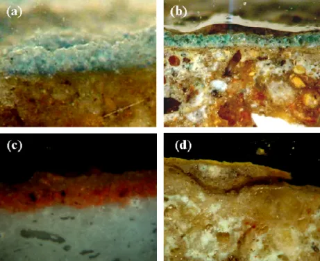
Figure 2 shows optical photomicrograph obtained on the polished cross-sections of the studied painted plasters. From the optical analysis, the investigation of the blue pigment sample shows the pale blue pigment in form of a thick single layer with some dark grains of cuprorivaite applied on a thin medium of white wash (Fig. 2a). The investigation of the green pigment sample shows that the pigment was applied on a thin layer of white wash of intonaco consisting of lime and pinkish particles and the colour of the pigment is a brilliant turquoise (Fig. 2b). The investigation of the red pigment sample shows thick layer of the paint layer with dark grains embedded in the layer (Fig. 2c). The investigation of the yellow pigment sample shows the canary hue of the pigment which is applied in form of a single layer on the plaster layer which is partially detached from the layers below (Fig. 2d).
Fig. 2. Optical photomicrographs obtained by the stereomicroscope and polarized light microscope (PLM) on the polished cross-sections of the painted plasters. a): blue pigment; b): green pigment;
c) red pigment, under the reflected light; d) yellow pigment, under the reflected light.
Microstructure and microanalysis (BSE−EDS)
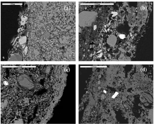
Figure 3 displays BSE micrographs obtained for the plaster layers. The BSE image obtained on a polished cross-section of the blue painted plaster shows clearly the brighter skeletal crystals, probably of cuprorivaite (CuCaSi4O10) (Fig. 3a). The EDS microanalysis
Fig. 3. Backscattered electrons (BSE) micrographs obtained on polished cross-sections of the painted plasters. a): blue painted plaster; b): green painted plaster; c): red painted plaster; d): yellow painted plaster.
The BSE image obtained on a polished cross-section of the green painted plaster shows large crystals of the massive quartz, silica-rich amorphous phase and crystals of parawollastonite [(Ca,Cu)3(Si3O9)] (Fig. 3b). The EDS microanalysis of the sample shows a
high ratio of silicon (34.38%) due to the high amount of glass-bearing materials in the sample, copper (3.81%) was detected with other elements of aluminium, magnesium, potassium, sulphur, iron and phosphorus which are coming from the plaster layer. The BSE image obtained on a polished cross-section of the red painted plaster shows massive granular aggregates and fine particles of the red ochre (Fig. 3c). The EDS spectrum obtained on the red pigment shows that the peak of iron (wt 11.79%) is present which indicates the existence of iron oxide as the possible material producing the red colour, other elements of sulphur and calcium refer to the presence of calcium sulphates, as well as the strong contribution of aluminium and silicon indicates possible existence of an aluminosilicate material. The BSE image obtained on a polished cross-section of the yellow painted plaster shows coarse grains of the pigment are scattered on the surface (Fig. 3d). The EDS microanalysis of the sample shows that silicon, aluminium, sulphur, arsenic, calcium, titanium and iron were detected. The beak of iron in the sample suggests that iron oxide was used to give the yellow colour, while the detection of arsenic (wt 7.03%) suggests that a blended layer of yellow ochre and orpiment (As2S3) was used for decorating. White crystals are noticed embedded in the plaster layer, the
spot microanalysis, performed with the EDS detector, on these white crystals, shows that they consist mainly of strontium (wt 41.04%), sulphur and silicon. Strontium commonly occurs in nature, and is found as the form of the mineral celestite (SrSO4) and the carbonate strontianite
µ- XRF results
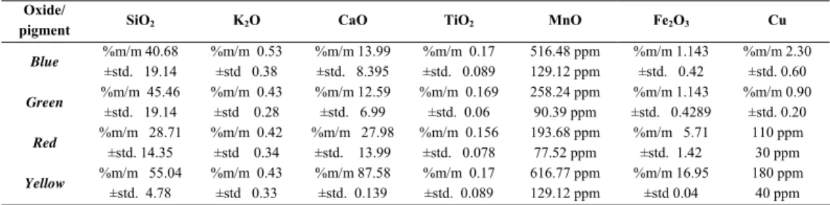
Table 1 and Figure 4 show the results of the µ-XRF analysis of the studied samples.
Table 1. Quantitative analysis obtained by micro XRF of pigments samples (% (m/m)±S.D.)
Cu Fe2O3
MnO TiO2
CaO K2O
SiO2 Oxide/ pigment %m/m 2.30 ±std. 0.60 %m/m 1.143
±std. 0.42 516.48 ppm
129.12 ppm %m/m 0.17
±std. 0.089 %m/m 13.99
±std. 8.395 %m/m 0.53
±std 0.38 %m/m 40.68
±std. 19.14
Blue
%m/m 0.90 ±std. 0.20 %m/m 1.143
±std. 0.4289 258.24 ppm
90.39 ppm %m/m 0.169
±std. 0.06 %m/m 12.59
±std. 6.99 %m/m 0.43
±std 0.28 %m/m 45.46
±std. 19.14
Green
110 ppm 30 ppm %m/m 5.71
±std. 1.42 193.68 ppm
77.52 ppm %m/m 0.156
±std. 0.078 %m/m 27.98
±std. 13.99 %m/m 0.42
±std 0.34 %m/m 28.71
±std. 14.35 Red 180 ppm 40 ppm %m/m 16.95 ±std 0.04 616.77 ppm 129.12 ppm %m/m 0.17
±std. 0.089 %m/m 87.58
±std. 0.139 %m/m 0.43
±std 0.33 %m/m 55.04
±std. 4.78
Yellow
The µ-XRF analysis of the green pigment sample showed that the sample is consists mainly of calcium (Ca) and silicon (Si), with copper (Cu) and iron (Fe) as minor elements in the sample (Fig. 4b). The µ-XRF analysis of the red pigment sample showed that it consists mainly of calcium (Ca), silicon (Si), and iron (Fe) (Fig. 4c). The µ-XRF analysis of the yellow pigment sample revealed that it consists mainly of calcium (Ca) and silicon (Si), and iron (Fe) with minor elements of titanium (Ti) and potassium (K) and traces of arsenic (As) (Fig. 4d).
The mineralogical characterization
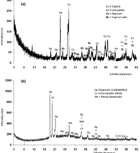
Figure 5 represents XRD patterns of some paint layers. XRD analysis of the blue pigment shows that it consists mainly of calcite (CaCO3) and anhydrite (CaSO4) with minor
amounts of gypsum (CaSO4·2H2O) and cuprorivaite (Fig. 5a). XRD analysis of the green
pigment shows that it consists mainly of gypsum and cristobalite (SiO2) and a secondary
component of parawollastonite in the sample (Fig. 5b). XRD analysis of the red pigment shows that it consists mainly of quartz, minor amounts of calcite, with traces of hematite (Fe2O3).
XRD analysis of the yellow pigment shows that it consists mainly of anhydrite and halite (NaCl) with traces of goethite FeO(OH) and orpiment (As2O3).
Discussions
Plaster layers
From the optical examination we observe two layers of the plasters. XRD analysis showed that the first one in the bottom is the coarse arriccio and it consists mainly of quartz, calcite and calcium sulphates. The ground layer (usually known as white wash) is based on calcite and anhydrite with minor amounts of gypsum.
Blue pigment
The analysis of the blue pigment showed that it consists of Egyptian blue (cuprorivaite, CaCuSi4O10). This pigment was manufactured by mixing calcium salt (carbonate, sulphate or
hydroxide), copper compound (copper oxide or malachite), silica and alkali flux (sources of alkali could have been either natron from areas such as Wadi Natroun and El-Kab, or soda-rich plant ashes) [9]. This mixture was heated to a temperature between 850 and 1000 °C to produce a coloured glass or frit and later ground to powder for use. In the studied sample, residues of arsenic were detected through µ-XRF analysis, which probably due to the use of an arsenic copper ore to produce the pigment.
Green pigment
The analysis of the green pigment shows that Egyptian green was used to produce the colour. Egyptian green is characterized by the presence of parawollastonite, the main phase responsible for the colour is a silica-rich copper glass and the reaction to produce the pigment takes place in a liquid phase [10].
Red pigment
In case of the red pigment sample, red ochre was identified. EDS microanalysis of the sample showed the extensive existence of iron in great proportions. Red ochre used from the 5th Dynasty till the Roman times [11]. Also, EDS microanalysis showed the strong contribution of Al and Si which indicated the existence of an aluminosilicate material, probably from the clay minerals associated with ochres. The presence of Ti in the studied samples could be a result of the presence of ilmenite (FeTiO3) which is found in the Egyptian sand. In red
pigments, the presence of ilmenite possibly forming intergrowths with hematite [12].
Yellow pigment
Conclusions
To accomplish the compositional analysis of the painted limestone blocks discovered at the excavations of Cairo University at Saqqara area, different analytical techniques such as optical microscopy (OM) µ-XRF, scanning electron microscopy (BSE−EDS) and X-ray diffraction analysis were used.
The results showed that the blue pigment was identified as Egyptian blue (Cuprorivaite) and the µ-XRF analysis of the sample showed the detection of following elements: Si, Cu, Ca, K, Fe, Ti and As. The green pigment was identified as Egyptian green, and the µ-XRF analysis of the sample showed the detection of following elements: Si, Cu, Ca, K, Fe and Ti. The red pigment was identified as red pigment, and the µ-XRF analysis of the sample showed the detection of following elements: Ca, Fe, Si, K and Ti. The yellow pigment was identified as a blended layer of ochre and orpiment, and the µ-XRF analysis of the sample showed the detection of following elements: Ca, Fe, Si, Ti, K and As. Moreover, an extensive study to characterize additional samples will help to document the archaeological finds at the excavations of Cairo University at Saqqara area and to establish a database of the chromatic palette used during this period of ancient Egypt.
Acknowledgements
The authors would like to thank Mr. S. Oikonomidis and Mr. G. Mechalidis School of Geology, Aristotle University for SEM investigation and the embedding of the cross-sections.
References
[1] M. Schreiner, M. Melcher, K.Uhlir, Scanning electron microscopy and energy dispersive analysis: applications in the field of cultural heritage, Analytical and Bioanalytical Chemistry, 387, 3, 2006, pp. 737–747.
[2] T. Čechák, L. Musílek, T.Trojek, I. Kopecká, Application of X-Ray Fluorescence Analysis in Investigations of Historical Monuments, Acta Polytechnica, 45, 5, 2005, pp. 48–51. [3] S. Fitzgerald, Non–destructive micro analysis of art and archaeological objects using
micro-XRF, Archeometriai Műhely, 3, 2008, pp. 75–80.
[4] B. Hochleitner, V. Desnica, M. Mantler, M. Schreiner, Historical pigments: a collection analyzed with X-ray diffraction analysis and X-ray fluorescence analysis in order to create a database, Spectrochimica Acta B, 58, 2003, pp. 641–649.
[5] D.N. Papadopoulou, G.A. Zachariadis, A.N. Anthemidis, N.C. Tsirliganis, J.A. Stratis, Development and optimisation of a portable micro-XRF method for in situ multi-element analysis of ancient ceramics, Talanta, 68, 2006, pp. 1692–1699.
[6]M.C. Corbeil, Applications of X-ray diffraction in conservation science and Archaeometry, JCPDS-International Centre for Diffraction Data, Advances in X-ray Analysis,47, 2004, pp. 18–29.
[8] H. Marey Mahmoud, N. Kantiranis, J. Stratis, Salt Damage on the Wall Paintings of the Festival Temple of Thutmosis III Karnak Temples Complex, Upper Egypt. A Case Study,
International Journal of Conservation Science, 1, 3, 2010, pp. 133-142.
[9] G.H. Hatton, A.J. Shortland, M.S. Tite, The production technology of Egyptian blue and green frits from second millennium BC Egypt and Mesopotamia, Journal of Archaeological Science, 35, 6, 2008, pp. 1591–1604.
[10] S. Pagés-Camagna, S. Colinart, The Egyptian Green pigment: Its Manufacturing process and links to Egyptian blue, Archaeometry, 45, 4, 2003, pp. 637–658.
[11] L. Lee, S. Quirke, Painting materials, Ancient Egyptian Materials and Technology
(Editors: Nicholson, T.P. and Shaw, I.),UK, 2001, pp. 104–119.
[12] M. Berry, A study of pigments from a Roman Egyptian shrine, AICCM Bulletin, 24, 1999, pp. 1–9.
[13] A. El Goresy, H. Jaksch, M. Abdel Razek, K.L. Weiner, Ancient pigments in wall paintings of Egyptian tombs and temples: an archaeometric project, Preprint of the Max Planck Institute of Nuclear Physics MPI H - V12, Heidelberg, 1986.