of
ridA
Mutant Phenotypes in
Salmonella enterica
Melissa R. Christopherson, Jennifer A. Lambrecht, Deanna Downs, Diana M. Downs*
Department of Bacteriology, University of Wisconsin–Madison, Madison, Wisconsin, United States of America
Abstract
The RidA (YjgF/YER057c/UK114) family of proteins is broadly conserved in the three domains of life yet the functional understanding of these proteins is at an early stage. Physiological studies ofridA mutant strains ofSalmonella enterica
provided a framework to informin vitrostudies and led to the description of a conserved biochemical activity for this family.
ridA mutant strains of S. enterica have characteristic phenotypes including new synthesis of thiamine biosynthetic intermediate phosphoribosylamine (PRA), inability to grow on pyruvate as a sole carbon and energy source or when serine is present in the minimal growth medium, and a decreased specific activity of transaminase B (IlvE). Secondary mutations restoring growth to aridAmutant in the presence of serine were indapA(encoding dihydrodipicolinate synthase) andthrA
(encoding homoserine dehydrogenase). These mutations suppressed multipleridAmutant phenotypes by increasing the synthesis of threonine. The ability of threonine to suppress the metabolic defects of aridAmutant is discussed in the context of recent biochemical data andin vivoresults presented here.
Citation:Christopherson MR, Lambrecht JA, Downs D, Downs DM (2012) Suppressor Analyses Identify Threonine as a Modulator ofridAMutant Phenotypes in Salmonella enterica. PLoS ONE 7(8): e43082. doi:10.1371/journal.pone.0043082
Editor:Dipshikha Chakravortty, Indian Institute of Science, India
ReceivedMay 9, 2012;AcceptedJuly 16, 2012;PublishedAugust 10, 2012
Copyright:ß2012 Christopherson et al. This is an open-access article distributed under the terms of the Creative Commons Attribution License, which permits unrestricted use, distribution, and reproduction in any medium, provided the original author and source are credited.
Funding:This work was supported by competitive grants GM47296 and GM95837 from the National Institutes of Health (NIH) to DMD and a National Science Foundation Graduate Research Fellowship to MRC. JAL was supported as a trainee on the Molecular Biosciences Training Grant from the NIH (T32 GM07215). DD was the recipient of undergraduate research fellowships from the Department of Bacteriology, the Hilldale Competition, and Merck and Co. The funders had no role in study design, data collection and analysis, decision to publish, or preparation of the manuscript.
Competing Interests:Deanna Downs was supported by an undergraduate fellowship provided by Merck and Co. This fellowship was to the student and had no impact on the design or scope of the work. The inclusion of this fellowship does not alter the authors’ adherence to all the PloS ONE policies on sharing data and materials.
* E-mail: [email protected]
Introduction
The RidA (formerly YjgF/YER057c/UK114) family of proteins is well conserved throughout the three domains of life. Members of this protein family have been implicated in a diverse number of phenotypes in a variety of organisms [1–12]. However, a common mechanism to explain these phenotypes was not obvious. Strains of Salmonella enterica lacking RidA display several characteristic phenotypes, including: synthesis of thiamine biosynthetic in-termediate phosphoribosylamine (PRA), inability to grow on pyruvate as a sole carbon and energy source or in the presence of serine [13], and a decreased specific activity of transaminase B (IlvE) [14]. Each of these phenotypes required the presence of a functional threonine dehydratase (IlvA; EC 4.3.1.19). These phenotypic analyses inSalmonella entericaled to a general model in which RidA eliminated reactive products that were generated in normal metabolic reactions involving IlvA [5].
In vitrostudies, which were informed by the phenotypic analyses, identified a biochemical function for the RidA protein family. RidA deaminated reactive enamine/imine metabolites generated by IlvA [15]. These enamine/imine compounds were normal intermediates in the pyridoxal-59-phosphate-dependent dehydra-tion of both threonine and serine. Further, reconstitudehydra-tion of the PRA formation phenotype required a short-lived intermediate produced by IlvA from threonine. This molecule, presumed to be the 2-aminocrotonate enamine was utilized by anthranilate phosphoribosyltransferase (TrpD; EC 2.4.2.18) to generate PRA
[16]. RidA inhibited the formation of PRA in vitro by this mechanism, which was consistent with the phenotype observed only in aridAmutant.
Aside from the IlvA-, TrpD-dependent formation of PRA, the in vivoconsequences of aridAmutation are not understood in the context of the biochemical activity of RidA. The in vitro bio-chemical work characterizing RidA did not address the signifi-cance of the enamine deaminase activity in vivo or relate the previously observed phenotypes to the in vitro activity. Herein suppressor analyses dissected the basis of the other phenotypes caused by the loss of RidAin vivo. The data showed that threonine reversed many of the phenotypes of aridAmutant ofS. enterica. We propose that threonine outcompetes serine for the active site of threonine dehydratase (IlvA) thus preventing the formation of a deleterious serine-derived reactive intermediate that is normally removed by RidA.
Materials and Methods
Bacterial Strains, Media, and Chemicals
Strains used in this study are derivatives of S. enterica serovar Typhimurium LT2 and are listed with their respective genotypes in Table 1.
(NB) medium. Luria broth was used for experiments involving plasmid isolation. Super Broth containing tryptone (32 g/L), yeast extract (20 g/L), NaCl (5 g/L), and NaOH (5 mM) was used to grow cultures for protein purification. Difco BiTek agar was added (15 g/L) for solid medium. When present in the culture medium the final concentrations of serine and isoleucine were 5 and 0.3 mM, respectively. The final concentrations of the antibiotics in rich and minimal medium, respectively, were: tetracycline, 20, 10 mg/L, chloramphenicol, 20, 5 mg/L, and ampicillin, 150, 15 mg/L. Unless otherwise noted, all chemicals were from Sigma-Aldrich. Aspartate 4-semialdehyde was custom synthesized com-mercially at the University of Canterbury by the Gerrard Laboratory.
Growth Quantification
Cells from overnight cultures in NB medium were pelleted and resuspended in an equal volume of saline (0.85% NaCl), and an aliquot (0.2 mL) was used to inoculate 5 mL of the appropriate minimal medium. Cell growth was monitored as optical density (OD) at 650 nm over time at 37uC with shaking. Growth rates (in h21) were determined as
m= ln(X/X0)/T where X = OD at 650 nm and T = time in hours during logarithmic growth.
Genetic Techniques
Transductional crosses were performed using the high-frequen-cy general transducing mutant of bacteriophage P22 (HT105/1,
int-201) [19]. Methods for transductional crosses, purification from phage, and identification of phage-free transductants have been described elsewhere [20]. Multiply-mutant strains were con-structed using standard genetic techniques. When necessary, genetic backcrosses were performed to confirm the presence of a respective allele.
To isolate mutants, independent cultures of ridA3::MudJ (DM3480) were grown overnight in NB, centrifuged, and resuspended in the same volume of saline. 107cells were spread on solid minimal glucose medium with 5 mM serine. Spontane-ously arising mutations (,1027) that allowedridAmutants to grow
on serine were isolated after 36 hours at 37uC. A transposon (Tn10d(Tc)) genetically linked to the causative mutation in one strain was isolated by standard genetic techniques and used to reconstruct the mutant for phenotypic confirmation. The chro-mosomal location of relevant insertions was determined by sequencing using a PCR-based protocol [21]. A DNA product was amplified with degenerate primers and primers derived from the Tn10d(Tc) insertion sequence and sequenced at the University of Wisconsin Biotechnology Center. Strains carrying suppressor mutations were reconstructed by transducing the relevant allele into dapA::cat (DM10460) and selecting for growth without diaminopimelic acid.
Molecular Techniques
ThedapAgenes from strains DM3480, DM7604, DM7606, and DM11019 were amplified by PCR using Herculase II Fusion DNA Polymerase (Stratagene) and primers 59 DapANdeI (GGGGCATATGTTCACGGGAAGTATTC) and 39DapAXhoI (GGGGCTCGAGTTACAGCAGGCCAGC) and cloned into the pET20b vector (Novagen) at NdeI and XhoI restriction sites. Sequence analysis of each clone confirmed the presence of the N-terminal hexahistidine tag and the relevant lesion. The construct carrying the wild-type allele (pLD-dapA) complemented a dapA mutant (DM10460), indicating that the gene was expressed in this construct (data not shown).
Protein Purification
The wild-type and variant DapA proteins were overexpressed in E. coli BL21(AI) according to the manufacturer’s protocol (Invitrogen). Cells from the resulting cultures were broken at 15,000 psi in a French Pressure cell at 4uC. Cell debris was removed by centrifugation (42,0006g) for 30 min at 4uC. Proteins were purified using a column containing Ni-NTA superflow resin (QIAGEN) according to manufacturer’s protocol. Fractions containing DapA were concentrated at 30 psi under Argon gas using a 10,000 Da molecular weight cut-off membrane (Amicon). The protein was dialyzed in 0.5 M NaCl, 20 mM Tris-HCl, 5 mM imidazole, pH 7.9 and stored at280uC. DapB was purified according to standard protocol using a hexahistidine-taggeddapB clone from the ASKA collection [22]. IlvE was purified as a hexahistidine-tagged protein as has been described [14]. Protein concentration was estimated with bovine serum albumin as the standard using a Bradford assay [23].
Biochemical Assays
i) Dihydrodipicolinate synthase (DapA) assay. DapA activity was measured in a coupled assay with DapB (dihydrodi-picolinate reductase; E.C. 1.3.1.26) following a published protocol [24]. A typical 1 mL reaction contained,2mg DapB, 100 mM HEPES pH 8.0, 0.125 mM NADPH, 40 mM pyruvate, and 0.05– 1.0mg DapA (.95% pure) and was initiated by the addition of ASA at concentrations ranging from 0–2 mM. Enzyme-dependent oxidation of NADPH was quantified at 340 nm.
Table 1.Bacterial strains.
Strain Relevant Genotype* Source
DM3480 ridA3::MudJ Lab collection
DM3871 ridA3::MudJpurF2085 Lab collection DM6309 ridA3::MudJpurF2085 thrA1371 This study DM7608 ridA3::MudJilvA3211 [4] DM7610 ridA3::MudJilvA3210 [4]
DM9404 Wild type (isogenic to DM3480) Lab collection
DM9521 ridA3::MudJdapA356 zxx4116::Tn10d(Tc) This study DM10009 ridA3::MudJilvY3212::Tn10d(Tc)ilvA3210 [4] DM10010 ridA3::MudJilvY3212::Tn10d(Tc) [4] DM10331 ilvY3212::Tn10d(Tc)ilvA3210 [4] DM10332 ilvY3212::Tn10d(Tc) [4]
DM10460 dapA362::cat This study
DM11412 ridA3::MudJpurF2085 dapA356 This study DM11558 ilvY3212::Tn10d(Tc)ilvA3211 [4] DM11609 ridA3::MudJthrA1371 stm0014-13::Tn10d(Tc) [4] DM11635 ridA3::MudJdapA357 This study DM11636 ridA3::MudJdapA358 This study DM11637 ridA3::MudJdapA356 This study DM11638 ridA3::MudJdapA361 This study DM11639 ridA3::MudJdapA359 This study DM11640 ridA3::MudJdapA360 This study DM11877 ridA3::MudJthrA1371 stm0014-13::Tn10d(Tc) This study DM11878 ridA3::MudJstm0014-13::Tn10d(Tc) This study l3520 DasdA1zhf4::Tn10 R. Curtiss III [36]
*MudJ refers to Mud1734 [37]. Tn10d(Tc) refers to the transposition-defective mini-Tn10(Tn10D16D17tetR
ii) Threonine dehydratase (IlvA) assay. IlvA was assayed as previously described [4,25], or alternatively, by quantification of [14C]-2-ketobutyrate (2-KB) formed from [14C(U)]-L-threonine. 200mL reactions containing 100 mM Tris pH 8.0, 50mM pyridoxal-59-phosphate, 20 mM ammonium chloride, 1 mM dithiothreitol (DTT), and 2mg purified IlvA were initiated with a final concentration of 40 mM [14C(U)]-L-Threonine (12.5mCi mmol21), incubated for 12 minutes at 37uC, and stopped with 0.5 mL 0.1% 2,4-dinitrophenylhydrazine in 2 N HCl. Derivatized [14C]-2-KB was extracted with 0.5 mL toluene and radioactivity from 200mL toluene phase, representing quantity of [14C]-2-KB generated, was counted in 5 mL scintillation fluid using a scintil-lation counter (Packard).
iii) Transaminase B (IlvE) assay. The transaminase B activity assay was based on previously described protocols [14,26]. Cells were permeabilized by sonication. Known concentrations of product 2-keto-3-methylvalerate were subjected to the extraction procedure to generate a standard curve.
iv) Homoserine dehydrogenase (ThrA) assay. The homo-serine dehydrogenase activity assay was adapted from a previously described protocol [27]. Cells were grown in 100 mL minimal medium to an OD650 nm of 0.8, pelleted, and resuspended in 0.5 mL 100 mM HEPES pH 8.0 with 0.125 mM DTT. Cells were disrupted by sonication, extract was clarified by centrifuga-tion, and total protein concentration was estimated by the method of Bradford [23]. Assay mixtures contained 100 mM HEPES pH 8.0, 0.125 mM DTT, 200 mM potassium chloride, 0.3 mM NADP+, and,300mg cell extract, in a final volume of 200mL. Assays were initiated by the addition of 15 mM homoserine and activity was monitored by the increase in absorbance at 340 nm at 30uC, representing NADPH production. Inhibitor L-threonine was added to a final concentration of 0.5 mM when indicated.
Results
Alleles ofdapARestore Growth of aridAMutant Strain on Glucose Serine
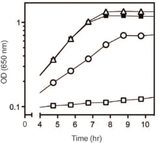
AridAnull mutant (DM3480) cannot grow on minimal glucose medium in the presence of 5 mM serine [13]. Six independent mutant derivatives ofridAthat grew in the presence of serine were isolated. Using Tn10d(Tc) insertions to map the location of the mutations, each of the causative mutations was subsequently found to affect thedapAlocus, encoding dihydrodipicolinate synthase (EC 4.2.1.52). Table 2 summarizes the six lesions that allowed growth of theridAmutant in the presence of serine. Four lesions generated variant DapA proteins (DapAA563G was isolated twice), one affected the Shine-Dalgarno sequence and one was in the dapA promoter. Strains with each of the mutant alleles were reconstructed (DM11635–40) and were analyzed in liquid media for growth in the presence of serine.ridAmutant strains containing allelesdapA356, dapA357,ordapA358grew similar to a wild-type strain in the presence of serine and are represented by strain DM11637 in Figure 1. The parentridAstrain (DM3480) failed to grow after 12 hours as expected. The strain carrying a lesion 36 nucleotides upstream ofdapA(DM11640) had limited growth with serine and was concluded to decrease transcription of the dapA gene. (The promoter ofdapAfromE. coliresides within a 70-base region upstream ofdapAcontaining an extended210 and235 site [28].) Growth of the suppressor-containing strains, with the exception of strainridA dapA359(DM11639), was indistinguishable from the parental strain on minimal glucose medium (data not shown). The dapA359allele encoded a variant with two deleted amino acid residues and despite growth on solid medium with
serine, growth was not detected in liquid media after 24 hours in the absence of exogenous diaminopimelic acid (DAP).
Suppressor Alleles ofdapAEncode Variants with Decreased Specific Activity
The wild-type gene and each of three suppressor alleles ofdapA were cloned into the pET20b vector to generate C-terminal hexahistidine tagged proteins, creating pLD-dapA, pLD-da-pAD188G, pLD-dapAS48Fand pLD-dapAD84–85. The recombinant proteins were purified by affinity chromatography. Wild-type and variant proteins were assayed for dihydrodipicolinate synthase activity using a coupled assay [24]. The variant proteins all had more than a 30-fold decrease in specific activity when compared to the wild-type protein, as shown in Table 2.
A simple interpretation of the above results was that decreased activity of DapA allowed growth of aridAmutant in the presence of serine. Complementation analysis eliminated the formal possibility that an altered function of DapA was responsible for Figure 1. Mutations indapArestore growth toridAmutants in the presence of serine.Growth was monitored over time as optical density at 650 nm. Strains were grown at 37uC in minimal glucose medium with no additions (closed symbols) or 5 mM serine (open symbols). Shown are strains ridA (DM3480), squares; ridA dapA356
(DM11637), triangles; and ridA dapA360 (DM11640), circles. Curves displayed were representative of 3 biological replicates.
doi:10.1371/journal.pone.0043082.g001
Table 2.Suppressing DapA variants have decreased specific activities.
Strain Allele* DNA change
Protein change
Specific activity{
DM9404 WT – – 5.1061.60
DM11637 dapA356 A563G D188G 0.1260.04 DM11635 dapA357 C143T S48F 0.1560.04 DM11636 dapA358 A(210)T – N.D.{
DM11637 dapA359 DG249–C254 DE84–A85 0.026,0.01
DM11640 dapA360 T(236)C – N.D.
DM11638 dapA361 A563G D188G N.D.
*AridAstrain carrying any of the listed alleles is able to grow in the presence of serine.
{
Specific activity of DapA inmmol NADPH oxidized/sec/mg of purified protein.
{
N.D. = not determined.
allowing growth of aridA mutant. When providedin trans, wild-typedapAeliminated growth of theridA dapA356mutant strain in the presence of serine and did not affect growth of aridAmutant (data not shown).
Aspartate 4-semialdehyde Accumulation Mediated Phenotypic Suppression by thedapAAlleles
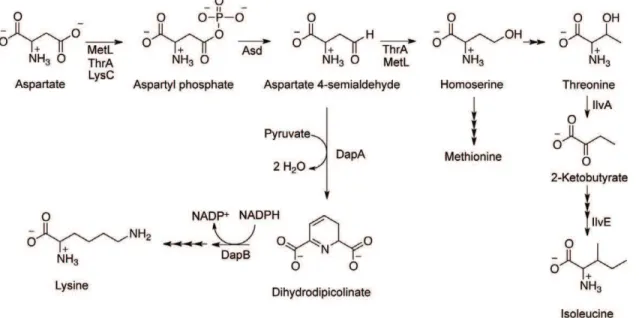
DapA functions in the synthesis of some aspartate-derived amino acids and uses aspartate 4-semialdehyde (ASA) as a substrate (Figure 2). In one scenario, a recessive lesion in dapA results in accumulation of ASA that allows a ridA mutant to grow in the presence of serine. ASA itself restored the growth of aridAmutant in the presence of serine, supporting a role for this molecule in suppression of theridAphenotype. As little as 0.5 mM ASA in the medium allowed aridA mutant to reach full density in medium with 5 mM serine. Growth rate (m) of ridA (DM3480) in the presence of serine (m= 0.0660.01) was restored by 1 mM ASA (m= 0.5560.03) and was the same as the growth rate of the same strain grown on minimal medium without serine (m= 0.5460.03). The nutritional requirements of anasdmutant (methionine, lysine, DAP, and threonine), which cannot make ASA, [29] were satisfied with ,1.3 mM exogenous ASA, indicating the cells have the
ability to transport and incorporate ASA into the biosynthetic pathways (data not shown).
In addition to suppressing serine sensitivity, the dapA alleles restored IlvE activity in aridAmutant. The IlvE activity in theridA strain carrying the dapA356 allele (23067 nmol/min/mg) was restored to an intermediate level between the wild-type (303613 nmol/min/mg) and ridA mutant strain (14067 nmol/ min/mg). This result suggested intracellular accumulation of ASA could impact the activity of IlvE in aridAmutant. No evidence of a direct role for ASA in mediating phenotypic suppression was found. The activity of purified IlvE was not significantly affected by 10 min incubation with 10 mM ASA (26.167mmol/min/mg without ASA, 18.666mmol/min/mg with ASA). Further, ASA had no detectable effect on the activity of threonine deaminase (IlvA)in vitro.While as little as 500mM isoleucine inhibited IlvA, ASA failed to inhibit IlvA in vitro at a range of concentrations
(0.1mM –1.0 mM) (data not shown). These data showed that the effect of ASA was not due to mimicking the effect of isoleucine as a feedback inhibitor [14], and suggest that further metabolism of this molecule was required.
Analysis of a Second Suppressor Locus Provides Insight into Role of ASA
In addition to the alleles ofdapAdescribed above, a mutation in thrA(thrA1371), encoding aspartokinase I/homoserine dehydroge-nase I, previously reported to suppress serine sensitivity of aridA mutant [4] was sequenced and found to encode variant ThrAG403D. The homoserine dehydrogenase activity in a strain with the ThrAG403Dvariant was indistinguishable from the wild-type parental strain. The location of the G403D substitution suggested the variant could be altered in allosteric interaction properties [30–32]. Data in Table 3 showed that the homoserine dehydrogenase activity of the ThrAG403Dvariant was resistant to inhibition by threonine. Significantly, this effect was evident at a low of concentration of threonine, as would be expected under in vivoconditions where the threonine concentration was reported to be 0.2 mM [33]. Taken together, the data suggested the ThrAG403D variant could increase conversion of ASA to
homo-Figure 2. Pathway for synthesis of aspartate-derived amino acids.Aspartate is a precursor to lysine, methionine, threonine, and isoleucine, as depicted here. Aspartate 4-semialdehyde (ASA) is a branchpoint metabolite controlled by the activities of DapA, ThrA, and MetL.
doi:10.1371/journal.pone.0043082.g002
Table 3.The ThrAG403Dvariant is insensitive to feedback inhibition by threonine and serine.
Homoserine dehydrogenase activity*
thrAallele Protein variant No inhibitor +Thr (0.5 mM)
thrAWT WT 4465 1863
thrA1371 ThrAG403D 3765 3866
*Homoserine dehydrogenase activity was measured in crude extracts from isogenic strains DM11877 (ridA thrA1371) and DM11878 (ridA) by following reduction of NADP+and was reported asDA420 nm/min/mg protein.
serinein vivo, consistent with the above conclusion that metabolism of ASA is required for suppression.
Threonine, not Isoleucine is the Metabolite Responsible for Suppression
ASA is a biosynthetic precursor to isoleucine, which is known to allow aridAmutant to grow in the presence of serine [13], so it was a formal possibility that ASA was correcting growth by leading to increased levels of isoleucine. Two IlvA variants with decreased threonine dehydratase activity were used to constrict flux between ASA and isoleucine. Neither of theilvAalleles caused a detectable growth defect on minimal glucose medium (Table 4). However, they each resulted in derepression of theilvoperon [4] indicating the strains were limited for isoleucine. Despite the constriction of flux between ASA and isoleucine, the double mutants ridA ilvA3210(DM10009) andridA ilvA3211(DM11558) had the same growth rates as a ridA mutant (DM10010) (m= 0.5360.10, 0.5460.04, and 0.5660.01, respectively) when grown in a minimal medium containing 5 mM serine and 1 mM ASA. These data suggested that ASA did not correct growth by increasing intracellular isoleucine levels.
Other metabolites in the pathway from ASA to the branch chain amino acids were considered and tested for their ability to suppress growth of a ridA mutant with serine. Nutritional tests showed qualitative suppression of multiple phenotypes with both homoserine and threonine. Addition of exogenous threonine to the growth medium of a ridA mutant restored growth on serine (m= 0.0960.01 without threonine, 0.5060.01 with threonine), growth on pyruvate (m= 0.0660.01 without threonine, 0.3760.02 with threonine), and IlvE activity (160631 nmol/min/mg in minimal medium without threonine versus 287633 nmol/min/ mg in minimal with threonine).
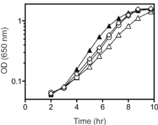
Threonine is a precursor in PRA formation in aridA mutant [16]. This fact provided a means to directly test whether the suppressor mutations indapAandthrAgenerated increased cellular threonine levels. If thedapAandthrAmutations acted by increasing flux to threonine, they would be expected to increase the PRA formed in aridA mutant. A purFmutant strain background was used to detect PRA, as it requires PRA to make thiamine and allow growth. The data in Figure 3 showed that thethrAanddapA suppressors increased growth of apurF ridAstrain, and exogenous threonine further increased growth. These results supported the conclusion that flux to threonine was increased by these mutations. Additionally, since isoleucine has been shown to have the opposite effect and inhibit PRA synthesis in aridAmutant [13], these data were consistent with the interpretation that the dapA mutations
were not increasing the synthesis of isoleucine. Considering the results of nutritional and suppressor analyses in total, threonine was identified as the metabolite that had a direct effect in suppressing the phenotypes caused by lack of RidA.
Discussion
The RidA (YjgF/YER057c/UK114) family of proteins is highly conserved, but the diverse cellular defects caused by its absence are not understood [1–11]. Recently it was shownin vitro that RidA family members deaminate reactive enamine/imine intermediates generated by threonine dehydratases (e.g., IlvA) [15]. This study investigated the relationship between the characterized biochem-ical activity of RidA and thein vivophenotypes observed in aridA mutant inS. enterica. Suppressor analyses identified an important role for threonine in attenuating multiple phenotypes of a ridA strain, including sensitivity to exogenous serine, lack of growth on pyruvate, and a decreased specific activity of IlvE.
When considering the results of this study in combination with the biochemical activity of RidA, we proposed a mechanism by which threonine could suppress the mutant phenotypes. Our model predicted that threonine relieved the sensitivities of aridA mutant by outcompeting serine in the IlvA active site. Threonine dehydratase (IlvA) was required for a number ofridAphenotypes [4,13,14,16]. The fact that threonine reversed those phenotypes suggested the metabolic defects required IlvA to use a different substrate. To our knowledge, the only other reported physiological substrate of IlvA is serine, and IlvA has a much higher Km for serine than for threonine (90 mM versus 4.5 mM, respectively [34]). Threonine and serine use the same active site in IlvA [35], and the presence of additional threonine would preclude IlvA from binding and dehydrating serine instead. This model suggested that the intermediate derived from serine, but not threonine, was deleterious to the cell unless it was removed by RidA.
The significance of threonine as a key metabolite that can modulate the ridA serine-sensitivity phenotype was further emphasized by the saturation of the suppressor analyses. Repeated attempts to isolate serine-resistant mutants only produced the decreased activitydapA (dihydrodipicolinate synthase) alleles and the feedback-resistant thrA (homoserine dehydrogenase) allele
Table 4.IlvA variants have reduced activity.
ilvA allele
Protein
variant Activity* m{(Glc) m{(Glc Ile)
ilvA WT WT 0.2260.01 0.5460.05 0.5360.01
ilvA3210 IlvAA142T B.D.{ 0.6260.01 0.6060.03
ilvA3211 IlvAG191S 0.0560.01 0.5660.03 0.5460.01
*Threonine dehydratase (IlvA) activity measured in crude extracts from DM3480 (ridA), DM7610 (ridA ilvA3210) and DM7608 (ridA ilvA3211) and reported as DA540 nm/min/mg protein.
{
Growth rate (in h21) (
m= ln(X/X0)/T where X = optical density at 650 nm and T = time in hours during logarithmic growth) for strains DM10332 (WT), DM10331 (ilvA3210), and DM11558 (ilvA3211) determined from growth in minimal medium with glucose (Glc) and glucose with isoleucine (Glc Ile).
{
Below Detection.
doi:10.1371/journal.pone.0043082.t004
Figure 3. Suppressor mutations increase growth inpurF ridA strain background.Strains were grown at 37uC in minimal glucose medium with adenine (open symbols) or further supplemented with 0.3 mM threonine (closed symbols). Growth was monitored over time as optical density at 650 nm. Shown are strainspurF ridA(DM3871), triangles; purF ridA thrA1371 (DM6309), diamonds; and purF ridA dapA356(DM11412), circles. Error bars represent standard deviations of three biological replicates.
described here. These mutants not only demonstrated that increased flux to threonine was key to reversing the serine-sensitivity of a ridA mutant, but they also suggested that the primary control of threonine levels in the cell occurs at the homoserine dehydrogenase step and can be affected by increasing substrate (ASA) or decreasing the allosteric control of ThrA. This finding has important implications for metabolic engineering and groups endeavoring to generate organisms that overproduce threonine or downstream metabolites.
The findings herein emphasized the central role of threonine in compensating for the lack of RidA. In combination with past results, these data refine a model to explain the phenotypes ofridA mutants. It has been shown that IlvA generates reactive enamine/ imines that are removed by RidA [15]. We suggest that serine is used as a substrate by IlvA to generate a reactive intermediate that attacks cellular components if it is not quenched by RidA. This is in contrast to the reactive intermediate derived from threonine reported to serve as a substrate for an alternative mechanism of PRA synthesis [16]. Thus, the IlvA-generated intermediates that accumulate in vivo in the absence of RidA can have either deleterious or productive consequences, depending on the
sub-strate used (e.g.,serine versus threonine). Together these results suggest a complex role for IlvA in thein vivo phenotypes ofridA mutants. Continued studies are needed to identify the diversity of both the reactive metabolites eliminated by RidA and the targets of these reactive intermediates to better understand the breadth of metabolic consequences that result from the lack of the conserved RidA protein.
Acknowledgments
We thank Dr. George Schmitz for isolating theridAsuppressor mutants in the presence of serine and for the initial characterization of thethrA1371 allele, Benjamin Bice for assaying IlvA variants, and Rebecca Schomer for performing the IlvE assay in the presence of threonine.
Author Contributions
Conceived and designed the experiments: DMD JL MC. Performed the experiments: JL MC DD. Analyzed the data: DMD JL MC DD. Contributed reagents/materials/analysis tools: JL MC DD. Wrote the paper: DMD JL MC.
References
1. Schmiedeknecht G, Kerkhoff C, Orso E, Stohr J, Aslanidis C, et al. (1996) Isolation and characterization of a 14.5-kDa trichloroacetic-acid-soluble trans-lational inhibitor protein from human monocytes that is upregulated upon cellular differentiation. Eur J Biochem 242: 339–351.
2. Oxelmark E, Marchini A, Malanchi I, Magherini F, Jaquet L, et al. (2000) Mmf1p, a novel yeast mitochondrial protein conserved throughout evolution and involved in maintenance of the mitochondrial genome. Mol Cell Biol 20: 7784–7797.
3. Kim JM, Yoshikawa H, Shirahige K (2001) A member of the YER057c/YjgF/ UK114 family links isoleucine biosynthesis and intact mitochondria mainte-nance inSaccharomyces cerevisiae. Genes Cells 6: 507–517.
4. Christopherson MR, Schmitz GE, Downs DM (2008) YjgF is required for isoleucine biosynthesis whenSalmonella entericais grown on pyruvate medium. J Bacteriol 190: 3057–3062.
5. Browne BA, Ramos AI, Downs DM (2006) PurF-independent phosphoribosyl amine formation inyjgFmutants ofSalmonella entericautilizes the tryptophan biosynthetic enzyme complex anthranilate synthase-phosphoribosyltransferase. J Bacteriol 188: 6786–6792.
6. Goupil-Feuillerat N, Cocaign-Bousquet M, Godon JJ, Ehrlich SD, Renault P (1997) Dual role of alpha-acetolactate decarboxylase inLactococcus lactissubsp. lactis. J Bacteriol 179: 6285–6293.
7. Farkas A, Nardai G, Csermely P, Tompa P, Friedrich P (2004) DUK114, the
Drosophilaorthologue of bovine brain calpain activator protein, is a molecular chaperone. Biochem J 383: 165–170.
8. Leitner-Dagan Y, Ovadis M, Zuker A, Shklarman E, Ohad I, et al. (2006) CHRD, a plant member of the evolutionarily conserved YjgF family, influences photosynthesis and chromoplastogenesis. Planta 225: 89–102.
9. Marchini A, Accardi R, Malanchi I, Schyr E, Oxelmark E, et al. (2002)
Schizosaccharomyces pombePmf1p is structurally and functionally related to Mmf1p ofSaccharomyces cerevisiae. Yeast 19: 703–711.
10. Morishita R, Kawagoshi A, Sawasaki T, Madin K, Ogasawara T, et al. (1999) Ribonuclease activity of rat liver perchloric acid-soluble protein, a potent inhibitor of protein synthesis. J Biol Chem 274: 20688–20692.
11. D’Inca R, Marteil G, Bazile F, Pascal A, Guitton N, et al. (2010) Proteomic screen for potential regulators of M-phase entry and quality of meiotic resumption inXenopus laevisoocytes. J Proteomics 73: 1542–1550.
12. Kim KS, Pelton JG, Inwood WB, Andersen U, Kustu S, et al. (2010) The Rut pathway for pyrimidine degradation: novel chemistry and toxicity problems. J Bacteriol 192: 4089–4102.
13. Enos-Berlage JL, Langendorf MJ, Downs DM (1998) Complex metabolic phenotypes caused by a mutation inyjgF, encoding a member of the highly conserved YER057c/YjgF family of proteins. J Bacteriol 180: 6519–6528. 14. Schmitz G, Downs DM (2004) Reduced transaminase B (IlvE) activity caused by
the lack ofyjgFis dependent on the status of threonine deaminase (IlvA) in
Salmonella entericaserovar Typhimurium. J Bacteriol 186: 803–810.
15. Lambrecht JA, Flynn JM, Downs DM (2012) Conserved YjgF protein family deaminates reactive enamine/imine intermediates of pyridoxal 59-phosphate (PLP)-dependent enzyme reactions. J Biol Chem 287: 3454–3461.
16. Lambrecht JA, Browne BA, Downs DM (2010) Members of the YjgF/ YER057c/UK114 family of proteins inhibit phosphoribosylamine synthesis
in vitro. J Biol Chem 285: 34401–34407.
17. Vogel HJ, Bonner DM (1956) Acetylornithinase of Escherichia coli: partial purification and some properties. J Biol Chem 218: 97–106.
18. Balch WE, Fox GE, Magrum LJ, Woese CR, Wolfe RS (1979) Methanogens: reevaluation of a unique biological group. Microbiol Rev 43: 260–296. 19. Schmieger H (1972) Phage P22-mutants with increased or decreased
trans-duction abilities. Mol Gen Genet 119: 75–88.
20. Downs DM, Petersen L (1994)apbA, a new genetic locus involved in thiamine biosynthesis inSalmonella typhimurium. J Bacteriol 176: 4858–4864.
21. Caetano-Anolles G (1993) Amplifying DNA with arbitrary oligonucleotide primers. PCR Methods Appl 3: 85–94.
22. Kitagawa M, Ara T, Arifuzzaman M, Ioka-Nakamichi T, Inamoto E, et al. (2005) Complete set of ORF clones ofEscherichia coliASKA library (a complete set ofE. coliK-12 ORF archive): unique resources for biological research. DNA Res 12: 291–299.
23. Bradford MM (1976) A rapid and sensitive method for the quantitation of microgram quantities of protein utilizing the principle of protein-dye binding. Anal Biochem 72: 248–254.
24. Yugari Y, Gilvarg C (1965) The condensation step in diaminopimelate synthesis. J Biol Chem 240: 4710–4716.
25. Burns RO (1971) L-Threonine deaminase–biosynthetic (Salmonella typhimurium). Methods Enzymol. 555–560.
26. Duggan DE, Wechsler JA (1973) An assay for transaminase B enzyme activity in
Escherichia coliK-12. Anal Biochem 51: 67–79.
27. Angeles TS, Smanik PA, Borders C, Jr, Viola RE (1989) Aspartokinase-homoserine dehydrogenase I fromEscherichia coli: pH and chemical modification studies of the kinase activity. Biochemistry 28: 8771–8777.
28. Acord J, Masters M (2004) Expression from theEscherichia coli dapApromoter is regulated by intracellular levels of diaminopimelic acid. FEMS Microbiol Lett 235: 131–137.
29. Jagusztyn-Krynicka EK, Smorawinska M, Curtiss R 3rd (1982) Expression of
Streptococcus mutans aspartate-semialdehyde dehydrogenase gene cloned into plasmid pBR322. J Gen Microbiol 128: 1135–1145.
30. Szczesiul M, Wampler DE (1976) Regulation of a metabolic systemin vitro: synthesis of threonine from aspartic acid. Biochemistry 15: 2236–2244. 31. Omori K, Imai Y, Suzuki S, Komatsubara S (1993) Nucleotide sequence of the
Serratia marcescens threonine operon and analysis of the threonine operon mutations which alter feedback inhibition of both aspartokinase I and homoserine dehydrogenase I. J Bacteriol 175: 785–794.
32. Paris S, Viemon C, Curien G, Dumas R (2003) Mechanism of control of
Arabidopsis thaliana aspartate kinase-homoserine dehydrogenase by threonine. J Biol Chem 278: 5361–5366.
33. Bennett BD, Kimball EH, Gao M, Osterhout R, Van Dien SJ, et al. (2009) Absolute metabolite concentrations and implied enzyme active site occupancy in
Escherichia coli. Nat Chem Biol 5: 593–599.
34. Burns RO, Hofler JG, Luginbuhl GH (1979) Threonine deaminase from
Salmonella typhimurium. Substrate-specific patterns of inhibition in an activator site-deficient form of the enzyme. J Biol Chem 254: 1074–1079.
35. Hofler JG, Burns RO (1978) Threonine deaminase fromSalmonella typhimurium. Effect of regulatory ligands on the binding of substrates and substrate analogues to the active sites and the differentiation of the activator and inhibitor sites from the active sites. J Biol Chem 253: 1245–1251.
37. Castilho BA, Olfson P, Casadaban MJ (1984) Plasmid insertion mutagenesis and
lacgene fusion with mini-mubacteriophage transposons. J Bacteriol 158: 488– 495.
38. Way JC, Davis MA, Morisato D, Roberts DE, Kleckner N (1984) New Tn10