Mitochondrial Dysfunction Is Involved in the Toxic
Activity of Boric Acid against
Saprolegnia
Shimaa E. Ali1, Even Thoen1,2, Øystein Evensen2, Jannicke Wiik-Nielsen1, Amr A. A. Gamil2, Ida Skaar1*
1Norwegian Veterinary Institute, Oslo, Norway,2Norwegian University of Life Sciences, Oslo, Norway
Abstract
There has been a significant increase in the incidence ofSaprolegniainfections over the past decades, especially after the banning of malachite green. Very often these infections are associated with high economic losses in salmonid farms and hatcheries. The use of boric acid to control the disease has been investigated recently both under in vitro and in vivo conditions, however its possible mode of action against fish pathogenicSaprolegniais not known. In this study, we have explored the transformation inSaprolegniaspores/hyphae after exposure to boric acid (1 g/L) over a period 4–24 h post treatment. Using transmission electron microscopy (TEM), early changes inSaprolegniaspores were detected. Mitochondrial degeneration was the most obvious sign observed following 4 h treatment in about 20% of randomly selected spores. We also investigated the effect of the treatment on nuclear division, mitochondrial activity and function using confocal laser scanning microscopy (CLSM). Fluorescence microscopy was also used to test the effect of treatment on mitochondrial membrane potential and formation of reactive oxygen species. Additionally, the viability and proliferation of treated spores that correlated to mitochondrial enzymatic activity were tested using an MTS assay. All obtained data pointed towards changes in the mitochondrial structure, membrane potential and enzymatic activity following treatment. We have found that boric acid has no effect on the integrity of membranes ofSaprolegniaspores at concentrations tested. It is therefore likely that mitochondrial dysfunction is involved in the toxic activity of boric acid againstSaprolegniaspp.
Citation:Ali SE, Thoen E, Evensen Ø, Wiik-Nielsen J, Gamil AAA, et al. (2014) Mitochondrial Dysfunction Is Involved in the Toxic Activity of Boric Acid against Saprolegnia. PLoS ONE 9(10): e110343. doi:10.1371/journal.pone.0110343
Editor:Guillermo Lo´pez Lluch, Universidad Pablo de Olavide, Centro Andaluz de Biologı´a del Desarrollo-CSIC, Spain
ReceivedMay 13, 2014;AcceptedSeptember 20, 2014;PublishedOctober 29, 2014
Copyright:ß2014 Ali et al. This is an open-access article distributed under the terms of the Creative Commons Attribution License, which permits unrestricted use, distribution, and reproduction in any medium, provided the original author and source are credited.
Data Availability:The authors confirm that all data underlying the findings are fully available without restriction. All relevant data are within the paper.
Funding:The work has been funded by the European Commission through the EU Marie Curie ITN project SAPRO (238550). The funders had no role in study design, data collection and analysis, decision to publish, or preparation of the manuscript.
Competing Interests:The authors have declared that no competing interests exist. * Email: [email protected]
Introduction
Saprolegniosisis a common problem in cultured freshwater fish. The disease is caused by species in the genusSaprolegnia,which belongs to the class Oomycetes. Infection can cause significant mortality among developing salmonid eggs and fry and contributes to severe economic losses [1]. Moreover, this problem can persist in the presence of treatment due to biofilms as described recently [2]. Outbreaks of saprolegniosis in aquaculture have increased after the banning of malachite green [3]. Thus, there is an urgent need to find efficacious alternatives for prophylaxis and treatment against this pathogen. Boron is omnipresent in nature and forms inorganic borate compounds when it binds to oxygen [4]. Although boron and its compounds have been identified as growth stimulators for fish [5,6] in a relatively high concentration, they can also be used to control bacterial and fungal infection. Boric acid for example has been used as an effective and safe candidate for controlling yeast and fungal infections in humans and plants [7–9]. Generally, the exact mode of action of BA is still not fully known. However, some studies indicated that mitochon-drial degeneration and consequent inhibition of oxidative metab-olism were the most prominent features observed following boric acid treatment [7,10]. A recent study suggested that boric acid could be used effectively to limit saprolegniosis in Atlantic salmon eyed eggs and yolk sac fry [11]. Therefore, we aimed to reveal the mechanisms underlying the activity of the boric acid in the control
of saprolegniosis. The effect of boric acid on nuclear division in
Saprolegnia spores/hypha, mitochondrial activity, distribution and cell membrane integrity was studied by means of transmission electron microscopy (TEM), confocal laser scanning microscopy (CLSM) and fluorescence microscopy. The general metabolism, viability and proliferation of treated spores were also tested using a MTS tetrazolium compound (MTS assay).
Materials and Methods
Chemical treatments
Boric acid (BA), H3BO3, M 61.83 g/mol (Merck) was used as a source of borate for thein vitrotreatment ofSaprolegniaspores/ hyphae. Boric acid was dissolved in sterilized aquarium water (SAW) to give a concentration of 1 g/L. Bronopol (Pyceze, Novartis) was included as a positive treatment control for all tested candidates (100 mg/L). Samples with no treatment diluted in SAW were included in all analysis (non-treated water control).
Saprolegniaisolates and zoospore production
To investigate the internal changes in Saprolegnia spores/ hyphae elicited by boric acid treatment, three strains of
Saprolegniahyphae were excised from colonized GY agar plates and incubated in GY broth at 15uC for 2 days to obtain further hyphal growth. Subsequently, bundles of these young hyphae were washed twice in SAW, transferred to two glass bottles containing one liter of SAW and incubated at 21uC for 24 h to allow extensive zoospore production.
Alterations inSaprolegniaspores following boric acid treatment using transmission electron microscopy (TEM)
The early morphological changes in BA treated Saprolegnia
spores and non-treated controls were investigated by TEM. A similar method to that described by Shi et al (2011) was used. Briefly, spores were concentrated by centrifugation at 16,0006g for 15min. Boric acid treatment was applied at 1 g/L for 4 h. Treated spores and non-treated controls were fixed in a solution of 1.25% glutaraldehyde and 2% paraformaldehyde in SAW. The pellets were rinsed thoroughly in 0.1M sodium cacodylate buffer (SCB). Gels of 1–2 mm3were prepared by adding 3% low gelling
temperature agarose in distilled water to the pellet. The gels were post-fixed with 1% osmium tetroxide in SCB for 2 h at room temperature. After thorough rinsing with SCB, the gels were dehydrated with 10 min stages in an ascending ethanol series (50– 100%). The samples were embedded in LR White resin. Ultrathin sections were obtained with a Leica EM UC6 Ultramicrotome and stained by 4% uranyl acetate and 1% potassium permanganate for 10 min. Mitochondrial changes in the spores were detected using a FEI Morgagni 268 transmission electron microscope at 80 kV.
Effect of boric acid on the nuclear division and germination ofSaprolegnia spores
The fluorescent dye 49,6-diamidino-2-phenylindole (DAPI) dilactate (Invitrogen) was used to observe nuclear changes and division in BA treatedSaprolegniaspores and controls. DAPI is a nucleic acid specific dye [13]. It has been used to visualize nuclear changes in treated yeast [14] and fungi [8]. A stock solution was prepared by dissolving 10 mg of the probe in 2 mL of deionized Figure 1. Alterations in Saprolegnia spores following boric acid treatment.Transmission electron microscopy image of an untreated Saprolegniaspore (a) and aSaprolegniaspore treated with boric acid (1 g/L) for 4 h (b). Normal, well defined mitochondrial structure is seen in the non-treated spores (a1 and a2) compared to the spore that has been exposed to boric acid (b1 and b2). Different degrees of degenerative changes were observed in the mitochondria of the treated spore (circle). The condensed nucleus (N) with disintegrated nuclear membrane is seen in the treated spore (b1), but this was not a consistent finding and seen only in a few spores.
doi:10.1371/journal.pone.0110343.g001
water (dH2O) to have final concentration of 5 mg/ml. Freshly harvestedSaprolegnia spores were exposed to 1 g/L BA in the presence of 0.5% Glucose-yeast broth (GY broth) [15]. Treated spores and non-treated controls (spores in SAW) were incubated for 2, 4, 6 and 8 h at 15uC. Later on, spores were concentrated by centrifugation, 16,0006g for 10 min, fixed in 70% ethanol and
incubated overnight at 4uC. Spores were centrifuged again and washed with SAW to remove ethanol. Spores were then seeded into 4-well chambered slides, and incubated in a staining solution of 25 nM 49,6-diamidino-2-phenylindole (DAPI) [16]. Samples were examined by confocal laser scanning microscopy (CLSM) (Zeiss LSM710) with a 405 nm laser.
Effect of boric acid on mitochondrial activity, distribution and membrane potential using confocal laser scanning microscopy (CLSM)
MitoTracker Red CMTMRos probes (50mg; Invitrogen) were used to assess the mitochondrial changes in spores and hyphae after BA treatment. MitoTracker is a mitochondrion-specific stain and its accumulation depends on the membrane potential.
Stock solution was prepared by dissolving 50mg of the stain in anhydrous dimethylsulfoxide (DMSO) (Invitrogen) to obtain 1 mM concentration. Stock solution was diluted in GY broth to give a 50 nM MitoTracker working solution. Saprolegniaspores were treated with BA 1 g/L for 4, 12 and 24 h and suspended in 50 nM MitoTracker solution. After 10–15 min incubation in the dark, excess stain was washed out and the spores were seeded in chambered slides and examined directly with CLSM, generating single plane images using a 561 nm laser line to excite the MitoTracker Red. To follow the changes within treated
Saprolegniahyphae, mycelia were prepared as follows: Saproleg-niacultures grown on agar plugs were incubated in GY broth for more mycelial growth. The agar plug was removed from the broth
and re-suspended in BA (1 g/L). The same protocol as described for spores was applied for mycelia. Stained, treated mycelia were examined directly using ordinary microscopic slides. Spores/ mycelia in water served as non-treated control. The images were generated on a Zeiss LSM 710 confocal microscope using a 561 nm laser to excite the Mitotracker red, through a 63x plan apochromat oil immersion objective. The fluorescence intensity was measured in spores/hyphae, following 24 h treatment, as spot measurements (25/hyphal apex, spot size: approx. 850 pixels) within the hyphal apex or spore. Measurements were performed using ZEN software (Zeiss). Mitotracker red is a live-cell dye, and uptake of the dye is expected to be higher in intact mitochondria than in compromised ones. Fluorescence intensity was thus used as a quantitative indicator of damage, alongside the visual difference in distribution.
Mitochondrial membrane potential and reactive oxygen species (ROS) using fluorescence microscopy
The cell permeating dye tetramethyl rhodamine, ethyl ester (TMRE) 1 mM in DMSO (abcam) was used together with dichlorofluorescein diacetate (DCFDA) cellular reactive oxygen species detection assay kit 20 mM (abcam). TMRE dye was used for labeling mitochondrial membrane potential and function [17] and DCFDA as an indicator for the reactive oxygen species (ROS) activity within boric acid treated hyphae and the non-treated control. DCFDA is able to diffuse into the cell and is de-acetylated by cellular esterases to a non-fluorescent compound, which is later oxidized by ROS into 29,79–dichlorofluorescin (DCF). DCF is a highly fluorescent compound which can be detected by fluores-cence microscopy. Briefly, hyphae on agar plugs grown in GY broth were exposed to BA treatment for 24 h and those without treatment were kept as a control. Preparations were suspended in 300ml of dye (50ml of TMRE working solution (100mm) and Figure 2. Effect of boric acid on the nuclear division and germination ofSaprolegniaspores.Confocal laser scanning microscopy images ofSaprolegniaspores stained with the nucleic acid dye DAPI. a1–a4) Spore germination in non-treated water control group. Note the movement of the nucleus towards the newly developing germ tube following 2 and 4 h incubation (a1 and a2). Development of multinuclear hyphae indicating growth and viability is shown in image a3 and a4. b1–b4) Gradual reduction of fluorescence intensity ofSaprolegniaspores treated with boric acid following 2, 4, 6 and 8 h of incubation, b1, b2, b3 and b4 respectively. No nuclear division was observed in the treated group.
doi:10.1371/journal.pone.0110343.g002
250ml of DCFDA diluted in SAW (10mm)). They were then incubated in the dark for 45–60 min before the excess unbound dyes were washed out and examined directly with fluorescence microscopy (Olympus IX81 motorized inverted microscope).
Assessing mitochondrial activity and viability of boric acid treatedSaprolegniaspores using MTS assay
CellTiter 96 aqueous one solution cell proliferation assay (Promega) was used. This assay involves the reduction of tetrazolium compound [3-(4,5-dimethylthiazol-2-yl)-5-(3-carboxy-methoxyphenyl)-2-(4-sulfophenyl)-2H-tetrazolium, inner salt; MTS] by viable cells to form formazan products from MTS reduction. The reaction occurs when mitochondrial enzymes are active (Mitochondrial dehydrogenase) [18], and could be used as an indicator for the metabolic rate of the mitochondria [19]. Briefly, 96-well plates were seeded with 100ml of Saprolegnia
spores in 10% GY broth. 50ml of boric acid was added to each tested well (except controls) to have a final concentration of 1 g/L. Bronopol was used as a positive treatment control and SAW as a negative control. MTS reagent was added to all tested wells and controls (50ml/well). Wells with the MTS reagent without spores were used as a background control. The plates were incubated at 15–20uC. Reading was first recorded on 96-well plate reader following 4 h incubation, then after 8 and 24 h of treatment application. The amount of the formazan product was measured by the absorbance at 490 nm. The MTS reduction is directly proportional to the number and activity of the living spores. Background absorbance was also measured. Spore viability was calculated as follows: % viability = 1006(O.D490 value for the sample – mean background O.D490)/(mean O.D490 for non-treated water control – mean background O.D490).
Figure 3. Effect of boric acid onSaprolegniamitochondria using confocal laser scanning microscopy.Confocal laser scanning microscopy showing the effect of the boric acid onSaprolegniaspore (a) and hyphal (b) mitochondrial activity using MitoTracker Red. a1) Accumulation of the stain in the non-treated control. Gradual reduction in the number of mitochondria in treated spores 4 (a2), 12 (a3), and 24 (a4) hours after boric acid treatment. b1) Saprolegnia hyphae with densely distributed mitochondria indicating high activity in the non-treated control. Pronounced degradation of hyphal mitochondria 4 (b2), 12 (b3), and 24 (b4) hours post boric acid treatment. Figure 3 c and d show the average fluorescence intensity of BA treatedSaprolegniaspores (c) and hyphae (d) compared to the non-treated control following 24 h exposure.
doi:10.1371/journal.pone.0110343.g003
Effect of boric acid on the integrity ofSaprolegniaspore membranes
The direct effect of boric acid onSaprolegniaspore membranes was assessed by viability staining. Propidium iodide (PI) 1.0 mg / mL solution in water (Invitrogen) was used to test the membrane integrity and viability of treated spores. It is based on the principle that live cells with intact membranes are normally able to exclude dyes that easily penetrate damaged or necrotic cells [20]. PI has been widely used in different experimental models to determine the cell viability [21] and for staining the cell walls of fixed plant
material [22]. It was also used to distinguish live Saprolegnia
hyphae from dead ones in biofilm [2].Saprolegniaspores with 1% GY broth were distributed in chambered slides and BA was added to have a concentration of 1 g/L. Spores treated with bronopol were used as positive control and those in water were used as non-treated control. Following 24 h incubation, 2–4 drops of PI (2mg/ ml) were added. The fluorescent nucleic acid dye SYTO 9 (Invitrogen) was used as a counter stain to visualize live spores/ hyphae. Slides were kept in the dark for 5 min prior to examination with fluorescence microscopy.
Statistics
The differences in viability between boric acid, bronopol and non-treated control were assessed by multiple t-test using GraphPad Prism version 6.00 for Windows, GraphPad Software, La Jolla California USA, www.graphpad.com, and considered significant at p,0.05.
Results
Alterations inSaprolegnia spores following boric acid treatment using transmission electron microscopy (TEM)
The majority of treated spores showed mild alterations in mitochondrial morphology and structure at 4 h after BA treatment. Mitochondrial damage was observed in 20% of randomly selected BA treated spores compared to the non-treated water control spores (Fig. 1). The changes included swelling of the mitochondria with loss of cristae and tubules (Fig. 1b-2). There was a trend towards spores appearing with condensation of the nucleus with disintegration of the nuclear membrane, however, this was not a consistent finding (Fig. 1).
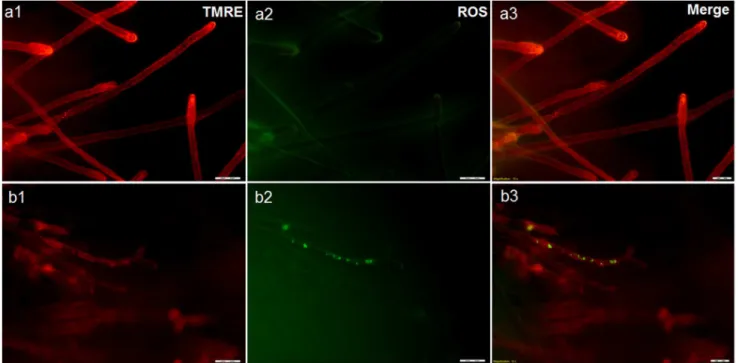
Figure 4. Effect of boric acid onSaprolegniamitochondria using fluorescence microscopy.Fluorescence microscope image showing the concentration of tetramethyl rhodamine (TMRE) staining in healthy non-treatedSaprolegniahyphae (a1) compared to boric acid treated hyphae (b1) where the depolarized mitochondria exhibit reduced red fluorescence. Figure (a2) is showing ROS level in the non-treated control compared to treatedSaprolegniahyphae (b2). TMRE and ROS staining are merged in a3 and b3.
doi:10.1371/journal.pone.0110343.g004
Figure 5. Viability of treated Saprolegnia spores at different time post treatment. Mitochondrial activity and viability in Sapro-legnia spores following boric acid (1 g/L) and bronopol (100 mg/L) treatment was compared to non-treated control using the MTS assay. The diagram shows the percent of viable spores relative to the non-treated ones calculated as described in the methodology section. Spores viability was significant reduced (p,0.001) in BA and bronopol treated samples at all time points (4–24 h) relative to non-treated control.
doi:10.1371/journal.pone.0110343.g005
Effect of boric acid on the nuclear division and germination ofSaprolegniaspores
The absence of nuclear division and germination was the most prominent features observed in BA treated spores (Fig. 2b) compared to non-treated water control (Fig. 2a). However, no fixed morphological pattern for the nuclear division was exhibited by those in the non-treated water control group. Some nuclei were able to divide inside the cyst body, while the majority migrated with the whole nucleic acid contents toward the germ tube leaving an almost empty cyst body structure. Other observations in the treated spores were an increase in the vacuolization and decrease in the fluorescence intensity of the extra-nuclear DNA (Fig. 2 b-3 and b-4), apparently due to the reduction in mitochondrial activity as the active mitochondria have a small amount of their own DNA.
Effect of boric acid on mitochondrial activity, distribution and membrane potential using confocal laser scanning microscopy (CLSM)
Saprolegniaspores treated with BA for 4 h (Fig. 3a-2) showed minor mitochondrial changes compared to the non-treated water control (Fig. 3a-1). There appeared a significant reduction in the spore mitochondrial numbers observed at 12 and 24 h following BA treatment (Fig. 3a-3 and a-4). Treated hyphae showed reduction in the number of hyphal apex mitochondria following 4 h exposure (Fig. 3b-2). After 12 h treatment, swollen hyphal apex and changes in the pattern of mitochondrial distribution were demonstrated (Fig. 3b-3). Different vacuolar forms without mito-chondrial activity were formed throughout the apical part of the hyphae 24h post initiation of treatment (Fig. 3b-4). In contrast, the mitochondria in the non-treated control showed dense red-colored cylindrical and round structures concentrated in the hyphal apex and distributed all over the hyphae (Fig. 3b-1). Figure 3 also shows Figure 6. Effect of boric acid on the integrity ofSaprolegniaspore membranes.Fluorescence microscopy analysis of Propidium Iodide (PI) uptake bySaprolegniaspores. a) non-treated spores kept in water, were able to germinate and to form hyphae that only flourecent green with SYTO 9 (a1) without PI uptake (a2). b) Boric acid treated spores, neither germinate (b1) nor absorb the PI dye (b2). c) Non-viable, bronopol treated Saprolegniaspores showing uptake of SYTO 9 (c1) and PI dye (c2).
doi:10.1371/journal.pone.0110343.g006
the difference in fluorescence intensity, in BA treatedSaprolegnia
spores (c) and hyphae (d) compared to the non-treated control following 24 h exposure.
Effect of boric acid on mitochondrial membrane potential and reactive oxygen species (ROS) using fluorescence microscopy
The drop in the fluorescence intensity and distribution of TMRE dye were the main differences observed in BA treated
Saprolegniahyphae (Fig. 4b-1) compared to the non-treated water control (Fig. 4 a-1). The dye accumulation in the hyphal apex of the non-treated control is an indication for their mitochondrial activity. ROS accumulation was detected in some of the BA treated hyphae (Fig. 4b-2). However, it was also observed in hyphae in the non-treated control with weak mitochondrial activity.
Assessing the metabolic activity and viability of treated spores using the MTS assay
The spores’ viability was significantly reduced after BA and bronopol treatment (Figure 5). At 4 h post treatment, 6% of the spores were found viable after BA treatment compared to 11% in bronopol-treated ones. The percent of viable spores remained unchanged at 8 h post treatment in BA treated groups (6%) while dropped to less than 5% in the bronopol treated samples. By 24 h post treatment, 2.4 and 1.5% of the spores were viable in BA and bronopol treated groups, respectively. BA and bronopol groups were significantly lower at all-time points while there was no difference between BA and bronopol (Figure 5).
Effect of boric acid on the integrity ofSaprolegniaspore membranes
Fluorescence microscopy revealed that Saprolegnia spores treated with BA neither absorbed the propidium iodide dye nor germinated, which indicate the integrity of plasma membrane was maintained as shown by staining with SYTO 9 dye (Fig. 6b). In contrast, bronopol treated spores fluoresced red and green as the propidium iodide dye was able to diffuse into their necrotic walls (Fig. 6c). Spores kept in water were able to germinate and to form hyphae that only fluoresce green (Fig. 6a–1).
Discussion
This study indicates that boric acid has a toxic effect on
Saprolegniaspore germination and hyphal growth underin vitro
condition. The degeneration of mitochondria or the inhibition of its enzymes and metabolism is probably the key for its mode of action.
The results obtained by the transmission electron microscopy following 4 h of exposure, indicated mild alterations in nuclear morphology in some of the BA treated spores (condensed nuclei with fragmentation of their membranes). Additionally, a consid-erable variation in the mitochondrial structure of BA treated spores compared to the non-treated water control was observed. Mitochondria are essential cellular organelles that play central roles in energy production, metabolism generation, regulation of reactive oxygen species (ROS) and apoptosis [23,24]. They are also responsible for more than 90% of cellular adenosine triphosphate (ATP) production which is essential for the cell to perform all essential activities for its survival and function [10,25,26]. Therefore, compounds able to affect the mitochondrial respiratory chain could be used as growth inhibitors [18,24,27]. Hyphal-growing organisms, includingSaprolegniausually have a
high density of apex-localized mitochondria [29]. They play a role in the high Ca2+gradient in the apex, which is important for the apical growth [28,29]. The mitochondria localized behind the tip are the ones responsible for the respiratory function and ATP production [29]. Using MitoTracker Red probes and CLSM, boric acid treated hyphae showed considerable variations in the number and distribution of mitochondria localized both in the hyphal apex and through the hyphae. The same result was recorded in the treatedSaprolegniaspores. The decrease in the mitochondrial density was directly proportional with the exposure time, when different time points were used. The reduction and disorganization in spore/hyphal mitochondria following boric acid treatment is supporting the hypothesis that mitochondrial toxicity might be one of its primary modes of action. Mitochondrial membrane potential (DYm) is critical for maintaining many mitochondrial processes including ATP synthesis and the control of ROS generation; its disturbance could diminish energy production [30]. Thus, changes inDYm were also followed using the fluorescent dye (TMRE). Hyphae from non-treated controls showed intense fluorescence as TMRE dye accumulates in active mitochondria due to their relative negative charge. On the other side, boric acid treated hyphae showed a drop in the fluorescence intensity as the inactive mitochondria with decreased membrane potential fail to sequester TMRE. Reactive oxygen species (ROS) are important signaling molecules normally present in cells, however, their accumulation under pathological conditions leads to oxidative stress. Therefore, ROS withDYm can be used as an indicator of the cell physiological status and the function of the mitochondria [30]. This might explain the high accumulation of ROS in some of the treated hyphae with a lower mitochondrial activity than the non-treated ones.
Regarding the nucleus, no obvious changes were observed on the nuclear morphology of the treated spores examined by TEM. The nuclear mitosis has been described before inSaprolegniaspp. [31], thus, the effect of the boric acid on the nuclear division was followed using the nucleic acid specific dye DAPI. Spores kept in water were able to develop germlings that elongated. Almost all the organelle-containing cytoplasm in the cyst body was able to relocate to the developed germlings as described before [32] and the nucleus was able to divide. In contrast, the nuclei of the treated spores failed to divide when they were examined with the CLSM. This effect of the boric acid on the nuclear division might be correlated to a reduction in DNA synthesis as suggested by Ku et al. [33], apparently due to the absence of sufficient energy related to the impairment of normal mitochondrial function.
The metabolic activity and proliferation of treated spores were also investigated using an MTS assay. This assay relies on the metabolism of the MTS reagent into formazan by dehydrogenase enzymes [34–36]. It thereby gives an indication on the functional state and integrity of the mitochondria. The significant reduction of the MTS reagent in the non-treated water control group following 24 h incubation indicates their high mitochondrial metabolic and proliferation activity. In contrast, the boric acid treated group has almost similar results as the positive control group treated with bronopol.
The integrity of the membrane of treatedSaprolegnia spores was interpreted by using a propidium iodide stain. The fact that the treated spores did not take up the dye nor germinate is suggesting that boric acid does not have a direct effect on treated spore membranes at the tested concentration which also suggests that BA could cause slow loss of viability.
possibility of combining boric acid with potential anti-Saprolegnia
candidates targeting cellular components other than mitochondria should be considered.
Conclusion
We can conclude that boric acid possess a toxic activity on
Saprolegniaspores germination and hyphal growth. The disrup-tion of metabolism and impairment of the normal mitochondrial function or some of its enzymes are probably included in its primary mode of action.
Acknowledgments
The authors would like to thank Hilde Kolstad (Imaging centre, A˚ s) for excellent assistance with the performance of the TEM analysis.
Author Contributions
Conceived and designed the experiments: SEA IS OE. Performed the experiments: SEA ET JW. Analyzed the data: SEA ET OE JW AG IS. Contributed reagents/materials/analysis tools: SEA IS OE. Contributed to the writing of the manuscript: SEA ET OE JW AG IS.
References
1. Bruno DW, Wood BP (1999) Saprolegnia and other Oomycetes. In: Woo PTK, Bruno DW, editors. Fish Diseases and Disorders III: Viral, Bacterial and Fungal Infections. Wallingford, Owon, United Kingdom: CABI Publishing. 599: 659. 2. Ali SE, Thoen E, Vralstad T, Kristensen R, Evensen O, et al. (2013)
Development and reproduction of Saprolegnia species in biofilms. Vet Microbiol 163: 133–141.
3. Torto-Alalibo T, Tian MY, Gajendran K, Waugh ME, van West P, et al. (2005) Expressed sequence tags from the oomycete fish pathogen Saprolegnia parasitica reveal putative virulence factors. Bmc Microbiology 5.
4. Coughlin JR (1996) Inorganic borates: Chemistry, human exposure, and health and regulatory guidelines. Journal of Trace Elements in Experimental Medicine 9: 137–151.
5. Eckhert CD (1998) Boron stimulates embryonic trout Growth. Journal of Nutrition 128: 2488–2493.
6. Takano J, Noguchi K, Yasumori M, Kobayashi M, Gajdos Z, et al. (2002) Arabidopsis boron transporter for xylem loading. Nature 420: 337–340. 7. De Seta F, Schmidt M, Vu B, Essmann M, Larsen B (2009) Antifungal
mechanisms supporting boric acid therapy of Candida vaginitis. Journal of Antimicrobial Chemotherapy 63: 325–336.
8. Shi XQ, Li BQ, Qin GZ, Tian SP (2011) Antifungal Activity and Possible Mode of Action of Borate Against Colletotrichum gloeosporioides on Mango. Plant Disease 95: 63–69.
9. Jovanovic R, Congema E, Nguyen HT (1991) Antifungal Agents Vs Boric-Acid for Treating Chronic Mycotic Vulvo-Vaginitis. Journal of Reproductive Medicine 36: 593–597.
10. Shi XQ, Li BQ, Qin GZ, Tian SP (2012) Mechanism of antifungal action of borate against Colletotrichum gloeosporioides related to mitochondrial degra-dation in spores. Postharvest Biology and Technology 67: 138–143. 11. Ali SE, Thoen E, Evensen O, Skaar I (2014) Boric Acid Inhibits Germination
and Colonization of Saprolegnia Spores In Vitro and In Vivo. PLoS ONE 9: e91878.
12. Stueland S, Hatai K, Skaar I (2005) Morphological and physiological characteristics of Saprolegnia spp. strains pathogenic to Atlantic salmon, Salmo salar L. Journal of Fish Diseases 28: 445–453.
13. Amberg DC, Burke DJ, Strathern JN (2006) Yeast Vital Stains: DAPI Stain of Nuclear and Mitochondrial DNA. CSH Protoc 2006.
14. Zuo XM, Djordjevic JT, Oei JB, Desmarini D, Schibeci SD, et al. (2011) Miltefosine Induces Apoptosis-Like Cell Death in Yeast via Cox9p in Cytochrome c Oxidase. Molecular Pharmacology 80: 476–485.
15. Hulvey JP, Padgett DE, Bailey JC (2007) Species boundaries within Saprolegnia (Saprolegniales, Oomycota) based on morphological and DNA sequence data. Mycologia 99: 421–429.
16. Hardham A (2001) Investigations of oomycete cell biology. In: Talbot N, editor. Molecular and Cellular Biology of Filamentous Fungi: A Practical Approach. USA: Oxford University Press. pp. 127–155.
17. Chazotte B (2011) Labeling mitochondria with TMRM or TMRE. Cold Spring Harb Protoc 7: 895–897.
18. Lopes G, Pinto E, Andrade PB, Valentao P (2013) Antifungal activity of phlorotannins against dermatophytes and yeasts: approaches to the mechanism of action and influence on Candida albicans virulence factor. PLoS One 8: e72203.
19. Wang P, Henning SM, Heber D (2010) Limitations of MTT and MTS-Based Assays for Measurement of Antiproliferative Activity of Green Tea Polyphenols Plos One 5: e10202.
20. Coder DM (2001) Assessment of cell viability. Curr Protoc Cytom Chapter 9: Unit 9 2.
21. AlRubeai M, Welzenbach K, Lloyd DR, Emery AN (1997) A rapid method for evaluation of cell number and viability by flow cytometry. Cytotechnology 24: 161–168.
22. Truernit E, Bauby H, Dubreucq B, Grandjean O, Runions J, et al. (2008) High-resolution whole-mount imaging of three-dimensional tissue organization and gene expression enables the study of phloem development and structure in Arabidopsis. Plant Cell 20: 1494–1503.
23. Szewczyk A, Wojtczak L (2002) Mitochondria as a pharmacological target. Pharmacological Reviews 54: 101–127.
24. Ruy F, Vercesi AE, Kowaltowski AJ (2006) Inhibition of specific electron transport pathways leads to oxidative stress and decreased Candida albicans proliferation. Journal of Bioenergetics and Biomembranes 38: 129–135. 25. Chan DC (2006) Mitochondria: Dynamic organelles in disease, aging, and
development. Cell 125: 1241–1252.
26. Mucha J, Zadworny M, Werner A (2009) Cytoskeleton and mitochondrial morphology of saprotrophs and the pathogen Heterobasidion annosum in the presence of Suillus bovinus metabolites. Mycological Research 113: 981–990. 27. Martins VD, Dinamarco TM, Curti C, Uyemura SA (2011) Classical and
alternative components of the mitochondrial respiratory chain in pathogenic fungi as potential therapeutic targets. Journal of Bioenergetics and Biomem-branes 43: 81–88.
28. Silverman-Gavrila LB, Lew RR (2003) Calcium gradient dependence of Neurospora crassa hyphal growth. Microbiology-Sgm 149: 2475–2485. 29. Levina NN, Lew RR (2006) The role of tip-localized mitochondria in hyphal
growth. Fungal Genetics and Biology 43: 65–74.
30. Joshi DC, Bakowska JC (2011) Determination of mitochondrial membrane potential and reactive oxygen species in live rat cortical neurons. J Vis Exp 51: 7588–7596.
31. Heath IB (1980) Behavior of kinetochores during mitosis in the fungus Saprolegnia ferax. Journal of Cell Biology 84: 531–546.
32. Beakes GW (1980) Electron microscopic study of oospore maturation and germination in an emasculate isolate of Saprolegnia ferax. 2. Wall differenti-ation. Canadian Journal of Botany 58: 195–208.
33. Ku WW, Shih LM, Chapin RE (1993) The Effects of Boric-Acid (Ba) on Testicular Cells in Culture. Reproductive Toxicology 7: 321–331.
34. Buttke TM, Mccubrey JA, Owen TC (1993) Use of an Aqueous Soluble Tetrazolium Formazan Assay to Measure Viability and Proliferation of Lymphokine-Dependent Cell-Lines. Journal of Immunological Methods 157: 233–240.
35. Cory AH, Owen TC, Barltrop JA, Cory JG (1991) Use of an Aqueous Soluble Tetrazolium Formazan Assay for Cell-Growth Assays in Culture. Cancer Communications 3: 207–212.
36. Smith SM, Wunder MB, Norris DA, Shellman YG (2011) A simple protocol for using a LDH-based cytotoxicity assay to assess the effects of death and growth inhibition at the same time. PLoS One 6: e26908.