Dissociation Dynamics of XPC-RAD23B from
Damaged DNA Is a Determining Factor of
NER Efficiency
Benjamin Hilton1☯, Sathyaraj Gopal2☯, Lifang Xu2, Sharmistha Mazumder1, Phillip R. Musich1, Bongsup P. Cho2*, Yue Zou1*
1Department of Biomedical Sciences, Quillen College of Medicine, East Tennessee State University, Johnson City, Tennessee, 37614, United States of America,2Department of Biomedical and
Pharmaceutical Sciences, College of Pharmacy, University of Rhode Island, Kingston, Rhode Island, 02881, United States of America
☯These authors contributed equally to this work. *[email protected](YZ);[email protected](BPC)
Abstract
XPC-RAD23B (XPC) plays a critical role in human nucleotide excision repair (hNER) as this complex recognizes DNA adducts to initiate NER. To determine the mutagenic potential of structurally different bulky DNA damages, various studies have been conducted to define the correlation of XPC-DNA damage equilibrium binding affinity with NER efficiency. How-ever, little is known about the effects of XPC-DNA damage recognition kinetics on hNER. Although association of XPC is important, our current work shows that the XPC-DNA disso-ciation rate also plays a pivotal role in achieving NER efficiency. We characterized for the first time the binding of XPC to mono-versusdi-AAF-modified sequences by using the real time monitoring surface plasmon resonance technique. Strikingly, the half-life (t1/2or the
retention time of XPC in association with damaged DNA) shares an inverse relationship with NER efficiency. This is particularly true when XPC remained bound to clustered adducts for a much longer period of time as compared to mono-adducts. Our results sug-gest that XPC dissociation from the damage site could become a rate-limiting step in NER of certain types of DNA adducts, leading to repression of NER.
Introduction
The human genome is constantly under assault from exogenous and endogenous causes of DNA damage. The formation and propagation of the resulting adducts can be particularly destructive when these mutations occur within tumor suppressing genes, leading to tumorigen-esis [1–4]. Consequently, human cells have several effective DNA repair pathways to protect against the plethora of genotoxic bombardments to the genome [5]; however, the mechanism by which damage-recognition proteins distinguish damage sites remains uncertain. Mutations that arise in genes associated with the nucleotide excision repair pathway (NER) result in a multitude of genetic disorders such asxeroderma pigmentosum, which is characterized by sen-sitivity to sunlight and, ultimately, the development of carcinomas [6].
a11111
OPEN ACCESS
Citation:Hilton B, Gopal S, Xu L, Mazumder S, Musich PR, Cho BP, et al. (2016) Dissociation Dynamics of XPC-RAD23B from Damaged DNA Is a Determining Factor of NER Efficiency. PLoS ONE 11 (6): e0157784. doi:10.1371/journal.pone.0157784
Editor:Robert W Sobol, University of South Alabama Mitchell Cancer Institute, UNITED STATES
Received:March 17, 2016
Accepted:June 3, 2016
Published:June 21, 2016
Copyright:© 2016 Hilton et al. This is an open access article distributed under the terms of the
Creative Commons Attribution License, which permits unrestricted use, distribution, and reproduction in any medium, provided the original author and source are credited.
Data Availability Statement:All relevant data are within the paper and its Supporting Information files.
Funding:This work was supported by the National Institutes of Health (Grants CA86927 to YZ and CA098296 to BPC). The funder had no role in study design, data collection and analysis, decision to publish, or preparation of the manuscript.
NER is utilized to remove primarily bulky adducts, plus cross-links, and various other lesions [7–9]. NER is either associated with transcription in transcription-coupled repair (TCR) or is independent of transcription in global genome repair (GGR). GGR inEscherichia coliconsists primarily of a collaborative effort of three proteins that both recognize and incise damaged bases: UvrA, UvrB, and UvrC [10]. Two UvrA molecules associate and then form a trimeric complex with UvrB. This trimeric complex is thought to be the DNA damage sensor. UvrA facilitates UvrB binding and positions UvrB to confirm the existence of a damage site. Once UvrB is in the correct position, UvrA utilizes its ATPase activity to dissociate from the preincision complex. UvrB then recruits UvrC endonuclease, which incises the damaged DNA strand by 3’and 5’cleavages flanking the damage site [11–14]. In human GGR the UvrA2B functional equivalent isXeroderma pigmentosumgroup C (XPC) in complex with
RAD23B (XPC-RAD23B, henceforth XPC). The XPC complex acts in the DNA damage rec-ognition step, thus initiating GGR [15]. XPC has been shown to bind at the site of many types of damagein vitroand in UV-treated cells arrives at damage sites before other NER fac-tors [9,16–18]. Once at the damage site XPC recruits the multi-subunit transcription factor TFIIH, including the helicase subunits of XPB and XPD, followed by XPA for damage confir-mation, fork binding and subsequent recruitment of replication protein A (RPA) for single-stranded DNA (ssDNA) stabilization, and XPG and XPF-ERCC1 for the dual incisions [19–
22].
Crystal structures of the yeast XPC-RAD23B ortholog, Rad4-Rad23, in association with undamaged or damaged DNA revealed a mechanism by which XPC hops along DNA until a thermodynamically stable recognition complex is formed, which effectively distinguishes damaged from non-damage sites [23,24]. Further studies have supported this hypothesis by suggesting that residence time of XPC on damages may play a role in the relationship between XPC binding and NER efficiency [25,26]. Binding affinity of XPC at the damage site has been suggested to be the rate-limiting step for NER [25,27,28]. Although various efforts have been made to correlate the equilibrium binding of damage recognition to overall NER efficiency, little is known about the role of the kinetics of damage recognition in the NER process.
Arylamines and heterocyclic amines are notorious environmental carcinogens. The DNA adduct-forming arylamines can be found naturally in the environment, in addition to a num-ber of unnatural sources such as cigarette smoke and hair dyes. Heterocyclic amines are most notably abundant in meat that has been cooked at high temperatures. It is inevitable that a per-son will be exposed to one or both of these carcinogens in his/her lifetime. Each of these muta-gens has been documented to cause many types of cancer, such as breast, liver, and bladder, to name a few [2]. Metabolic activation of these aminesin vivoproduces C8-substituted dG as the major bulky DNA adduct [29]. A well-known example is the human bladder carcinogen 4-ami-nobiphenyl [30]. The prototype environmental arylamine 2-aminofluorene produces two major DNA adductsvia in vivoactivation: N-(20-deoxyguanosin-8-yl)-2-aminofluorene (AF)
and N-(20-deoxyguanosin-8-yl)-2-acetylaminofluorene (AAF) (Fig 1A) [31]. Their fluorine
dependent on the flanking sequence, which modulates mutational and repair outcomes [27,39–41]. One such sequence is the mutational hotspot known as theNarI sequence (5’-. . .C G1G2CG3CC. . .-3’) (Fig 1B), which has been extensively studied [33,42].
Fig 1. Adduct structures and sequences.(A) Structure of AAF [N-(2’-deoxyguanosin-8-yl)-2-acetylaminofluorene], AF [N
-(2’-deoxyguanosin-8-yl)-2-aminofluorene] and fluoro models, FAAF [N-(2-deoxyguanosin-8-yl)-7-fluoro-2-acetylaminofluorene], FAF [N-(2’
-deoxyguanosin-8-yl)-7-fluoro-2-aminofluorene]; (B) Fully-paired 16-mer duplexes containing the centralNarI sequence (CGGCGCC) used in SPR, EMSA andin vitro
NER constructs illustrating the placement of the adducted bases at G1, G2, and G3positions; (C) Major groove views of the B-, S-, and
W-conformers of AAF. Modified-dG (red), dC (green) opposite the lesion site (orphaned C), fluorene (grey CPK),N-acetyl (magenta).
The reparability of adducts in theNarI sequence has been tested in both theE.coliUvrABC and human endonuclease systems and were found to be sequence dependent [27,33,43]. In addition, different repair efficiencies of the same lesions were observed between the two sys-tems [27,33]. Furthermore, recent work has attempted to correlate the binding affinities of repair proteins with adduct excision or NER efficiency [25–27]. Yeo and colleagues, imple-menting electrophoretic mobility shift (EMSA) and dual-incision assays, concluded that increased DNA thermodynamic destabilization, XPC-RAD23B binding, and overall NER effi-ciency of AAF adducts are directly correlated [25]. In contrast, Muet al. showed that NER effi-ciencies of the same AAF lesions are correlated with greater extents of base
sequence-dependent local untwisting and minor groove opening together with weaker stacking interac-tions [27]. Leeet al. have found minimal differences in XPC binding affinities of lesions derived from bulky polycyclic aromatic hydrocarbons while observing dramatic differences in NER efficiency [26]. These three individual reports employed different bulky adducts in their stud-ies; however, Shellet al. demonstrate that XPC acts as a general sensor for DNA damage, with a preferential binding to damage sites, but concluded that lesion identity is not a determinant of XPC binding affinity [44].
In the present study, we analyzed the kinetic aspects ofE.coliUvrA2and human XPC
pro-tein interactions with AAF adducts in theNarI sequence context by surface plasmon resonance (SPR) analysis and defined the relationship with NER. SPR has the significant advantage over other methods designed to observe protein-DNA interactions in that the interaction can be observed in a dynamic real-time environment, much closer to native conditions. We show that at lesion clusters the kinetic off-rate of XPC has an inverse correlation to repair efficiency. In other words, thet1/2(time required for 50% of the bound XPC to dissociate from the DNA,t1/2
(s) = ln(2)/kd,) of the damage recognition complex inversely correlates to NER efficiency. This
work reveals the significance of the dynamics of XPC recognition of conformationally diverse DNA adducts in NER. Here, we describe a new model for XPC activation of NER where the off-rate of XPC from the lesion site, particularly in the case of clustered lesions, is the rate-lim-iting step of NER, and propose applying this finding to design a more efficiently targeted approach to cancer therapy.
Materials and Methods
Caution
2-Aminofluorene derivatives are mutagens and suspected human carcinogens and, therefore, must be handled with caution.
Crude desalted oligodeoxynucleotides (1μmol) were purchased from Operon (Eurofin, Huntsville, AL) and purified by reverse phase HPLC. All HPLC solvents were purchased from Fisher Inc. (Pittsburgh, PA).
Substrate preparation and characterization
Preparation of arylamine-modified template
The modified 55-mer biotinylated DNA templates were prepared according to published pro-cedures [45–47]. Mono- and di-adduct oligodeoxynucleotides in theNarI modified strand were purified by HPLC (described above) and characterized by Shimazdu Axima MALDI-TOF mass spectrometry as previously reported [38] (Fig 1andS3 Fig). 5’-Biotinylated 55-mer (1 OD) was annealed with 55-mer complementary strand (1.05 ODs) in 1x HBS-EP+buffer for 5 min at 95°C. Identical unmodified duplexes were concurrently prepared as controls. The annealed oligodeoxynucleotides then were used for SPR experiments.
Oligonucleotide sequence used for surface plasmon resonance
5’-biotin-CCACTCCTATCCACCATCCATCTTACTCTCG1G2CG3CCATCACCACTCACCACCA
CA-3’
3’-GGTGAGGATAGGTGGTAGGTAGAATGAGAGC C GC GGTAGTGGTGAGTGGTGGTGT-5’
G1, G2, and/or G3: dG or dG-FAAF
Purification of XPC-RAD23B protein complex
XPC-RAD23B protein was prepared from Sf21 insect cells infected with recombinant baculo-virus expressing XPC and RAD23B proteins (graciously provided by A. Sancar, University of North Carolina, Chapel Hill). The XPC-RAD23B complex was purified as described previously [48,49]. Protein concentration was determined using the Bio-Rad protein assay. Following purification by size-exclusion chromatography, SDS-PAGE (10%) and Western blotting con-firmed the purity of the XPC-RAD23B complex.
Immobilization of streptavidin on CM5 chip and DNA coating
SPR measurements were conducted with a Biacore T200 (GE Healthcare). Streptavidin (SA) was immobilized on a CM5 dextran chip using an amine-coupling method [45,47]. Four flow cells were immobilized with streptavidin amine to ~2,200 resonance units (RU). Flow cell 1 was used as a reference. Before the coating of biotinylated DNA templates over SA, the surface was washed with 50 mM NaOH five times, each with 60s pulses at 100μl/min to remove any free SA until the change in response units was below 5 RU. The surface was further injected 3–4 times with HBS-P+ running buffer (10 mM HEPES, 150 mM NaCl, 0.05% non-ionic factant P20) to remove any residual NaOH in the microfluidics path and to stabilize the sur-face. Biotinylated unmodified and various FAAF-modified DNA duplexes (0.025 nM) were injected at 100μl/min for 240–300s over the flow cells 2, 3, or 4 to achieve 2–5 RU relative to flow cell 1, which was a blank reference. Any unbound DNA was washed away with running buffer.
Kinetics analysis
The binding kinetics for the interaction of UvrA or XPC with DNA was determined by inject-ing the UvrA (0–500 nM) or XPC (0–5 nM) in HBS-P+ running buffer containing 5 mM MgCl2, 1 mM DTT and BSA (100μg/mL). The flow rate was 100μL/min for 30 s followed by
a simple 1:1 Langmuir model (S1 Fig). Processing included zeroing and cropping data, aligning injection times, fitting of binding curves and off-rate analysis. The equilibrium dissociation constant (KD) for ternary systems was calculated using the steady-state affinity analysis in the
BIA-Evaluation software package v2.0 provided by the manufacturer, General Electric. The average of the data (with standard deviation) of KD, ka, and kdis shown in Tables1and2. The
Scrubber software package (BioLogic Software) was used to process off-rate analysis of raw XPC-H23B SPR binding sensograms. Curve fittings were not ideal for certain UvrA data (S2 Fig), which affected the reliability of rate constants (seeResults).
Electrophoretic mobility shift assay (EMSA)
Binding of XPC to various DNA substrates was analyzed by a gel mobility shift assay as described previously [49]. Typically, DNA substrates (0.5–1 nM) were incubated with varying concentrations of protein at 30°C in 20μL of binding buffer [20 mM Hepes-KOH, pH 7.9, 75
mM KCl, 5 mM MgCl2, 1 mM DTT, 5% glycerol, 100μg/mL acetylated BSA (Promega)].
Reac-tions then were placed on ice, 2μL of 80% (v/v) glycerol was added, and the mixture was
imme-diately loaded onto a 3.5% native polyacrylamide gel and electrophoresed at 80 V in 1× TBE buffer for 2 h at 4°C. The gels were dried and exposed to phosphoimage screens overnight. Quantification of the radioactivity was carried out using a Fuji FLA-5000 scanner with the Ima-geGuage software.
Table 1. Correlation of XPC-RAD23B binding and dissociation parameters, melting temperature, and hNER efficiencies of FAAF-modifiedNarI substrates.
NarI-FAAF SPR (KD) (M) SPR (ka) (M-1s-1) SPR (kd) (s-1) t1/2 (s) Tm(°C)(ΔTm) hNER
CCG1*G2CG3CC (G1-mono) 1.8 (±0.02) x 10−9 1.1 (±0.01) x 107 1.9 (±0.02) x 10−2 24 (±0.5) 68.6 (-5.3) 1 (±0.14)
CCG1G2*CG3CC (G2-mono) 0.9 (±0.01) x 10−9 6.9 (±0.17) x 107 6.3 (±0.16) x 10−2 44 (±0.4) 66.0 (-7.9) 0.69 (±0.03)
CCG1G2CG3*CC (G3-mono) 1.1 (±0.01) x 10−9 3.7 (±0.07) x 107 4.2 (±0.08) x 10−2 38 (±0.1) 65.6 (-8.3) 0.65 (±0.06)
CCG1*G2*CG3CC (G1G2-di) 7.6 (±0.26) x 10−11 1.0 (±0.008) x 108 2.5 (±0.01) x 10−3 102 (±1.3) 60.6 (-10) 0.69 (±0.08)
CCG1*G2CG3*CC (G1G3-di) 1.6 (±0.76) x 10−11 1.4 (±0.008) x 108 6.7 (±0.05) x 10−3 282 (±2.9) 56.5 (-14.1) 0.30 (±0.05)
CCG1G2*CG3*CC (G2G3-di) 0.42 (±0.34) x 10−11 1.4 (±0.01) x 108 1.4 (±0.01) x 10−3 492 (±4.5) 52.7 (-17.9) 0.12 (±0.02)
There is an inverse relationship between off-rate kinetics and human NER of the di-adducted dG-FAAF substrates. SPR (ka), SPR (kd) and SPR (KD) are
the association rate (ka), dissociation rate (kd) and equilibrium dissociation constant (KD) values determined by SPR analysis of the interaction of XPC with
mono- and di-adducts and t1/2(s) is the calculated half-life of the protein-DNA complex. The Tm(°C)ΔTmdata are the thermodynamic stability as previously
reported [33,38]. The hNER efficiency is relative to the data displayed inFig 2.
doi:10.1371/journal.pone.0157784.t001
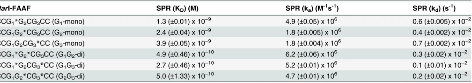
Table 2. Binding and dissociation parameters of UvrA2binding to FAAF-modifiedNarI substrates.
NarI-FAAF SPR (KD) (M) SPR (ka) (M-1s-1) SPR (kd) (s-1)
CCG1*G2CG3CC (G1-mono) 1.3 (±0.01) x 10−9 4.9 (±0.05) x 106 0.6 (±0.005) x 10−2
CCG1G2*CG3CC (G2-mono) 2.4 (±0.04) x 10−9 1.8 (±0.005) x 106 0.4 (±0.002) x 10−2
CCG1G2CG3*CC (G3-mono) 3.9 (±0.05) x 10−9 1.8 (±0.004) x 106 0.7 (±0.002) x 10−2
CCG1*G2*CG3CC (G1G2-di) 4.9 (±0.46) x 10−10 6.2 (±0.06) x 106 0.3 (±0.02) x 10−2
CCG1*G2CG3*CC (G1G3-di) 2.7 (±0.46) x 10−10 5.2 (±0.01) x 106 0.1 (±0.01) x 10−2
CCG1G2*CG3*CC (G2G3-di) 5.0 (±1.33) x 10−10 4.7 (±0.01) x 106 0.2 (±0.02) x 10−2
The association rate (ka), dissociation rate (kd) and binding affinity constant (KD) values of the interaction of UvrA with FAAF-modifiedNarI mono- and
di-adducts in the presence of ATP determined by SPR analysis of the interaction of UvrA2.
Construction of closed-circular plasmid with adducts
The following double-stranded oligonucleotide was cleaved by KpnI and Xba1 (both lower-case) for insertion into the multiple cloning site of the vector pTZ19U between theXbaIand KpnIrestriction sites using the QuikChange II Site-Directed Mutagenesis Kit (Agilent Technol-ogies). The 16-mer sequence containing theNarI hotspot ofFig 1Bis underlined.
NER_ins1 5’-CGGggtaccCCGCTCTCGGCGCCATCACTTAGtctagaCTAG-3’
NER_ins2 3’-GCCccatggGGCGAGAGCCGCGGTAGTGAATCagatctGATC-5’
The NER-pTZ19U plasmid was propagated inE.coli(DH5α) cells, which were infected with virus M13KO7 (NEB) to generate single-stranded plasmid which was purified using the M13 isolation maxi kit (Omega Biotek). Closed-circular double-stranded plasmid containing adduct was made by priming the single-stranded NER-pTZ19U plasmid with 5’ -phosphory-lated 16-mer oligos (Fig 1B) containing adduct in the presence of Sequenase 2.0 (Affymetrix) and T4 DNA ligase (Promega) following the manufactures protocol. Closed-circular plasmid DNA containing adduct then was purified by agarose gel electrophoresis and elution.
HeLa whole-cell extract preparation
Whole cell extracts were prepared from HeLa cell pellets purchased from the National Cell Culture Center. The thawed cell pellet was resuspended in four packed-cell volumes (PCV) of 10 mM Tris-HCl pH 8.0, 1 mM EDTA, 5 mM DTT, then incubated on ice for 20 min. The cells were lysed by homogenization in a Dounce homogenizer using eight strokes of the B pestle. Four PCV of 50 mM Tris-HCl pH 8.0, 10 mM MgCl2, 2 mM DTT, 25% sucrose (w/v),
50% glycerol (v/v) were added and the mixture was stirred gently. One PCV of saturated (NH4)2SO4(pH 7.0) was added slowly, then the mixture was stirred for 20 min at 4°C. The
lysate was clarified by centrifugation at 11,500xg for 30 min at 4°C. The supernatant was trans-ferred to a fresh tube and solid (NH4)2SO4(0.33g/ml of suspension) was added. The
suspen-sion was mixed for 30 min and 0.01 ml of 1M NaOH per 10 grams of (NH4)2SO4was added.
The precipitated proteins were collected by centrifugation at 11,500xg for 30 min and resus-pended in dialysis buffer (25 mM Hepes-KOH pH 7.9, 100 mM KCl, 12 mM MgCl2, 0.5 mM
EDTA, 2 mM DTT, 12% glycerol) and was dialyzed against the same buffer overnight. Follow-ing dialysis the extract was clarified by centrifugation at 10,000xg for 10 min and aliquots of the supernatant were stored at -80°C.
Dual incision assay
The dual incision assay was adapted from Shivjiet al. [50]. All incisions were carried out in a total reaction volume of 10μl. A reaction mixture with 100μg HeLa whole-cell extract protein in 5x repair buffer [200 mM Hepes-KOH, 25 mM MgCl2, 110 mM phosphocreatine (di-Tris
Results
Model systems
Mono- and di-FAAF adducted substrates were prepared within theNarI core sequence (5’-CT CTCG1G2CG3CCATCAC-3’,Fig 1B) as previously reported [33,38,39] and their
correspond-ing duplexes were used to examine their structural and thermodynamic properties (see below). The fluorinated FAAF has been used as a powerful19F NMR probe for investigating arylamine induced conformational heterogeneity [32–34]. This commonNarI sequence was ligated to prepare 55 bp substrates for SPR and double-incision assays on closed circular plasmid DNA. Depending on the location of the FAAF, the mono-adducts were designated asNarI-G1,
NarI-G2orNarI-G3, in which G1, G2and G3signify the position of modified guanine. The
di-adducts were designated asNarI-G1G2,NarI-G2G3, orNarI-G1G3, in which the numbers
sig-nify the positions of FAAF-modified guanines (Fig 1B). Note, these adducts can exist in the B, S or W conformations (Fig 1C) with the distribution between conformers strongly influenced by the sequence context and nature of other adducts within theNar1sequence [35,51].
Thermodynamics of mono- and di-FAAF adducts and
E
.
coli
NER
We previously studied the structures and thermodynamics of mono- and di-FAAF duplex adducts derived from the afore-mentionedNarI-16-mer sequence [33,38]. The thermodynamic results summarized inS1 Tablerevealed that di-adducts produced an additive effect on duplex destabilization relative to the mono-adducts. Briefly, the lesion-induced thermal instabilities (Tm,S1 Table) were substantially greater for di- (-10.0 ~ -17.9°C) over mono-adducts (-5.3 ~
-8.3°C). Within the di- adducts, the thermal stability was generally in the order ofNarI-G1G2 >NarI-G1G3>NarI-G2G3. Molecular dynamic simulation data indicated that the
perturba-tions of nucleotide base stacking are a major contributor to the observed sequence effect. The di-adducts were more reparable inE.colithan the corresponding mono-adducts [33,38]. More-over, we observed a dramatic trend in repair efficiency inE.coliwhich parallels the reductions in thermal stability, i.e.,NarI-G2G3>NarI-G1G3>>NarI-G1G2. Taken together, these results
indicate the importance of base stacking and related thermal and thermodynamic instability in the repair of bulky cluster arylamine DNA adducts. However, assessing the overall thermody-namic stability of di-adducts was complicated by the fact that the di-adducts sample a complex range of S/B/W-conformational heterogeneity [38]. The single FAAF adduct at the G3position
of theNarI sequence exhibited a preference for the S conformation (61%). Interestingly, greater instability was observed for the G3-containingNarI-G2G3andNarI-G1G3duplexes that
pro-duce a higher combined S population (~76 and ~95%, respectively) thanNarI-G1G2(~49%).
In addition, the greater proximity of two FAAF lesions inNarI-G2G3(e.g., just one base apart)
compared to that inNarI-G1G3(two bases apart) possibly could induce a greater helical
distortion.
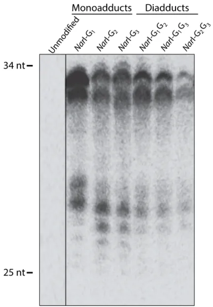
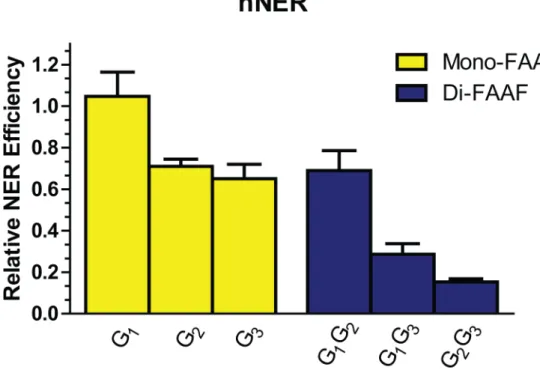
Human NER of mono- and di-FAAF adducted DNA
incision extension products in a denaturing urea gel as shown inFig 2. In contrast to the unmodified substrate the modified substrates generated products ranging in size from 25–34 bases.
Of the FAAF adducts,NarI G1-FAAF was observed to have the maximum incision;
there-fore, all otherNarI adducts were normalized to G1(Fig 3). The relative incision, therefore, was
1 for G1, 0.69 for both G2and G1G2, 0.65 for G3, 0.30 for G1G3, and 0.12 for G2G3. The
observed order for incision efficiency in the mono-adducts was G1>G2~ G3. The relative
hNER efficiencies of the di-FAAF adducts in theNarI sequence were G1G2>G1G3>G2G3.
Previous work on these same mono-adduct substrates has yielded a variety of conclusions [25,27,28]; however, this is the first report to include hNER incision analysis of cluster di-AAF
Fig 2. NER dual incision at adducts in theNarI sequence in the human NER system.Plasmids containing site-specific mono-FAAF (lanes G1, G2, G3) or di-FAAF adducts (lanes G1G2, G2G3, G1G3), were
incubated with HeLa whole-cell extracts. Detection of excision products was monitored by 3’-end-labeling using a complementary oligonucleotide containing a 5’-GGGG base overhang. The reaction products were resolved on a 12% denaturing polyacrylamide gel run under constant current. The range of excision products is indicated on the left of the gel.
adducts. One interesting observation is that the most thermodynamically stable di-adduct, G1G2, has no base separating the lesions and, in this respect, is structurally comparable to the
UV-induced cyclopyrimidine dimer (CPD) adduct. Work from the Min lab has allowed a bet-ter understanding of how XPC binds undamaged and damaged DNA, specificallyviathe CPD adduct [23,24]. Other observations indicate comparability between the cluster di-adduct G2G3,
which has FAAF adducts separated by one intact nucleotide base, and di-nuclearcis-platinum complexes [52]. These observations, discussed in further detail below, potentially have signifi-cant biological implications.
E
.
coli
and human NER produce different incision efficiency patterns on
common substrates
Interestingly, there is no direct correlation when comparing the NER incisions between theE. coliand human systems (Fig 3) [33,38]. Our previous work revealed that in theE.colisystem mono-FAAF adducts were excised in the order of G3~ G1>G2while di-FAAF adducts were
excised in the order of G2G3>G1G3>G1G2[33,38]. In theE.colisystem, the di-adducts
over-all are more readily incised by the UvrABC system than the mono-adducts; in contrast, in the human system the mono-adducts have significantly greater incisionversusthe di-adducts. Another interesting observation is that the mono- and di-adducts that produced the most inci-sion product in theE.colisystem (G3and G2G3), led to the least incision product in the human
system [33,38].
Although many processes in eukaryotic cells are conserved from prokaryotic systems and operate by similar mechanisms, in the case of NER the issue becomes more complex in that the UvrABC system is made up of only three proteins while in the human system nearly 30
Fig 3. The efficiencies of NER at adducts in theNarI sequence in the human NER system.The relative incision rates of mono-FAAF and di-FAAF adducts in the histogram were calculated by normalizing the mono- (yellow) and di-adducts (blue) relative to theNarI-G2G3FAAF value for theNarI-G1FAAF value for the
human system. Quantification of NER efficiencies was from at least three independent experiments.
proteins carry out the same function. Also, in humans multiple pathways arose to deal with the repair of a much more complex genome that also exists in a chromatin structure. On the con-trary, adducts that destabilize and disorder the DNA are the best substrates for theE.coli sys-tem [33,38] but are poorer substrates in the human system. This leads to the assertion that there are factors other than damage recognition that influences activation of NER in the human system.
UvrA2
and XPC binding to FAAF adducts in the
Nar
I sequence context
InE.colior human NER, UvrA2or XPC is required for the recognition of adduct-induced
destabilization of DNA structures as the initial step [9]. The incision efficiency of the UvrABC system on mono- and di-FAAF-adducted DNA has been thoroughly studied in our past work [33,38] and the incision efficiency of the human system on these adducts is presented in the present study. To determine the KD, ka, and kdof the UvrA2, and XPC interactions with the
FAAF adducts we employed SPR molecular interaction analysis. By real-time monitoring of UvrA2and XPC binding, a more informed conclusion can be drawn of their binding to and
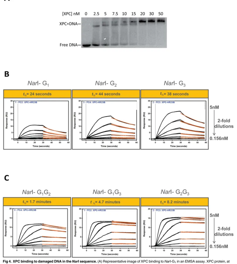
dissociation from adducted DNA. First, as a preliminary test, a traditional method, EMSA, was employed to demonstrate complex formation of XPC and FAAF-adducted DNA (Fig 4A). The gel shift binding pattern is consistent with the previous reports that XPC can bind an adducted oligonucleotide as a multimer at high concentrations, as shown by the two slower migrating bands [25,26]. It was thought that the formation of the slower migrating band could be through biochemical manipulation of the enzyme [25,26]. This EMSA result is of XPC binding to the G1adduct and is representative of the results obtained for all other adducted substrates (data
not shown).
Next, mono- and di-FAAF-adducted DNA was used to determine XPC and UvrA2
associa-tion and dissociaassocia-tion rates by SPR. The SPR binding results are shown inFig 4andS2 Fig, in which the average of the triplicate data is displayed. Use of the usual simple 1:1 Langmuir-type binding model did not produce desirable closeness of fit between the protein and modified DNA (S1 Fig). The fitting for XPC binding was improved somewhat (for di-adducts especially) by applying the‘heterogeneous ligand’model which is designed to accommodate the interac-tion of one protein analyte with two ligand sites on the surface. The usual remedy such as reducing the magnitude of the concentration gradient did not significantly mitigate the lack of ideal curve fittings, nor did decreasing the density of the immobilized DNA construct on the surface or increasing the flow rate. It is clear that the adduct-induced conformational heteroge-neity of our substrates contributes many non-ideal molecular complexities, e.g., molecular dif-fusion affects, analyte heterogeneity [53,54], steric hindrance, and protein-DNA binding cooperativity and stoichiometry (e.g., seeEMSAinFig 4A). These factors affected the kinetic rate constants, which made them less reliable. However, we observed a dramatic difference in equilibrium dissociation constant (KD) values with XPC and association and dissociation rates
between mono- and di-adducts (Table 1). A similar trend was observed with UvrA (Table 2
andS2 Fig). In the SPR experiments, ATP was added to the binding buffer for UvrA2because
ATP appeared to stabilize the dimer. This observation is consistent with the known ATPase activity of UvrA2for adduct binding [55].
The binding of UvrA2and XPC to lesions in theNarI sequence is more stable with the
di-FAAF adducts than the mono-di-FAAF adducts. Importantly, our SPR binding results are similar to previous work demonstrating the binding affinity of XPC to cisplatin-damaged DNA [56,57]. It is of note that the thermostability (ΔTm) of these adducts varies and that the
di-adducts have the lowest thermostability (G1>G2>G3>>G1G2>G1G3>G2G3) (S1 Table).
mono-Fig 4. XPC binding to damaged DNA in theNarI sequence.(A) Representative image of XPC binding toNarI-G1in an EMSA assay. XPC protein, at
FAAF-adducts (Table 2). This is in agreement with the finding that the NER efficiency inE.coliis higher for the di-adducts over the mono-adducts. The tightest UvrA2-FAAF binding was to
G1G3where the adducts are separated by two intact nucleotide bases with a dissociation rate of
2.7 x 10⁻¹⁰. Interestingly, the correlation between equilibrium constants and NER efficiency for UvrA2and XPC with mono-versusdi-FAAF-adducted DNA are conversely related. In the case
of XPC-FAAF binding, the di-adducts bound with significantly slower dissociation (off) rates relative to the mono-adducts. These results are consistent with conformational changes accom-panying the protein-DNA interactions, which could provide the basis for deciphering the bind-ing/repair mechanisms. Comparing KDvalues (Table 1), the di-FAAF-adducted DNAs show
more stable XPC binding than the mono-adducts. The tightest XPC binding was to G2G3with
a dissociation rate of 0.42 x 10⁻¹¹. The low K
Dfor XPC di-adduct binding signifies a much
tigh-ter association of XPC protein to clustigh-tered lesions.Table 1summarizes these findings of XPC binding compared to the thermodynamic stability, the XPC-DNA complex half-life, and the incision efficiencies of FAAF adducts in the human NER system. It should be noted that the G1G2adduct has a longer half-life but similar incision to the mono-adducts, G1and G3.
Inter-estingly, this is the only cluster adduct studied in which the adducts are on adjacent nucleotide bases and its binding half-life is much shorter than those of G1G3and G2G3with the di-adducts
separated by at least one intact nucleotide bases.
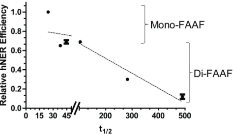
By calculating the half-life we observed that XPC-DNA complexes containing the di-FAAF adducts were on average at least 8 times more stable than complexes with the mono-FAAF adducts (Fig 4B and 4C,Table 1). In the extreme case the half-life of XPC-DNA complexes containingNarI di-G2G3-FAAF was 20 times longer than that ofNarI mono-G1-FAAF. The
half-life demonstrates an inverse relationship with the hNER efficiency (Fig 5). This indicates that the longer XPC stays bound to the adduct site, the less productive the human NER process. These data are provocative as they suggest that a strong DNA damage recognition or binding itself does not necessarily guarantee efficient NER.
modified full DNA duplexes. The recorded data are displayed as black lines while red lines represent curve fitting. The half-life (t1/2) is indicated above the
curves (in yellow box) and is defined as the time it takes for half of the XPC-DNA complex to dissociate. The fitted curves obtained from fittings using a one-independent site model (“Scrubber”) are displayed (SeeMethods).
doi:10.1371/journal.pone.0157784.g004
Fig 5. Comparison of hNER efficiency and half-life (t1/2) of the XPC-DNA complex.The data points were analyzed independently as mono- or di-FAAF adduct groups and the two dashed lines indicates the group trends. Mono- and di-adducts are indicated on the right.
Discussion
Various studies have attempted to relate the protein binding interactions involved in DNA dam-age recognition with NER excision efficiency utilizing a wide array of DNA damdam-ages [26,58]. This work proposes a novel mechanism beyond the conventional concept that the binding capability of DNA damage recognition proteins directly relates to the ability of the repair process to remove the damage from DNA. The presented SPR/hNER results suggest strongly that di-FAAF adducts fail to produce a productive complex for hNER even though the damage recognition binding is strong. In other words, robust XPC binding (KDof 10−9~ 10−10M) may be required for initiation
of hNER. However, unusually strong XPC binding (KD<10−11) and, more importantly, the
extremely slow dissociation of di-FAAF adducts, and thus the long residence time (t1/2) of XPC at
the damage site, could be detrimental to recruitment of subsequent downstream proteins to com-plete the hNER process (Fig 5). We demonstrate that in addition to the equilibrium binding affin-ity of XPC for DNA damage, kinetics and the off-rate of the interaction also play critical roles in determining the NER efficiency. This is particularly true for certain types of DNA adducts, such as the di-FAAF examined here, which have a long XPC residence time during DNA damage rec-ognition. Since dissociation of XPC from the damage site after initial recognition is necessary for subsequent binding of other repair factors in the mechanism of NER [15,58,59], it is possible that such an extended residence of XPC at the damage site would likely make dissociation the rate-lim-iting step of NER. This could lead to inefficient DNA repair even though the XPC-damaged DNA binding affinity is high. Our findings may help us better understand the complex mechanisms relating protein binding and adduct clearanceviaNER.
For the mono-FAAF adducts we observed that the repair efficiency of the G1adduct was
sig-nificantly higher than that of the G2and G3adducts, in agreement with the shorter half-life of
XPC on DNA adducted at the G1rather than the G2or G3positions (Table 1). This contrasts
with different reported preferences for repair in HeLa cell extracts of adducts at the G2position
[27,28] or at the G3position [25]. These differences in NER preferences may stem from the
nature of the DNA constructs used in the respective studies. The long-range sequence context of the DNA in which the adductedNarI site is embedded differs significantly between our con-struct and that of Yeoet al. as do the plasmids carrying these constructs (pTZ19U vs. pBlue-script II SK+, respectively) [25]. In addition, the studies reporting a preference for repair of adducts at the G2position employed relatively short (<150 bp), linear, and internally labeled
constructs that may display different torsional stresses on the DNA double helix in the HeLa extracts. Most studies on sequence context on NER efficiencies focus on the position effects within theNarI sequence rather than the influence of the long-range sequence in which it is embedded or the effect of super-helical stresses within the circular plasmids. Further studies are needed to resolve the influence of these experimental parameters on the NER efficiencies. However, the SPR, thermodynamic, EMSA and repair efficiency data reported here are inter-nally consistent in highlighting the importance of the half-life of the XPC-DNA complex in determining NER efficiency.
such cellular factors might reduce the binding and retention time of XPC on DNA containing mono- and di-FAAF adducts, and to extend these analyses to UV-induced damage. However, our data are consistent with their observation that the prolonged retention of XPC in chroma-tin or an increase t1/2reduces the NER efficiency [61], though the prolonged retention in the
previous UV-induced damage study was due to the deficiency of p97 segregase.
TheE.coliNER system has been studied for decades, leading to many breakthroughs in our understanding of how cells can repair DNA damage. These studies have provided vital infor-mation that is applicable to our understanding of the human NER system; however, these two systems are not directly comparable when considering damage recognition. Tight binding of recognition proteins may imply a better incision substrate, and this is true forE.coli, but in the human system much tighter binding can lead to a longer residence time, which results in a decrease in substrate processing. Analysis of previously reported structures of XPC- and UvrA2B (or UvrA2B2)-damaged DNA interactions may provide some understanding of our
observed differences of the FAAF adduct incisions between hNER and UvrABC systems [62–
64]. In bothE.coliand human systems a damage recognition protein is required to initiate the NER. UvrA2B and XPC-RAD23B, respectively, fill this role in the GGR sub-pathway of NER.
Interestingly, although both protein complexes recognize DNA damage, dissociation of these proteins from the damage site after recognition is quite different. In the case of UvrABC, UvrA2dissociates, while UvrB remains bound to the damage. In contrast, in hNER, RAD23B
dissociates shortly after binding; however, XPC remains bound [65]. Interestingly, structural evidence revealed that UvrA2makes contact with the DNA that flanks the damage site and has
no contact with the lesion itself [64], while UvrB establishes lesion contact, utilizing aβ-hairpin domain to insert into the DNA strands and flip-out bases opposing the lesion [63,66]. On the other hand, XPC appears to be responsible for both roles carried out by UvrA2and UvrB of the
UvrA2B complex since XPC also inserts itsβ-hairpin domain between DNA strands at the
damage site [23,24]. Theβ-hairpin insertion is likely to make protein-DNA interaction more stable and, thus, the protein less likely to dissociate from DNA. Since UvrA2does not carry out β-hairpin insertion while XPC does, it is possible that UvrA2would be energetically easier to
dissociate from DNA than XPC whose dissociation is more damage-type dependent. Thus, the type of di-FAAF adduct may increase the affinity of XPC at the damage site, leading to a longer residence time. For mono-FAAF adducts, in contrast, a normalt1/2keeps the dissociation from
being a rate-limiting step, remaining close to the regular binding-repair efficiency correlation. Our data supports the notion that XPC has an increased residence time at clustered lesion sites, making the dissociation from the lesion the rate-limiting step.
Fig 6Ashows a‘hypothetical’3D model of the G1mono-FAAF adduct that was printed (not
simulated), derived from the published Rad4-CPD DNA structure (PDB ID# 2QSG) [24]. The G1mono-FAAF and G2G3di-FAAF (not shown, additional FAAF site designated as red)
adducts exhibit distinctive differences in hNER (1vs. 0.12 relative efficiencies) (Fig 3) and XPC residence time (24vs. 495 s) (Table 1), as well as DNA thermal stability (ΔTm-5.3vs. -17.9°C)
(S1 Table). The CPD mismatch site was simply replaced by FAAF (yellow,) for visualization
of the G1mono-FAAF. The mono-lesion is expected to produce a similar NER complex as the
CPD; that is, Rad4 inserts aβ-hairpin into the damaged site with most of the modified strand being fully exposed and the mismatched bases (cyan) on the complimentary strand flipped out the double helix. The di-G2G3adduct, however, contains two FAAF adducts on separated by
may affect the logistics of the subsequent verification step. One potential future study is to crys-talize Rad4 or XPC complexed with the afore-mentioned mono-G1and di-G2G3-FAAF
adducts used in this study. If successful, the results are likely to provide valuable structural insights on XPC-DNA interactions that contribute to the large discrepancy in their residence time and reveal important clues regarding the structural requirements for recruitment of other NER proteins and subsequent lesion verification.
Tight binding during damage recognition may imply a better substrate for incisions, and this is true forE.coliNER, but in the human system binding too tightly can lead to a longer res-idence time, which decreases DNA repair (Fig 6B). In addition to the new insights into under-standing the mechanisms of hNER, the inverse relationship between thet1/2of tight binding
and NER efficiency suggests a novel strategy as a new therapeutic approach in cancer therapy. Given that DNA damaging agents have been widely exploited for anticancer activities, target-ing properly spaced di-adducts or a cluster-like drug that effectively stalls XPC or other damage recognition proteins could lead to strong resistance to repair and, thus, a higher efficiency in killing cancer cells. Recent studies have introduced new models that utilize residence time in drug design and to increase the efficacy of known drugs [67,68]. These models could be applied to our system allowing for design of novel drugs that could increase the residence time of XPC on damaged DNA for cancer therapeutics.
In summary, the present study suggests that dissociation of XPC from adducted DNA is the determining factor for successful NER elimination of adducts. In recognizing the types of adducts that are comparable toNar1-G2G3andNarI-G1G3cluster adducts, XPC can be stalled
on these damage sites, preventing clearance of induced adducts. Exploiting the high prolifer-ative rate of cancer cells and the slow dissociation rate of XPC from clustered adducts allows for a more efficiently targeted approach to cancer therapy. Furthermore, the current work also advances our understanding of the intricacies of the NER mechanism.
Supporting Information
S1 Fig. Variation of curve fitting between the heterogeneous ligand and langmuir fitting models.Representative SPR sensograms of mono- (A) and di-adducted (B) duplexes demon-strating the variation of curve fitting using the heterogeneous ligand fitting model (top) and the 1:1 Langmuir fitting model (bottom). The XPC protein concentrations used were 2.5, 1.25, 0.62, 0.31, and 0.15 nM.
(TIF)
S2 Fig. The effects of ATP on the UvrA binding to FAAF-damaged DNA in theNarI
sequence.Sensograms showing UvrA binding kinetics to mono-adducted substrates in the absence of ATP (A) or in the presence of ATP (B) and to di-adducted substrates in the absence of ATP (C) or in the presence of ATP (D). SPR responses were recorded to of the binding of UvrA NER protein (250, 125, 62.5, 31.2, 15.6, and 7.8 nM) to modified full DNA duplexes. The recorded data are displayed as black lines while red lines represent curve fitting. The fitting curves obtained from fittings using a one-independent site model are displayed.
(TIF)
(yellow*, as shown). The site of additional FAAF in the di-G2G3-adduct is designated in red asterisk. The insertion of the BHD3 β-hairpin was accompanied with flipping of the mismatched bases (cyan) on the complimentary sequence. (B) A schematic illustrating the proposed mechanism of action where XPC is loosely bound to mono-FAAF adducted DNA (left) or tightly bound to di-FAAF adducted DNA (right). Following dissociation of XPC from the damage site subsequent NER factors are recruited to complete the excision of the damaged base; however, in the di-adduct situation XPC is retained on the damaged DNA, delaying successful NER completion.
S3 Fig. Representative MALDI-TOF mass spectra.MALDI-TOF mass spectra analysis of unmodified (orange) or FAAF-modified (red) substrates (55-mer).
(TIF)
S1 Table. Thermal and thermodynamic parameters of mono- and di-FAAF modifiedNarI
duplexes.Comparative thermodynamic parameters are listed for the FAAF-modified sub-strates. This is a summary of previously reported data for mono- and di-FAAF substrates [33,38]. The average standard deviations for−ΔΔH,−ΔΔG, andΔΔTmare ±3.0, ±0.4, and
±4.0, respectively [33,38].ΔΔH =ΔH(modified duplex)–ΔH (control duplex).ΔΔG =ΔG (modified duplex)–ΔG (control duplex).ΔΔTm=ΔTm(modified duplex)–ΔTm(control
duplex). (TIF)
Acknowledgments
We thank Dr. Matthew Blome of GE HealthCare for helpful comments on the SPR experiments.
Author Contributions
Conceived and designed the experiments: BH SG SM BPC YZ. Performed the experiments: BH SG LX SM. Analyzed the data: BH SG PRM BPC YZ. Contributed reagents/materials/analysis tools: BPC YZ. Wrote the paper: BH SG PRM BPC YZ.
References
1. Melchior WB, Marques MM, Beland FA. Mutations induced by aromatic amine DNA adducts in pBR322. Carcinogenesis. 1994; 15: 889–899. PMID:8200092
2. Luch A. Nature and nurture—lessons from chemical carcinogenesis. Nat Rev Cancer.; 2005; 5: 113– 125. doi:10.1038/nrc1546PMID:15660110
3. Neumann H-G. Aromatic amines in experimental cancer research: tissue-specific effects, an old prob-lem and new solutions. Crit Rev Toxicol.; 2007; 37: 211–236. doi:10.1080/10408440601028603PMID: 17453932
4. Poirier MC. Chemical-induced DNA damage and human cancer risk. Discov Med. 2012; 14: 283–288. PMID:23114584
5. Friedberg EC, Walker GC, Siede W, Wood RD. DNA Repair and Mutagenesis.; 2005.
6. Lehmann AR. DNA repair-deficient diseases, xeroderma pigmentosum, Cockayne syndrome and tri-chothiodystrophy. Biochimie. 2003; 85: 1101–1111. PMID:14726016
7. Truglio JJ, Croteau DL, Van Houten B, Kisker C. Prokaryotic nucleotide excision repair: the UvrABC system. Chem Rev.; 2006; 106: 233–252. doi:10.1021/cr040471uPMID:16464004
8. de Laat WL, Jaspers NG, Hoeijmakers JH. Molecular mechanism of nucleotide excision repair. Genes Dev. 1999; 13: 768–785. PMID:10197977
9. Gillet LCJ, Scharer OD. Molecular mechanisms of mammalian global genome nucleotide excision repair. Chem Rev. 2006; 106: 253–276. doi:10.1021/cr040483fPMID:16464005
10. Van Houten B. Nucleotide excision repair in Escherichia coli. Microbiol Rev. 1990; 54: 18–51. PMID: 2181258
11. Zou Y, Van Houten B. Strand opening by the UvrA(2)B complex allows dynamic recognition of DNA damage. The EMBO Journal. 1999; 18: 4889–4901. doi:10.1093/emboj/18.17.4889PMID:10469667 12. Zou Y, Ma H, Minko IG, Shell SM, Yang Z, Qu Y, et al. DNA damage recognition of mutated forms of
UvrB proteins in nucleotide excision repair. Biochemistry. 2004; 43: 4196–4205. doi:10.1021/ bi035992aPMID:15065863
14. Zou Y, Bassett H, Walker R, Bishop A, Amin S, Geacintov NE, et al. Hydrophobic forces dominate the thermodynamic characteristics of UvrA-DNA damage interactions. J Mol Biol. 1998; 281: 107–119. doi: 10.1006/jmbi.1998.1903PMID:9680479
15. Sugasawa K, Ng JM, Masutani C, Iwai S, van der Spek PJ, Eker AP, et al. Xeroderma pigmentosum group C protein complex is the initiator of global genome nucleotide excision repair. Mol Cell. 1998; 2: 223–232. PMID:9734359
16. Scharer OD. Nucleotide excision repair in eukaryotes. Cold Spring Harb Perspect Biol. 2013; 5: a012609–a012609. doi:10.1101/cshperspect.a012609PMID:24086042
17. Ray A, Milum K, Battu A, Wani G, Wani AA. NER initiation factors, DDB2 and XPC, regulate UV radia-tion response by recruiting ATR and ATM kinases to DNA damage sites. DNA Repair (Amst). 2013; 12: 273–283. doi:10.1016/j.dnarep.2013.01.003
18. Volker M, Moné MJ, Karmakar P, van Hoffen A, Schul W, Vermeulen W, et al. Sequential assembly of the nucleotide excision repair factors in vivo. Mol Cell. 2001; 8: 213–224. PMID:11511374
19. Wood RD. DNA damage recognition during nucleotide excision repair in mammalian cells. Biochimie. 1999; 81: 39–44. PMID:10214908
20. Hilton B, Shkriabai N, Musich PR, Kvaratskhelia M, Shell S, Zou Y. A new structural insight into XPA-DNA interactions. Biosci Rep. 2014; 34: e00162–840. doi:10.1042/BSR20140158PMID:25385088 21. Zou Y, Liu Y, Wu X, Shell SM. Functions of human replication protein A (RPA): from DNA replication to
DNA damage and stress responses. J Cell Physiol. 2006; 208: 267–273. doi:10.1002/jcp.20622PMID: 16523492
22. Missura M, Buterin T, Hindges R, Hübscher U, Kaspárková J, Brabec V, et al. Double-check probing of DNA bending and unwinding by XPA-RPA: an architectural function in DNA repair. The EMBO Journal. 2001; 20: 3554–3564. doi:10.1093/emboj/20.13.3554PMID:11432842
23. Chen X, Velmurugu Y, Zheng G, Park B, Shim Y, Kim Y, et al. Kinetic gating mechanism of DNA dam-age recognition by Rad4/XPC. Nat Commun. 2015; 6: 5849. doi:10.1038/ncomms6849PMID: 25562780
24. Min J-H, Pavletich NP. Recognition of DNA damage by the Rad4 nucleotide excision repair protein. Nature.; 2007; 449: 570–575. doi:10.1038/nature06155PMID:17882165
25. Yeo J-E, Khoo A, Fagbemi AF, Scharer OD. The efficiencies of damage recognition and excision corre-late with duplex destabilization induced by acetylaminofluorene adducts in human nucleotide excision repair. Chem Res Toxicol. 2012; 25: 2462–2468. doi:10.1021/tx3003033PMID:23088760
26. Lee Y-C, Cai Y, Mu H, Broyde S, Amin S, Chen X, et al. The relationships between XPC binding to con-formationally diverse DNA adducts and their excision by the human NER system: is there a correlation? DNA Repair (Amst). 2014; 19: 55–63. doi:10.1016/j.dnarep.2014.03.026
27. Mu H, Kropachev K, Wang L, Zhang L, Kolbanovskiy A, Kolbanovskiy M, et al. Nucleotide excision repair of 2-acetylaminofluorene- and 2-aminofluorene-(C8)-guanine adducts: molecular dynamics sim-ulations elucidate how lesion structure and base sequence context impact repair efficiencies. Nucleic Acids Res.; 2012; 40: 9675–9690. doi:10.1093/nar/gks788PMID:22904073
28. Mu D, Bertrand-Burggraf E, Huang JC, Fuchs RP, Sancar A, Fuchs BP. Human and E.coli excinu-cleases are affected differently by the sequence context of acetylaminofluorene-guanine adduct. Nucleic Acids Res. 1994; 22: 4869–4871. PMID:7702657
29. Anderson KE, Hammons GJ, Kadlubar FF, Potter JD, Kaderlik KR, Ilett KF, et al. Metabolic activation of aromatic amines by human pancreas. Carcinogenesis. 1997; 18: 1085–1092. PMID:9163700 30. Juricek L, Bui L-C, Busi F, Pierre S, Guyot E, Lamouri A, et al. Activation of the aryl hydrocarbon
recep-tor by carcinogenic aromatic amines and modularecep-tory effects of their N-acetylated metabolites. Arch Toxicol.; 2014;: 1–10. doi:10.1007/s00204-014-1367-7
31. Heflich RH, Neft RE. Genetic toxicity of 2-acetylaminofluorene, 2-aminofluorene and some of their metabolites and model metabolites. Mutation Research/Reviews in Genetic Toxicology. 1994; 318: 73–174. doi:10.1016/0165-1110(94)90025-6
32. Cho BP, Zhou L. Probing the conformational heterogeneity of the acetylaminofluorene-modified 2'-deoxyguanosine and DNA by 19F NMR spectroscopy. Biochemistry. 1999; 38: 7572–7583. doi:10. 1021/bi990182dPMID:10360955
33. Jain V, Hilton B, Patnaik S, Zou Y, Chiarelli MP, Cho BP. Conformational and thermodynamic proper-ties modulate the nucleotide excision repair of 2-aminofluorene and 2-acetylaminofluorene dG adducts in the NarI sequence. Nucleic Acids Res.; 2012; 40: 3939–3951. doi:10.1093/nar/gkr1307PMID: 22241773
35. Patnaik S, Cho BP. Structures of 2-acetylaminofluorene modified DNA revisited: insight into conforma-tional heterogeneity. Chem Res Toxicol. 2010; 23: 1650–1652. doi:10.1021/tx100341uPMID: 20954689
36. Kalam MA, Basu AK. Mutagenesis of 8-oxoguanine adjacent to an abasic site in simian kidney cells: tandem mutations and enhancement of G—>T transversions. Chem Res Toxicol. 2005; 18: 1187– 1192. doi:10.1021/tx050119rPMID:16097791
37. Shikazono N, Pearson C, O'Neill P, Thacker J. The roles of specific glycosylases in determining the mutagenic consequences of clustered DNA base damage. Nucleic Acids Res. 2006; 34: 3722–3730. doi:10.1093/nar/gkl503PMID:16893955
38. Jain V, Hilton B, Lin B, Jain A, Mackerell AD, Zou Y, et al. Structural and thermodynamic insight into Escherichia coli UvrABC-mediated incision of cluster diacetylaminofluorene adducts on the NarI sequence. Chem Res Toxicol. 2013; 26: 1251–1262. doi:10.1021/tx400186vPMID:23841451 39. Jain V, Hilton B, Lin B, Patnaik S, Liang F, Darian E, et al. Unusual sequence effects on nucleotide
exci-sion repair of arylamine leexci-sions: DNA bending/distortion as a primary recognition factor. Nucleic Acids Res. 2013; 41: 869–880. doi:10.1093/nar/gks1077PMID:23180767
40. Mekhovich O, Tang MS, Romano LJ. Rate of incision of N-acetyl-2-aminofluorene and N-2-aminofluor-ene adducts by UvrABC nuclease is adduct- and sequence-specific: comparison of the rates of UvrABC nuclease incision and protein-DNA complex formation. Biochemistry.; 1998; 37: 571–579. doi: 10.1021/bi971544pPMID:9425079
41. Zou Y, Shell SM, Utzat CD, Luo C, Yang Z, Geacintov NE, et al. Effects of DNA adduct structure and sequence context on strand opening of repair intermediates and incision by UvrABC nuclease. Bio-chemistry. 2003; 42: 12654–12661. doi:10.1021/bi034446ePMID:14580212
42. Seeberg E, Fuchs RP. Acetylaminofluorene bound to different guanines of the sequence -GGCGCC- is excised with different efficiencies by the UvrABC excision nuclease in a pattern not correlated to the potency of mutation induction. Proc Natl Acad Sci USA. 1990; 87: 191–194. PMID:2296578 43. Liu Y, Reeves D, Kropachev K, Cai Y, Ding S, Kolbanovskiy M, et al. Probing for DNA damage withβ
-hairpins: similarities in incision efficiencies of bulky DNA adducts by prokaryotic and human nucleotide excision repair systems in vitro. DNA Repair (Amst). 2011; 10: 684–696. doi:10.1016/j.dnarep.2011. 04.020
44. Shell SM, Hawkins EK, Tsai M-S, Hlaing AS, Rizzo CJ, Chazin WJ. Xeroderma pigmentosum comple-mentation group C protein (XPC) serves as a general sensor of damaged DNA. DNA Repair (Amst).; 2013;: 1–7. doi:10.1016/j.dnarep.2013.08.013
45. Xu L, Vaidyanathan VG, Cho BP. Real-time surface plasmon resonance study of biomolecular interac-tions between polymerase and bulky mutagenic DNA lesions. Chem Res Toxicol. 2014; 27: 1796– 1807. doi:10.1021/tx500252zPMID:25195494
46. Vaidyanathan VG, Xu L, Cho BP. Binding kinetics of DNA-protein interaction using surface plasmon resonance. Nature Protocol Exchange. 2013. doi:10.1038/protex.2013.054
47. Vaidyanathan VG, Xu L, Cho BP. Binary and ternary binding affinities between exonuclease-deficient Klenow fragment (Kf-exo(-)) and various arylamine DNA lesions characterized by surface plasmon res-onance. Chem Res Toxicol.; 2012; 25: 1568–1570. doi:10.1021/tx300289dPMID:22804627 48. Reardon JT, Mu D, Sancar A. Overproduction, purification, and characterization of the XPC subunit of
the human DNA repair excision nuclease. Journal of Biological Chemistry. 1996; 271: 19451–19456. PMID:8702634
49. Yang Z, Roginskaya M, Colis LC, Basu AK, Shell SM, Liu Y, et al. Specific and efficient binding of xero-derma pigmentosum complementation group A to double-strand/single-strand DNA junctions with 3“ -and/or 5-”ssDNA branches. Biochemistry. 2006; 45: 15921–15930. doi:10.1021/bi061626qPMID: 17176115
50. Shivji MK, Moggs JG, Kuraoka I, Wood RD. Dual-incision assays for nucleotide excision repair using DNA with a lesion at a specific site. Methods Mol Biol. 1999; 113: 373–392. doi: 10.1385/1-59259-675-4:373PMID:10443435
51. Jain V, Vaidyanathan VG, Patnaik S, Gopal S, Cho BP. Conformational insights into the lesion and sequence effects for arylamine-induced translesion DNA synthesis: 19F NMR, surface plasmon reso-nance, and primer kinetic studies. Biochemistry. 2014; 53: 4059–4071. doi:10.1021/bi5003212PMID: 24915610
52. Zou Y, Van Houten B, Farrell N. Sequence specificity of DNA-DNA interstrand cross-link formation by cisplatin and dinuclear platinum complexes. Biochemistry. 1994; 33: 5404–5410. PMID:8180163 53. Kortt AA, Gruen LC, Oddie GW. Influence of mass transfer and surface ligand heterogeneity on
quanti-tative BIAcore binding data. Analysis of the interaction of NC10 Fab with an anti-idiotype Fab'. J Mol Recognit.; 1997; 10: 148–158. doi:10.1002/(SICI)1099-1352(199705/06)10:3<148::AID-JMR360>3.0.
54. Cooper MA. Label-free screening of bio-molecular interactions. Anal Bioanal Chem. 2003; 377: 834– 842. doi:10.1007/s00216-003-2111-yPMID:12904946
55. Wagner K, Moolenaar GF, Goosen N. Role of the two ATPase domains of Escherichia coli UvrA in bind-ing non-bulky DNA lesions and interaction with UvrB. DNA Repair (Amst). 2010; 9: 1176–1186. doi:10. 1016/j.dnarep.2010.08.008
56. You J-S, Wang M, Lee S-H. Biochemical analysis of the damage recognition process in nucleotide exci-sion repair. Journal of Biological Chemistry.; 2003; 278: 7476–7485. doi:10.1074/jbc.M210603200 PMID:12486030
57. Trego KS, Turchi JJ. Pre-steady-state binding of damaged DNA by XPC-hHR23B reveals a kinetic mechanism for damage discrimination. Biochemistry. 2006; 45: 1961–1969. doi:10.1021/bi051936t PMID:16460043
58. Sugasawa K, Akagi J-I, Nishi R, Iwai S, Hanaoka F. Two-step recognition of DNA damage for mamma-lian nucleotide excision repair: Directional binding of the XPC complex and DNA strand scanning. Mol Cell.; 2009; 36: 642–653. doi:10.1016/j.molcel.2009.09.035PMID:19941824
59. Wakasugi M, Sancar A. Assembly, subunit composition, and footprint of human DNA repair excision nuclease. Proc Natl Acad Sci USA. 1998; 95: 6669–6674. PMID:9618470
60. Luijsterburg MS, Bornstaedt von G, Gourdin AM, Politi AZ, Moné MJ, Warmerdam DO, et al. Stochastic and reversible assembly of a multiprotein DNA repair complex ensures accurate target site recognition and efficient repair. J Cell Biol. 2010; 189: 445–463. doi:10.1083/jcb.200909175PMID:20439997 61. Puumalainen M-R, Lessel D, Rüthemann P, Kaczmarek N, Bachmann K, Ramadan K, et al. Chromatin
retention of DNA damage sensors DDB2 and XPC through loss of p97 segregase causes genotoxicity. Nat Commun. 2014; 5: 3695. doi:10.1038/ncomms4695PMID:24770583
62. Pakotiprapha D, Inuzuka Y, Bowman BR, Moolenaar GF, Goosen N, Jeruzalmi D, et al. Crystal struc-ture of Bacillus stearothermophilus UvrA provides insight into ATP-modulated dimerization, UvrB inter-action, and DNA binding. Mol Cell. 2008; 29: 122–133. doi:10.1016/j.molcel.2007.10.026PMID: 18158267
63. Pakotiprapha D, Samuels M, Shen K, Hu JH, Jeruzalmi D. Structure and mechanism of the UvrA-UvrB DNA damage sensor. Nat Struc and Mol Bio. 2012; 19: 291–298. doi:10.1038/nsmb.2240
64. Jaciuk M, Nowak E, Skowronek K, Tańska A, Nowotny M. Structure of UvrA nucleotide excision repair protein in complex with modified DNA. Nat Struc and Mol Bio. 2011; 18: 191–197. doi:10.1038/nsmb. 1973
65. Bergink S, Toussaint W, Luijsterburg MS, Dinant C, Alekseev S, Hoeijmakers JHJ, et al. Recognition of DNA damage by XPC coincides with disruption of the XPC-RAD23 complex. J Cell Biol.; 2012; 196: 681–688. doi:10.1083/jcb.201107050PMID:22431748
66. Malta E, Moolenaar GF, Goosen N. Base flipping in nucleotide excision repair. Journal of Biological Chemistry. 2006; 281: 2184–2194. doi:10.1074/jbc.M508901200PMID:16282327
67. Bradshaw JM, McFarland JM, Paavilainen VO, Bisconte A, Tam D, Phan VT, et al. Prolonged and tun-able residence time using reversible covalent kinase inhibitors. Nat Chem Biol. 2015; 11: 525–531. doi: 10.1038/nchembio.1817PMID:26006010
![Fig 1. Adduct structures and sequences. (A) Structure of AAF [ N -(2’-deoxyguanosin-8-yl)-2-acetylaminofluorene], AF [ N -(2’-deoxyguanosin- -(2’-deoxyguanosin-8-yl)-2-aminofluorene] and fluoro models, FAAF [ N -(2-deoxyguanosin-8-yl)-7-fluoro-2-acetylamin](https://thumb-eu.123doks.com/thumbv2/123dok_br/17177687.241670/3.918.109.864.101.909/structures-deoxyguanosin-acetylaminofluorene-deoxyguanosin-deoxyguanosin-aminofluorene-deoxyguanosin-acetylamin.webp)