Jemds.com
Case Report
J. Evolution Med. Dent. Sci./eISSN- 2278-4802, pISSN- 2278-4748/ Vol. 5/ Issue 65/ Aug. 15, 2016 Page 4678
PRIMARY MUCINOUS ADENOCARCINOMA OF APPENDIX: A RARE CASE REPORT
Lavanya Narasingamoorthy1, Padmavathi Rajagopalan2, Dhanalakshmi Subramaniam3
1Postgraduate, Department of Pathology, Institute of Pathology, Madras Medical College, Chennai. 2Professor, Department of Pathology, Institute of Pathology, Madras Medical College, Chennai.
3Assistant Professor, Department of Pathology, Institute of Pathology, Madras Medical College, Chennai.
ABSTRACT
Primary mucinous adenocarcinoma of appendix is extremely a rare tumour. Clinically, these tumours present as appendicitis and very rarely as a mass in right iliac fossa; hence, preoperative recognition of adenocarcinoma is very difficult and is usually diagnosed after histopathological examination of specimen operated for appendicitis. Here we report such a rare case of mucinous adenocarcinoma of appendix in a 49-year-old male presented as appendicitis and underwent emergency appendicectomy.
KEYWORDS
Adenocarcinoma, Appendicitis, Appendix, Appendicectomy.
HOW TO CITE THIS ARTICLE: Narasingamoorthy L, Rajagopalan P, Subramaniam D. Primary mucinous adenocarcinoma of appendix: a rare case report. J. Evolution Med. Dent. Sci. 2016;5(65):4678-4680, DOI: 10.14260/jemds/2016/1066
INTRODUCTION
Malignancy of the appendix is very rare and it is commonly found incidentally in approximately <1% of appendicectomies.(1) Clinically, these tumours present as
appendicitis and very rarely as a mass in the right iliac fossa and intestinal obstruction. Therefore, preoperative recognition of adenocarcinoma is not possible and is usually diagnosed after histopathological evaluation of specimens after removal of suspected appendicitis.(2-4)
In our institution, two appendiceal neoplasms have been reported among 2043 appendicectomy specimens in the last two years. One is carcinoid and the other is mucinous adenocarcinoma. We report this case of mucinous adenocarcinoma of the appendix presented with symptoms of acute appendicitis.
CASE REPORT
A 49-year-old male presented to our emergency department with complaints of right lower abdominal pain for three days associated with low-grade fever. On examination, he had tenderness in Right Iliac Fossa (RIF). Severe probe tenderness was elicited on ultrasonographic examination. With a probable diagnosis of acute appendicitis, emergency laparotomy was done on the same day. Preoperatively, appendix was seen adherent with the posterior abdominal wall and ileum. Appendicectomy was done. Grossly, appendix specimen measured 5 cm length, external surface was covered with exudate, and cut section showed thickened appendiceal wall with focal mucinous areas and the lumen was occluded. Microscopic examination section showed appendicular mucosa with ulceration and a neoplasm arranged predominantly in submucosa and muscularis propria composed of glands and clusters of tumour cells. The cells are round to polyhedral with intracytoplasmic mucin
Financial or Other, Competing Interest: None. Submission 07-07-2016, Peer Review 01-08-2016, Acceptance 06-08-2016, Published 13-08-2016. Corresponding Author:
Dr. Lavanya Narasingamoorthy, New No.38, Old No.18, Rathinam Street, Gopalapuram, Chennai-600086.
E-mail: [email protected] DOI: 10.14260/jemds/2016/1066
and pleomorphic dark-staining nuclei. The neoplasm was seen to infiltrate through the muscularis propria into the serosa.
Extravasated mucin was also made out. The entire appendiceal wall showed acute inflammatory cell infiltrate. In Immunohistochemistry (IHC), the tumour cells showed diffuse strong cytoplasmic positivity for Cytokeratin 20 (CK20).
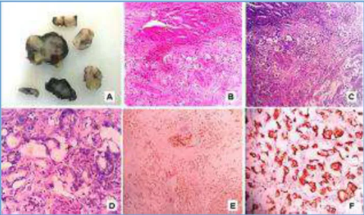
Fig. A. Cut Surface of Appendix showed Thickened Appendiceal
Wall with Mucinous Area and Occluded Lumen, B. Sections from Appendix showing Ulcerated Appendicular Mucosa and a Neoplasm Arranged in Glands and Clusters of Tumour Cells Predominantly in Submucosa and Muscularis Propria. (H&E, 40X), C. Glands and Clusters of Tumour Cells with Extracellular Pools of Mucin Infiltrating through the Muscularis Propria into the Serosa. (H&E, 100X), D. The Tumour Cells are Round to Polyhedral with Intracytoplasmic Mucin and Pleomorphic Dark-Staining Nuclei. (H&E, 400X), E. Immunohistochemistry-the Tumour Cells showed Diffuse Strong Cytoplasmic Positivity for Cytokeratin 20. (40x), F. IHC (400X).
Epithelial Tumours
Adenoma Carcinoma Adenocarcinoma Mucinous adenocarcinoma
Jemds.com
Case Report
J. Evolution Med. Dent. Sci./eISSN- 2278-4802, pISSN- 2278-4748/ Vol. 5/ Issue 65/ Aug. 15, 2016 Page 4679 Carcinoid
Tubular carcinoid
Goblet cell carcinoid (Mucinous carcinoid) Mixed carcinoid-adenocarcinoma
Others
Non-Epithelial Tumours
Neuroma, Lipoma, Leiomyoma Gastrointestinal stromal tumour Leiomyosarcoma, Kaposi sarcoma
Malignant Lymphoma
Secondary tumours and Hyperplastic (Metaplastic) polyp Table 1: WHO Classification of Tumours of the Appendix
Courtesy-WHO Classification of Tumours (Ref 9)
DISCUSSION
Appendiceal neoplasms are rare and most often found incidentally in an appendicectomy specimen.(1) Primary
Mucinous Adenocarcinoma of the Appendix is very rare accounting for 0.05% to 0.2% of all appendicectomies and only 6% of all malignant tumour of the appendix.(5) It
constitutes less than 0.5% of all GI neoplasm.(6) The incidence
of adenocarcinoma has been stated to be from 0.004% to 0.08%.
The mean age at presentation for mucinous adenocarcinoma is about 50 years with a male preponderance of 4:1. Adenocarcinoma of appendix most often present as acute appendicitis, (this may be due to distension of the appendix causing pain or from a superadded infection) or as a palpable abdominal mass or with intestinal obstruction, but some are entirely asymptomatic. Mucinous neoplasms of appendix can also present with uncommon anatomical abnormalities such as intestinal malrotation or situs inversus.(7) Since distant metastasis and viscera
involvement are very rare, death occurs mostly due to loss of intestinal function and obstruction by peritoneal implants. Carcinoma appendix is usually well-differentiated mucinous adenocarcinoma, which tend to perforate and produce Pseudomyxoma peritonei and do not metastasise until late in the disease process. The term for extensive spread of these tumours in the abdomen is Primary Mucinous Carcinomatosis (PMCA).(8) According to WHO, the neoplasms of the appendix
have been classified as epithelial and non-epithelial tumours (Table 1).(9) Recently, Misdraji et al(10) classified the mucinous
tumours of the appendix into three subtypes. They are: 1. Low-Grade Appendiceal Mucinous Neoplasm (LAMN),
which are adenomas confined to appendix or various alterations of the muscularis mucosa or wall).
2. LAMN with peritoneal spread having low malignant potential, and
3. Invasive adenocarcinoma.
Histopathological evidence of invasion of the appendiceal wall by the atypical glands and identification of epithelial cell in any intraperitoneal mucinous collection are the characteristic features of malignancy.(9) Mucinous histology
has a better prognosis than the colonic and goblet cell type.(11) The optimal management for mucinous
adenocarcinoma of appendix is right hemicolectomy. Immunohistochemically, these tumour cells are positive for
CDX2, MUC2, β-catenin, and CK20.
Molecular biology is similar to that of colorectal adenocarcinoma. Precursor lesions are villous polyp and serrated polyp. Similar to the large intestine, an adenoma-carcinoma sequence is assumed to occur in the appendix as well. KRAS mutations have been identified in 70% of cases and Loss of Heterozygosity (LOH) at 5q22, 6q, 17p13, and 18q21 are also identified. LOH at 5q is frequently linked Adenomatous Polyposis Coli (APC). LOH at one or two polymorphic microsatellite loci are seen in approximately half of the cases.(9)
Five year overall survival rate for localised adenocarcinoma to be 95% compared with 5-year survival rate of 80% for mucinous or cystadenocarcinoma. When distant metastases were present, the 5-year survival rate was 0% and 51% respectively.(9) This reflects the low aggressive
potential of mucinous tumour that spread to the peritoneum. It must be emphasised that up to 35% of the patients with appendiceal adenocarcinoma is more likely to have a second GI malignancy(12) that underlines the significant risk for both
synchronous and metachronous neoplasms.(13)
CONCLUSION
Appendiceal mucinous adenocarcinomas are rare and usually diagnosed when patient presents with pain, mass in RIF, or intestinal obstruction. Histopathological examination of all appendicectomy specimens is mandatory to rule out malignant pathology. The incidence of primary appendiceal adenocarcinoma in our institute is 0.005%, which correlates with that described in literature.
REFERENCES
1. McCusker ME, Cot´e TR, Clegg LX, et al. Primary malignant neoplasms of the appendix: a population-based study from the surveillance, epidemiology, and end results program, 1973-1998. Cancer 2002;94(12):3307-12. 2. Komm M, Kronawitter-Fesl M, Kopp R, et al. Primary
mucinous adenocarcinoma of the vermiform appendix with high-grade microsatellite instability. Journal of Cancer 2011;2:302-6.
3. Grover AS, Mittal S, Singla P, et al. Cystadenocarcinoma of appendix with cutaneous fistula - an unusual case presentation. Indian J Surg 2005;67(5):267-9.
4. Terada T. Non-invasive adenocarcinoma of the vermiform appendix: incidence and report of four cases among 512 appendectomies. Gastroenterol Res 2009;2(4):238-41.
5. O' Donnell ME, Badger SA, Beattie GC, et al. Malignant
neoplasms of the appendix. Int J Colorectal Dis 2007;22(10):1239-48.
6. Ozakyol AH, Saricom T, Kabukceaglu S, et al. Primary
appendiceal adenocarcinoma. Am J Clin Oncol
1999;22(5):458-9.
7. Petrou A, Papalambros A, Katsoula N, et al. Primary appendiceal mucinous adenocarcinoma alongside with situs inversus totalis: a unique clinical case. World Journal of Surgical Oncology 2010;8:49.
8. Sington JD, Warren BF, Manek S. Reduplication cyst of appendix with mucinous carcinoma and Mullerian metaplasia: a case report. J Clin Pathol 2002;55(7):551-3. 9. WHO classification of tumours. Pathology and genetics of
Jemds.com
Case Report
J. Evolution Med. Dent. Sci./eISSN- 2278-4802, pISSN- 2278-4748/ Vol. 5/ Issue 65/ Aug. 15, 2016 Page 4680 10.Misdraji J. Appendiceal mucinous neoplasms:
controversies. Arch Pathol Lab Med 2010;134:864-70.
11.Kabbani W, Houlihan PS, Luthra R, et al. Mucinous and
non-mucinous appendiceal adenocarcinoma, different
clinic pathological features, and similar genetic
alterations. Mod Pathol 2002;15(6):599-605.
12.Ito H, Osteen RT, Bleday R, et al. Appendiceal adenocarcinoma: long-term outcomes after surgical therapy. Dis Colon Rectum 2004;47(4):474-80.