SONOGRAPHIC FINDINGS IN NON-NEOPLASTIC TESTICULAR
LESIONS*
Ricardo Jorge Vital1
, Leandro Accardo de Mattos1
, Luís Ronan Marquez Ferreira de Souza2
, Sizenildo da Silva Figueirêdo3
, Jacob Szejnfeld4
Pathological processes of the testis are very frequently found, and include non-tumoral and tumoral lesions. Ultra-sonography performed with a high-frequency transducer has become the imaging modality of choice for examination of these organs. This method is of help to improve intratesticular lesions characterization, and in many instances the findings suggest a more specific diagnosis, especially in the presence of similar clinical manifestations such as pain, swelling and local increase in volume. Color Doppler is an invaluable tool for demonstrating anomalous patterns of testicular perfusion and aids in the diagnosis of acute clinical conditions. In the present iconographic essay, the authors summarize the most frequent clinical, pathologi-cal findings as well as the main diagnostic features of benign intratesticular lesions such as microlithiasis, simple cysts, spermatocele, varicocele, tubular ectasia of the rete testis, orchitis, hematomas and more rare conditions. Familiarity with clinical and sonographic features is essential for the correct diagnosis and start-ing of the most effective therapy, as necessary.
Keywords: Testis; Benign testicular lesions; Ultrasonography; Color Doppler.
Aspectos ultra-sonográficos das alterações não-neoplásicas do testículo.
Processos patológicos dos testículos são muito comuns, incluindo-se lesões tumorais e não-tumorais neste contexto. A ultra-sonografia com transdutor de alta freqüência tornou-se a modalidade de imagem de esco-lha para a avaliação desses órgãos. Este método ajuda a melhor caracterizar lesões intratesticulares e em muitas situações sugere um diagnóstico mais específico, principalmente nos casos em que há manifesta-ções clínicas similares, tais como dor, inchaço e aumento volumétrico locais. O mapeamento com Doppler colorido é importantíssimo para demonstrar padrões anormais de perfusão testicular e auxilia no diagnóstico de condições clínicas agudas. Neste ensaio iconográfico os autores sumarizam os mais comuns achados clínicos, patológicos e as principais características diagnósticas de lesões testiculares, tais como microlitíase, cisto simples, espermatocele, varicocele, ectasia tubular da rete testis, orquite, hematomas e condições mais raras. A familiaridade com as características ecográficas e clínicas destas alterações é essencial para o es-tabelecimento do diagnóstico correto e início da terapêutica mais eficaz, quando necessária.
Unitermos: Testículos; Afecções testiculares benignas; Ultra-sonografia; Color Doppler. Abstract
Resumo
* Study developed at Department of Diagnostic Imaging – Universidade Federal de São Paulo/Escola Paulista de Medicina, São Paulo, SP, Brazil.
1. MDs, Radiologists, Titular Members of Colégio Brasileiro de Radiologia e Diagnóstico por Imagem.
2. Assistant Professor Doctor of the Discipline of Diagnostic Imaging – Universidade Federal do Triângulo Mineiro, Titular Mem-ber of Colégio Brasileiro de Radiologia e Diagnóstico por Imagem. 3. Assistant Professor of the Discipline of Imaginology – Fa-culdade de Medicina da Universidade Federal de Rondônia, Ti-tular Member of Colégio Brasileiro de Radiologia e Diagnóstico por Imagem.
4. Adjunct Professor, Private Docent at Department of Diag-nostic Imaging – Universidade Federal de São Paulo/Escola Paulista de Medicina.
Mailing Address: Prof. Dr. Sizenildo da Silva Figueirêdo. Rua Natal, 371. Porto Velho, RO, 78905-120 – Brazil. E-mail: sizenildo@uol.com.br
Received October 28, 2004. Accepted after revision February 16, 2005.
TESTICULAR MICROLITHIASIS
Microlithiasis has been recently de-scribed and is a relatively rare entity, re-ported to be incidentally observed in 0.6% to 0.75% of patients submitted to testicu-lar ultrasound(1–4). Frequently, both testicles
are affected, however there are reports on unilaterality in patients from 10 months to 70 years of age(4,5).
The disease is clinically asymptomatic(2)
and its cause remains unknown. The inci-dental diagnosis should indicate a complete clinical evaluation for detecting subfertil-ity/infertility or systemic diseases.
Histologically,microliths represent scat-tered laminated calcium deposits in the lu-mina of the seminiferous tubules, called calcospherites(6).
It may be associated with other patho-logical entities like Klinefelter’s syndrome, cryptorchism, Down syndrome, male pseudohermaphroditism, pulmonary alveo-lar microlithiasis, previous radiotherapy and infertility conditions(7).
Initially, testicular microlithiasis was thought to be a benign process not requir-ing follow-up(7). However, a strong
asso-ciation has been seen with the presence of germ cell tumors, particularly semino-mas(5), and intratubular germ cell
neo-plasms which progress to carcinoma 50% of times (Figure 3).
At ultrasound, the most characteristic aspect of microlithiasis is of multiple, small, hyperechogenic foci ranging be-tween 1 mm and 3 mm in diameter, which typically do not shadow(2,8). Calcifications
appear diffusely scattered throughout the testicular parenchyma, but may be seg-mented or distributed peripherically (Fig-ures 1 and 2). Although not presenting posterior acoustic shadowing, occasionally they might generate comet-tail artifacts or a scintillating pattern at color Doppler(2).
There are scientific evidences justifying the ultrasound follow-up of patients with microlithiasis, aiming at the early diagno-sis of testicular tumors. Some authors have recommended an annual ultrasound fol-low-up, and that self-examination is en-couraged for all patients affected(2,4,10).
SIMPLE INTRATESTICULAR CYST
Intratesticular cysts are incidentally de-tected lesions in men with 40 or more years of age, measuring from 2 mm to 2 cm in diameter. They may be uni- or multilocu-lated(2), usually are single and non-palpable,
and may be multiple and bilateral(11,12).
The pathogenesis is still to be com-pletely known; three theories have been proposed: a) congenital, where simple intratesticular cysts would be a result from an increase in epithelial cells secretion or cystic distention from remnants of Müllerian and Wolffian ducts; b) post-trau-matic; c) post-inflammatory.
Microscopically, they are lined by flat or cuboid epithelium, surrounded by tes-ticular parenchyma, with serous content(2).
They may be found in any portion of the testes, but are more frequently seen near the mediastinum testis and adjacent to the tu-nica albuginea, associated with extratesti-cular spermatocele.
At ultrasound, the simple cyst present as an anechoic, well-defined lesion, with im-perceptible walls and posterior acoustic shadowing, localized inside the testicular parenchyma(11) (Figure 4).
When complicated with hemorrhage and infection, these cysts loose their
anechoic feature, and start showing inter-nal suspended echoes, resulting in differ-ential diagnosis with epidermoid cyst and teratoma(2).
Simple intratesticular cysts may be as-sociated with other congenital anomalies like Von Hippel-Lindau syndrome and cys-tic dysplasias(13).
INTRATESTICULAR SPERMATOCELE
Spermatocele refers to an intraparen-chymatous cystic lesion adjacent to the mediastinum testis, in the area of rete tes-tis, and may be septate and communicating with seminiferous tubules.
At histological evaluation, they present a fibromuscular wall surrounded by cuboid epithelium, pseudostratified or ciliated epithelium being rarely observed(14).
Ultrasound usually demonstrates anechoic lesions measuring between 1 cm and 2 cm, and that may reach more than 15 cm, with posterior acoustic enhancement. Spermatoceles might be confused with simple cysts, although its walls use to be more irregular(11,15,16).
INTRATESTICULAR VARICOCELE
While extratesticular varicocele is a common clinical condition, its intratesti-cular form is a rare entity which is seen in less than 2% of a population affected by scrotal symptoms(1,17). Intratesticular
varicoceles usually are left-sided, in asso-ciation with ipsilateral extratesticular Figure 1. Mild presentation of testicular
microlithi-asis. Sparse, small echogenic spots (arrow) distrib-uted in the parenchyma (calcospherites).
Figure 2. A more accentuated pattern of microli-thiasis. The calcospherites appear in higher num-ber and diffusely scattered.
Figure 3. Association of microlithiasis with testicular tumor. Note a partially cystic, round-shaped, het-erogeneous and hypoechogenic lesion (arrow), with lobulated margins (teratoma) in testicular paren-chyma with abundant calcospherites.
varicoceles, but their independent exist-ence is more common.
The most frequent claim of patients is local pain, while infertility, edema or scro-tal mass also present as typical symptoms. Its sonographic appearance consists of multiple serpiginous tubular and anechoic structures, varying in sizes, usually larger than 3 mm in diameter, localized around the mediastinum testis(18). These vessels are
more frequently found at left, during Val-salva maneuver, preferably in the orthos-tatic position.
At the mapping with color Doppler, the venous reflux is seen at the Valsalva ma-neuver, which is pathognomonic(1,19). The
differential diagnosis includes testicular cysts, tubular ectasia of the rete testis, he-matoma, focal orchitis and cystic intrates-ticular neoplasms(17).
raphic diagnostic characteristic of initial/ mild varicocele. A consensus is still to be reached about this matter, but most of urologists, in our practical experience, con-sider the venous reflux as a more reliable feature than the venous dilatation.
TUBULAR ECTASIA OF THE RETE TESTIS
Tubular ectasia of the rete testis is a rela-tively rare benign entity resulting from a partial or complete obstruction of efferent ducts(16). This obstruction may occur at
dif-ferent levels, and may be classified into intra- or extratesticular.
In testicular lesions, the obstruction is situated in the seminiferous tubules or in the rete testis, generally resulting from se-vere inflammatory episodes like orchitis. In extratesticular lesions, the proximal ob-structions result from luxation or traumatic tearing of epididymis, testicular varicoceles and cystic dysplasia. Middle obstructions are due to typical epididymal conditions like epididymitis, spermatoceles and tunica albuginea cysts. On the other hand, distal obstructions occur post-vasectomy or after scrotal surgery(8,20). Usually, these lesions
affect patients above 55 years of age, and frequently are bilateral (45%) and asymp-tomatic(2,21).
The typical presentation of the rete tes-tis consists of a serpiginous, hypoechoic structure communicating the mediastinum testis with the epididymis head usually lo-calized in the posterolateral region of the testes(20).
In cases of ectasia, ultrasound depicts numerous small tubular and cystic struc-tures with fluid and hypoechoic contents in the region of the rete testis, proximal to the mediastinum testis. At the mapping with color Doppler no flow is detected within the rete testis(16), differentiating it from the
intratesticular varicoceles. Occasionally, a pseudocystic pattern may be identified(2)
(Figures 5 and 6).
It is important to differentiate tubular ectasia of the rete testis from testicular tu-mors such as adenocarcinoma of the rete testis, epididymal cystadenoma, teratoma, non-Hodgkin lymphoma, and seminiferous tubules dilatation secondary to testicular neoplasms(16,22).
In dubious cases, magnetic resonance might be of help, showing non-enhance-able tubular structures after intravenous gadolinium injection, with homogeneous low-intensity signal in T1-weighted im-ages, and high-intensity or intermediary signal on T2-weighted images(22).
TESTICULAR TRAUMA
Testicular traumas usually result from car accidents, or contact sports, and may lead to contusion, hematoma, fracture or even testicular rupture which is considered as a surgical emergency(21). In closed
trau-mas, there is a formal indication for ultra-sound to evaluate the testicular viability, vascularization, and to serve as a basis for follow-up(6).
Hematomas are identified at ultrasound as heterogeneous testicular lesions, and may become complex, with cystic compo-nents. Considering the frequency of post-traumatic atrophy, the ultrasound follow-up is essential until the complete resolution of
testicular alterations. This atrophy occurs as a result of ischemia caused by increased pressure inside the tunica albuginea sec-ondary to hematoma formation(23) (Figures
7 and 8).
Tunica albuginea fracture is another condition characterized by a fault (interrup-tion) on the organ borders, an even exter-nal protrusion of the testicular pulp due to the segmental interruption of the echogenic testicular coat (tunica albuginea)(1).
On the other hand the diagnosis of tes-ticular fracture is made when there is a lin-ear, irregular, hypoechoic intratesticular abnormality, dividing the testis into two or more parts. The differential diagnosis with a transmediastinal artery is not easy. Frac-tures with severe testicular injury present with a heterogeneous echotexture(1).
TESTICULAR INFARCTION
Testicular infarction is associated with episodes of subacute torsion or infectious phenomenon determining zones of
is-Figure 6. Ectasia of the rete testis. Other more ex-tensive case demonstrating clustered anechoic tu-bular structures (arrow) in the testis anterior region.
Figure 5. Ectasia of the rete testis. There is a net-work of anechoic, thin tubular structures in the testicular hilar region.
Figure 7. Testicular hematoma. Anechoic paren-chymal collection surrounded by irregular hypo-echoic areas.
chemia in the testicular parenchyma(2).
More rarely, segmental infarcts are dis-closed during a picture of accentuated pain due to several etiologies such as trauma, arterial embolism, venous thromboses and various angiopathies(2). Other iatrogenic
origin is the resection or connection of the spermatic cord vascular elements in an in-guinal hernioplasty.
Its sonographic appearance depends on the patient’s age and severity of the in-jury(23). In an early phase, ultrasound does
not demonstrate any change in echogen-icity. Posteriorly, ischemic zones may be single or multiple, and appear round-shaped and hypoechoic on a plane, and in a more triangular shape with poorly defined bor-ders on the perpendicular plane (Figure 9). The mapping with color Doppler shows perfusion of hypervascularized perilesional regions and/or absent flow in the lesions(2).
With the clinical progression, lesions tend to decrease in size, calcify, and increase its echogenicity as a result of fibrosis(23).
Small, more frequent infarction zones are hardly differentiated from small tumors (which may be equally avascular), however the triangular-shaped infarction is a dis-criminative semiological element(2).
Amongst the differential diagnosis, one should consider testicular tumor and ab-scess which present a diverse clinical his-tory(11). In dubious cases, magnetic
reso-nance imaging shows the gadolinium en-hancement in tumor processes, and absent enhancement in infarction(2).
Hemorrhagic infarcts may rarely occur as a complication of inguinal hernioplasty. The anatomopathological substrate is the
venous flow interruption, generating local congestion and hemorrhagic necrosis.
At ultrasound, an increase in testis is observed with diffuse heterogeneity asso-ciated with permeated hyperechogenic ar-eas, suggesting zones of liquefaction look-ing like a thick fluid.
At magnetic resonance imaging, T1-weighted sequences demonstrate an in-creased signal typical of hemorrhagic col-lections, because of products from hemo-globin degradation(2).
ACUTE ORCHITIS
Usually orchitis results from a direct extension of epididymis inflammation to testis, which becomes acutely edematous and painful(1,23). Primary orchitis is
fre-quently viral and associated with parotidi-tis. Contrarily to bacterial orchiepididy-mitis, the viral infections usually are bilat-eral(1).
In adolescents, most common causing agents are those sexually transmitted like
Chlamydia trachomatis and Neisseria gonorrhoeae. In prepubertal boys and men aged above 35 years, most frequently the disease is caused by Escherichia coli and
Proteus mirabilis(24).
Ultrasound shows indirect signs of in-flammation such as hydrocele or pyocele associated with scrotal wall thickening, these forms being present in most of cases. The diffuse testicular involvement is con-firmed by increase in parenchymal volume as a result of testicular edema, presenting a heterogeneous echotexture(21). In focal
processes, presentation is characterized by
multiple hypoechoic lesions scattered throughout the parenchyma(21) (Figure 10).
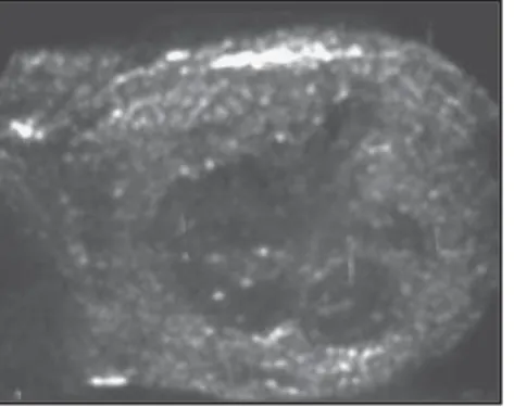
In viral orchitis there is a diffuse increase in volume, and homogeneous or heteroge-neous hypoechogenicity with peripheral hypoechoic zones(1) (Figure 11).
Since these findings are not specific of orchitis, they must be correlated with clini-cal history and laboratory tests. The patient should be followed-up until complete reso-lution. Additionally, findings must be docu-mented by ultrasound aiming at ruling out tumor, infarction and metastasis.
At color Doppler-mapping, one ob-serves an increase in the blood flow into epididymis and testis, characterized by in-creased number of vascular structures iden-tifiable per view field which must always be compared with the contralateral testis. The analysis of the resistivity index and spectral pattern may bring useful informa-tion, since in these cases there is a decrease in the vascular resistance in relation to healthy individuals(25) (Figure 12).
In this context, ultrasound also becomes essential for researching the process sever-ity, the complications, and elements of differential diagnosis like a subacute tes-ticular torsion(1).
A relatively frequent infectious compli-cation is the abscess, corresponding to a hypoechogenic fluid collection with poorly defined contours and a hypo/hyperecho-genic halo, sometimes associated with echogenic débris.
The color Doppler-mapping shows an increase in the perilesional vascularization and absent blood flow inside the abscess. Rarely, gas bubbles can be observed,
ap-Figure 9. Testicular infarction.
A: Hypoechoic, homogeneous, round-shaped focus (arrow-head), with precise contours, occupying a great part of the organ (between arrows). B:
Other case showing two hypo-echoic foci similar to that on Figure 9A, but with smaller di-mensions, corresponding to the infarction area.
pearing like hyperechoic, supernatant punctate foci(1) (Figures13, 14 and 15).
Another complication is the acute is-chemia generated by compression of the vascular pedicle of the spermatic cord with the resulting formation of intratesticular venous thrombi. At the color Doppler-map-ping, decreased or absent vascularization is observed, in contrast to the adjacent epid-idymis hypervascularization(1). With the
clinical progression, the affected testis at-rophy is observed (Figure 16).
CRYPTORCHISM AND ECTOPIA
The frequency of cryptorchism in the population ranges between 0.5% and 0.8%. The undescended testis may be situated
Figure 13. Testicular abscess. Orchitis complica-tion. Heterogeneous parenchymal collection with irregular limits, occupying practically the whole tes-tis.
Figure 12. Acute orchitis. At color Doppler-map-ping (although under black and white photo-docu-mentation) an increase in caliber and quantity of intratesticular vessels is observed, characterizing a pattern of intraparenchymal vascularization.
Figure 14. Testicular abscess. Orchitis complica-tion. In this case, the patient had already presented purulent secretion on the skin. The testes presented enlarged, heterogeneous and hypoechogenic (edema), in association with thickening of the tu-nica vaginalis.
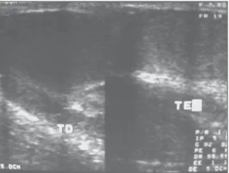
Figure 11. Bilateral acute orchitis. Diffusely hypoechoic testes present-ing increased volume in higher proportion at left (patient affected by viral parotiditis). (TD, right testis; TE, left testis).
Figure 10. Unilateral acute orchitis. Note hypoechogenicity and diffuse increase in volume of the left testis in relation to its contralateral.
Figure 15. Bilateral testicular necrosis. Orchitis complication in a diabetic patient. Bilateral testes parenchymal destruction with interspersed necrotic debris in the scrotal sac (a bilateral orchiectomy was performed). (TD, right testis; TE, left testis). from the renal hilus to the inguinal canal.
In one-third of cases is bilateral, but in cases of unilaterality the right side is most fre-quently affected. Most common localiza-tion is in the inguinal canal (72% of cases), followed by the prescrotal (20%) and ab-dominal or pelvic topography (8%)(1).
Testicular ectopia is a quite rare condi-tion, and the most frequent localization is the subcutaneous region situated between the internal and external inguinal fossas(21).
RARE TESTICULAR DISEASES
Ectopic rests of adrenal tissue
During the embryonal development, some adrenocortical cells may remain in the testicular parenchyma, and, unless they receive sufficient hormonal stimulation, remain undetectable. In children with con-genital adrenal hyperplasia, there is a de-fect in the glucocorticoid synthesis and possible intratesticular adrenal rests are abnormally stimulates, suffering from hy-pertrophy. The lesions tend to be detected by ultrasound as multiple, round-shaped and hypoechoic(6,16). These lesions are not
considered as potentially malignant(23).
Sarcoidosis
Sarcoidosis rarely affects the testis, with genital involvement occurring in only 1% of patients with systemic involvement. Usually, it causes a painless enlargement of the testis, and at ultrasound irregular, solid, hypoechoic and potentially calcifying le-sions are identified(2,23).
and at color Doppler demonstrates a blood flow pattern with low impedance, high di-astolic peak velocity, and presence of a drainage vein(27).
REFERENCES
1. Roy C, Tuchmann C. Échografie scrotale – patho-logies non tumorales courantes (part 1). J Radiol 2003;84:581–595.
2. Roy C, Tuchmann C. Échografie scrotale – patho-logies kystiques, tumorales et non tumorales ra-res (part 2). J Radiol 2003;84:667–680. 3. Hobarth K, Susani M, Szabo N, Kratzik C.
Inci-dence of testicular microlithiasis. Urology 1992; 40:464–467.
4. Cast JEI, Nelson WM, Early AS, et al. Testicular microlithiasis: prevalence and tumor risk in a population referred for scrotal sonography. AJR Am J Roentgenol 2000;175:1703–1706 5. Backus ML, Mack LA, Middleton WD, King BF,
Winter TC III, True LD. Testicular microlithiasis imaging appearances and pathologic correlation. Radiology 1994;192:781–785.
6. Dambro TJ, Stewart RR, Barbara CA. The scro-tum. In: Rumack CM, Wilson SR, Charboneau JW, editors. Diagnostic ultrasound. 2nd ed. St Louis: Mosby, 1998;791–821.
7. Frush DP, Kliewer MA, Madden JF. Testicular microlithiasis and subsequent development of metastatic germ cell tumor. AJR Am J Roentgenol 1996;167:889–890.
Tuberculosis and syphilis may present similar sonographic manifestations, and should be considered as differential diag-nosis(23). When this disease presents with
a focal pattern, the differential diagnosis with other tumor-like lesions is not feasible with ultrasound(2).
Splenogonadal fusion
The testicular fusion with splenic tissue is an unusual finding, and may be discon-tinuous (intratesticular ectopic splenic tis-sue), or continuous (direct connection be-tweenthe spleen and gonad by a fibrous cord)(26). Clinically, the patients present
with an inguinal or scrotal mass; lesions larger than 5 cm have already been de-scribed. Frequent associated findings are: hernia, cryptorchism and micrognathia(16).
Intratesticular arteriovenous malforma-tion
An intratesticular arteriovenous malfor-mation is a rare entity, and the differential diagnosis is hemangioma. This condition usually presents like a hypoechoic nodule,
Figure 17. Cryptorchism. A:
Testis in left inguinal region, above the spermatic cord, with elongated shape and reduced dimensions (ar-row). B: Other case shows testis in the inguinal region (between markers), sur-rounded by a discrete halo of anechoic fluid.
B A
seminoma and embryonic carcinoma being the most frequent types of tumors(1).
At ultrasound, the cryptorchidic testis appears atrophic, hypoechoic, homogene-ous or heterogenehomogene-ous (Figure 17). In its most frequent localization, the external inguinal canal opening is identifiable by echography. In the cases where this opening is localized in the deep opening of the inguinal canal, the presence of gas reverberation artifacts from the digestive apparatus complicate the visualization of a small testis; so in these cases magnetic resonance imaging is the indicated modality(1).
8. Diego AC, Rasines GL, Rodriguez RH. Lesiones testiculares que no hay que tocar. Arch Esp Urol 2003;56:255–266.
9. Janzen DL, Mathieson JR, Marsh JI, et al. Tes-ticular microlithiasis: sonographic and clinical features. AJR Am J Roentgenol 1992;158:1057– 1060.
10. Abbehusen C, Oliveira JMA, Granero L, Ajzen AS, Szejnfeld J. O valor do ultrassom na micro-litíase testicular. Urologia on line 1998;2(5):sp. [citado agosto 2004]. Disponível em: http://www. unifesp.br/dcir/urologia/uronline
11. Dogra VS, Gottlieb RH, Rubens DF, Liao L. Be-nign intratesticular cystic lesions: US features. RadioGraphics 2001;21 Spec No:S273–S281. 12. Rifkin MD, Jacobs JA. Simple testicular cyst
di-agnosed preoperatively by ultrasound. J Urol 1983;129:982–983.
13. Rezende DEL. Cisto simples de testículo: relato de caso e revisão da literatura. Radiol Bras 2001; 34:251–252.
14. Yalowitz BR, Eble JN, Wilks DC.
Spermatozoa-containing simple cysts of the rete testis. J Urol 1989;142:1572–1573.
15. Davis RS. Intratesticular spermatocele. Urology 1998;51(SA Suppl):167–169.
16. Rubenstein RA, Dogra VS, Seftel AD, Renick MI. Benign intrascrotal lesions. J Urol 2004;171: 1765–1772.
17. Das KM, Prasad K, Szmigielski W, Noorani N. Intratesticular varicocele: evaluation using con-ventional and Doppler sonography. AJR Am J Roentgenol 1999;173:1079–1083.
18. O’Donell PG, Dewbury KC. The ultrasound ap-pearances of intratesticular varicocele. Br J Radiol 1998;71:324–325.
19. Atasoy C, Fitoz S. Gray-scale and color Doppler sonographic findings in intratesticular varicocele. J Clin Ultrasound 2001;29:369–373.
20. Burrus JK, Lockhart ME, Kenney PJ, Kolettis PN. Cystic ectasia of the rete testis: clinical and radio-graphic features. J Urol 2002;168(4 Pt 1):1436– 1438.
21. Dogra VS, Gottlieb RH, Oka M, Rubens DJ.
Sonography of the scrotum. Radiology 2003;227: 18–36.
22. Rouviere O, Bouvier R, Pangaud C, Jeune C, Dawahra M, Lyonnet D. Tubular ectasia of the rete testis: a potential pitfall in scrotal imaging. Eur Radiol 1999;9:1862–1868.
23. Howlett DC, Marchbank NDP, Sallomi DF. Pic-torial review. Ultrasound of the testis. Clin Radiol 2000;55:595–601.
24. Luker GD, Siegel MJ. Color Doppler sonography of the scrotum in children. AJR Am J Roentgenol 1994;163:649–655.
25. Middleton WD, Siegel BA, Melson GL, Yates CK, Andriole GL. Acute scrotal disorders: prospective comparison of color Doppler US and testicular scintigraphy. Radiology 1990;177:177–181. 26. Putschar WG, Manion WC. Splenic-gonadal
fu-sion. Am J Pathol 1956;32:15–33.