PRELIMINARY STUDY OF CELL METABOLISM, BY USE OF NBT TEST,
DETERMINATION THE INTENSITY
OF LIPID PEROXIDATION AND ANTIOXIDANT ACTIVITY
Annamaria PALLAG*, Ladislau RITLI**, Ildikó SZABÓ*, Mariana MUREŞAN*, Diana BEI*
* University of Oradea, Faculty of Medicine and Pharmacy, Department of Pharmacy, Oradea, Romania **University of Oradea, Faculty of Medicine and Pharmacy, Department of Medicine, Oradea, Romania
Corresponding author: Annamaria Pallag, University of Oradea, Faculty of Medicine and Pharmacy, Department of Pharmacy, 10 P-ta 1 Decembrie, 410068 Oradea, Romania, tel.: 0040259415680, fax: 0040259418266, e-mail: [email protected]
Abstract. Otto Warburg, in the early part of the 20th century, originated a hypothesis, that the cause of cancer is primarily a
defect in energy metabolism.
A decrease in the capacity of mitochondria to reduce NAD(P), together with a decline in the NAD(P)H/NAD(P) redox couple, uncouples oxidative phosphorylation, lead to depletion of ATP and decrease the cell viability.
Nitro-bleu tetrazolium have been used to assay cell proliferation and viability. The method to measure cell proliferation is based on enzymatic cleavage of the tetrazolium salts to a water-soluble formazan dye.
Succinate-tetrazolium reductase, is an enzymatic sistem, which belongs to the respiratory chain of the mitochondria and it is active only in viable cells. The reagent diffuses into the cells and it is cleaved to formazan. The absorption change is measured and analysed.
Free radicals such as superoxide, can cause a damage in cellular components, but several antioxidants inhibiting the lipid peroxidation and limiting the level of free radicals in cells.
In the present study we had in view the proliferation and viability of leukemia cells during antineoplastic treatment along with the alteration of the serum level of malondialdehyde (MDA) and ceruloplasmin (CP). With serum level of malondialdehyde we monitored the presence of the lipid peroxidation by the reactive oxygen species, and with the oxidized ceruloplasmin level in blood serum we evidenced the activity of antioxidant system in blood.
Keywords: nitro-bleu tetrazolium, leukemia, cell proliferation, MDA. CP
INTRODUCTION
In the early part of the 20th century, Otto Warburg originated a hypothesis that the cause of cancer is primarily a defect in energy metabolism This hypothesis was based on the observation, since repeated and verified many times, that cancer cells show clear differences in energy metabolism when compared to normal cells [12].
In the respiratory chain, during transfer of electrons to molecular oxygen, an estimated 1 to 5% of electrons lose their way, most participating in formation of reactive oxygen species [3].
However, generation of reactive oxygen species (ROS), may be a relatively late event, occurring after cells have embarked on a process of caspase activation. A decrease in the capacity of mitochondria to reduce NAD(P), together with a decline in the NAD(P)H/ NAD(P) redox couple, permeabilizes the inner mitochondrial membrane. This favors the release of Ca2+ from the organelle and uncouples oxidative phosphorylation and these effects lead to depletion of ATP [13].
The reduced ATP levels also lead to further impairment of other Ca2+ regulation system, in the plasma membrane and the endoplasmatic reticulum. In addition, the decrease in NAD(P)H compromises the activity of protective enzymes, which further increases the deleterious effects of ROS [6, 12].
Cancer cells exhibit unlimited proliferative potential, resistance to cell death stimuli and abnormal energy metabolism [7].
Several tetrazolim salts, including NBT (nitro bleu tetrazolium) have been used to assay cell proliferation and viability.
Succinate-tetrazolium reductase, is an enzymatic
mitochondria and it is active only in viable cells [19, 12].
The total activity of this mitochondrial dehydrogenase increases proportionally to the number of viable cells, leading to an increase in tetrazolium salt conversion to formazan dye, which is in turn quantified by absorbance. We can analyse cytotoxic and cytostatic compounds as anticancer drugs and other pharmaceuticals.
During the respiratory explosion, the increase of the oxigen metabolism and the production of ROS is accompanied by the increase of the reduction potential in phagolysosomes and the turning into blue of NBT [16].
The more the cellular metabolic activity is intensified, and consequently the oxidation reduction reactions take place at a higher intensity, the more a larger quantity of formazan will result, colouring the cells more intensly. In the more of the cell displays a decreased metabolic activity, or it is dead, the smaller the amount of formazan resulted.
The exogenous cellular stresses, such as chemotherapy or increased level of reactive oxygen species (ROS) are activators to mitochondrial distruption [5].
Free radicals such as superoxide and hydroxyl radicals, can cause a damage in cellular components via peroxidation of proteins, nucleic acids, free amino acids, and lipoproteins. Free radicals first attack fatty acids, because the C=C double bonds in these molecules are sensitive to oxidative damage [9].
(MDA) which is commonly used as an index of lipid peroxidation [8, 14].
Ceruloplasmin (CP) is an enzyme, which have antioxidant activity, inhibiting the lipid peroxidation and limiting the level of free radicals in cells. The oxidized ceruloplasmin level in blood serum is evidence the activity of antioxidant system in blood [16].
In the present study we had in view the proliferation and viability of leukemia cells during antineoplastic treatment along with the alteration of the seric level of malondialdehyde and ceruloplasmin.
MATERIALS AND METHODS
It have been studied leukemia cells. We were monitored in various stages of the antineoplasic treament, the cell proliferation and cell viability by use of nitro bleu tetrazolium, the serum level of malondialdehyde and ceruloplasmin.
Blood samples were taken from the patients before the treatment and after 24 after the beginning of polychemotherapy.
NBT test
The formazan salt (NBT) diffuses into the cell and it is cleaved to formazan, by tetrazolium succinate reductase, a system belonging to the mitochondrial respiratory chain and active in the viable cells.
NBT is turned into formazan by breaking the N-N bonds within the molecule, and the N adds a H+ atom, representing an intermediary electron acceptor.
H+ results from NADH, which will thus be oxidized:
NADH → NAD+ + H+
The more of cellular metabolic activity is intensified, and consequently the oxidation reduction reactions take place at a higher intensity, the more a larger quantity of formazan will result, colouring the cells more intensly. The more the cell displays a decreased metabolic activity, or it is dead, the smaller the amount of formazan resulted [14].
Materials needed
-Test tubes with Na2EDTA, used for blood sample taking;
-NBT (1%) stock solution, prepared from 10 mg NBT and 1 ml bidistilled water. The mixture obtained this way can be kept in the refrigerator at 4 ˚C for several months;
-Michaelis buffer, pH 7.4, prepared from Na hydro-chlorate 0.85 % neuter solution; 5 ml veronal acetate solution; 5 ml hydrochloric acid N/10;
-Methylic alcohol [4, 5].
Working method
1-2 ml blood is drawn into a test tube containing 2 mg Na2EDTA and shaken. 0.08 ml Michaelis buffer pH 7.8 is added into the test tube; 0.02 ml 1% NBT solution. 0.10 ml 0.2 % NBT solution is obtained this way. 0.10 ml blood drawn on Na2EDTA is added.
The test tube is incubated in the thermostat, at 370C, for 30 minutes. The numeration of cells it realized atmicroscope. The cells which are colored in blue have formazan sediment. With numeration of this cells we can determination the rate of cell viability and cell proliferation. When the percentage of viable cells are below 30% it considered the NBT test negative, but when above 30%, it considered the NBT test positive.
Colorimetric determination of malondialdehyde
Malondialdehyde is one of the products of lipid peroxidation, it’s determination represents a standard method of assessing the oxidative stress.
The dosage method is based on the reaction with thiobarbituric acid (TBA). The biological sample is heated with TBA, in acidic medium. As a result of the reaction, one molecule of MDA reacts with two molecules of TBA, with the production of a pink pigment, with a measured optical density at 530 nm, using Pharmacia LKB Ultraspec. III spectrophoto-meter.
Normal values of the MDA serum levels are between 0.27 – 1.02 nmol/ml. Increased values of the MDA serum levels confirm the presence of the oxidative stress [11].
Colorimetric determination of ceruloplasmin
Ceruloplasmin (ferroxidase) is a 150 kD, blue α
2-glycoprotein that is synthesized in the liver and it is accumulated in the matrix and the inner membrane of the mitochondria. Ceruloplasmin acts mainly as a ferroxidase, catalyzing the oxidation of Fe (II) to Fe (III), and as a Fe (II) carrier in the plasma in association with transferrin, the only protein which can carry iron in this state.
Beside its detoxifying activity in the blood, ceruloplasmin also presents a dismutase-like activity (lower than that of the superoxide dismutase), it inhibits the peroxidation of polyunsaturated fatty acids (in vitro demonstration) and it has immunologic activity. Ceruloplasmin limits the quantity of free radicals, acting as a plasmatic antioxidant [9, 15].
Normal ceruloplasmin serum levels are between 0.20-0.40 mg/ml (11-24 μM).
Abnormal ceruloplasmin level impedes the mitochondrial respiratory process [1, 16].
The principle of the method is based on the phenol oxidative property of ceruloplasmine, which catalyzes the oxidation of paraphenylenediamine, with the production of a violet compound.
The optical density against a reference blank was measured at 530 nm, using Pharmacia LKB. Ultraspec. III spectrophotometer. The ceruloplasmine quantity in 1 ml can be determined using the formula:
E x 0.87 = CP serum level (mg/ml)
RESULTS
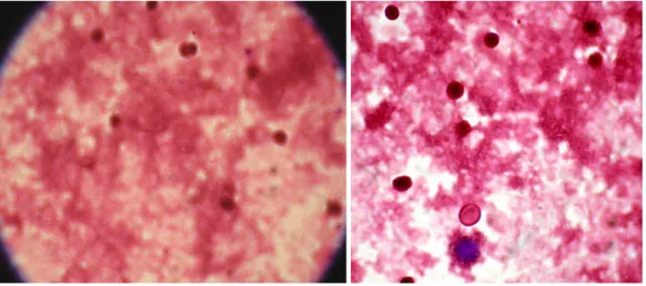
In Figure 1 and 2 we are showing two examples of NBT test before and after beginning the antineoplasic treatment.
A B
Figure 1. Positive NBT test, when A represent NBT test from blood derived by subject 1 before treatmen and B represent NBT test from blood derived by subject 2 before treatment (400X).
A B
Figure 2. Negative NBT test, when A represent NBT test from blood derived by subject 1 after 24 hours at the beginning of treatment and B represent
NBT test from blood derived by subject 2 after 24 hours at the beginning of treatment (400X)
We can see in Figure 1 that before the antineoplasic treatment the cells display proliferation, they are alive. This proliferation significantly decreases after 24 hours of treatment (Fig. 2), the cells displays a decreased
metabolic activity, together with a decline in the NAD(P)H/NAD(P) redox couple, resulted the smaller amount of formazan.
0 0,5 1 1,5 2 2,5 3 3.5
1 2 3 4 5 6 7 8 9 10 11 12 13 14 15
Figure 4. CP seum level before antineoplastic treatment and after 24 hours
In Figure 3 we are showing the serum level of MDA before treatment and after 24 hours. In Figure 4 we can observed that the serum level of ceruloplasmin decreased after 24 hours after the beginning of treatment.
The cytotoxicity of the antineoplastic agents is manifested by the increase in the level of free radicals, by the peroxidation of polyunsaturated fatty acids at mitochondrial level, this occurring without an immediate response of the organism, which would be manifested by an increase in the ceruloplasmine level.
DISCUSSIONS
Cancer cells exhibit unlimited proliferative potential, resistance to cell death stimuli and abnormal energy metabolism. This metabolic alteration has been observed in many cancer types, including leukemia [7]. It is now recognized that the Warburg effect (respiration chain deficient) represents a prominent metabolic characteristic of malignant cells.
An efficient chemotherapy depends on the inductive of the appearance of some modifications on the signal ways of the programmed death of cells [2].
Before the treatment the cells display proliferation, they are alive. This proliferation significantly decreases after 24 hours of treatment, the cells displays a decreased metabolic activity, together with a decline in the NAD(P)H/NAD(P) redox couple.
The presence of the oxidative stress, of the accumulation of the reactive oxigen species, due to lipid peroxidation is confirmed throught the study.
Increased level of ROS (reactive oxygen species) in cells cause mitochondrial disfunction, the disfunction of the mitochondrial respiratory chain, which can induce an more efficient response to antileukemic therapy through beginning the apoptosis process [3].
The ceruloplasmin serum level in patients under polychemotherapeutic treatment was monitored in order to determine the presence of plasmatic antioxidant during the antineoplastic treatment , as well as to indicate a possible mitochondrial disorder, because of too low ceruloplasmin serum levels. The
oxidative activity of ceruloplasmin towards paraphenylenediamine is used for dosing the oxidative activity, therefore for dosing ceruloplasmine [11].
The cytotoxicity of the antineoplasic agents is manifested by the increase in the level of free radicals, by the peroxidation of polyunsaturated fatty acids at the mitochondrial level, with no other response of the organism subjected to treatment. The decrease of cell proliferation along with an increased level of the oxidative stress in leukemia cells de
The present study aims to investigate the possible relationship between cell viability, serum malon-dialdehyde level, an index of lipid peroxidation, and ceruloplasmin level, a protective agent against lipid peroxidation.
A correlation was detected between cell viability and serum level of malondialdehyde. No correlation was detected between the serum malondialdehyde and ceruloplasmin levels. It was suggested that increase in the serum malondialdehyde level might be associated with an imbalance of other antioxidants rather than ceruloplasmin.
REFERENCES
[1] Aksoy, H., Koruk, M., Akcay, F., (2003): The relationship between seum malondialdehyde and ceruloplasmine in chronic liver disease, Turkish Journal of Biochemistry, 28 (2): 32-34.
[2] Amuthan, G., Biswas, G., Zhang, S.Y., Klein-Szanto, A., Vijayasarathy, C., Avadhani, N.G., (2001): Mitochondria to nucleus stress signaling induces phenotypic changes, tumor progression and cell invasion, EMBO, J., 20: 1910. [3] Carew, J.S., Huang, P., (2002): Mitochondrial defects in
cancer, Molecular Cancer, 1: 9.
[4] Carlson, M.A., (2006): Tehnical note: assay of cell quantity in the fibroblast-populated collagen matrix with tetrazolium reagent, European Cells and Materials, 12: 44-48.
[5] Carmichael, J., De Graff, W.G., Gazdar, A.F., Minna, J.D., Mitchell, J.B., (1987): Evaluation of a tetrazolium-based semiautomated colorimetric assay, assessment of radiosensitivity, Cancer Res. 47: 936-942.
[6] Cory, A.H., Owen, T.C., Barltrop, J.A., Cory, J.G., (1991): Use of an aqueros soluble tetrazolium 0
0.1 0.2 0.3 0.4 0.5 0.6
1 2 3 4 5 6 7 8 9 10 11 12 13 14 15
salt/formazan assay for cell growth assays in culture. Cancer Commun., 3: 207-212.
[7] Czarnecka, A.M., Gammazza, A., Felice, V.D., Zummo, G., Cappello, F., (2007): Cancer as a „Mitochondriophathy”, J. Cancer Mol. 3 (3): 79-95.
[8] Evereklioglu, C., Er, H., Turkoz, Y., Cekmen, M., (2002): Serum levels of TNF-alpha, sIL-2R, IL-6, and IL-8 are increased and associated with elevated lipid peroxidation in patients with Behcet’s disease. Mediators Inflamm. 11: 87-93.
[9] Gerasimov, I.G., Ignatov, D., (2005): Nitrobleu tetrazolium reduction by human blood neutrophils, Tsitologia, 47(6): 554-558.
[10] Ishiyama, M., Tominaga, H., Shiga, M., Sasamoto, K., Ohkura, Y., Ueno, K., (1996): A combined assay of cell viability and in vitro cytotoxicity with a highly water-soluble tetrazolium salt, neutral red and crystal violet, Biol. Pharm.. Bull., 19: 1518-1520.
[11] Muresan, M., Muresan, I., Burta, L., Burta, O., Micle, L., Micle, O., Dorofteiu, M., (2003): Stresul oxidativ în bolile medico-chirurgicale, University of Oradea Press, pp. 89-97.
[12] Pelicano, H., Xu, R., Du, M., Feng, L., Saski, R., Carew, J.S., Hu, Y., Ramdas, L., Hu, L., Keating, M.J., Zhano, W., Plunkett W., Huang, P., (2006): Mitochondrial respiration defects in cancer cells cause activation the Akt survival pathway through a redox-mediated mechanism, The Journal of Cell Biology Vol. 175: 6, 913-923. [13] Punchard, N.A., Kelly, F.J., (1996): Free Radicals, pp.
125-128.
[14] Sies, H., 1997: Oxidative stress: Oxidants and antioxidants, Experim. Physiol., 82: 291-295.
[15] Schimmer, A.D., Hedley, D.W., Penn, Z.L., Minden, M.D., 2001: Receptor and mitochondrial-mediated apoptosis in acute leukemia: a translational view, Blood, 98: 3541-3553.
[16] Vasin, A.V., Platonova, N.A., Povalikhin, R.G.,
Klotchenko, S.A., Samsonov, S., Tsymbalenko,
N., Puchkova, L., 2005: Mitochondrial ceruloplasmin of mammals, Molecular Biology, 39 (1): 48-60.