CASE REPORT
J of Evidence Based Med & Hlthcare, pISSN- 2349-2562, eISSN- 2349-2570/ Vol. 2/Issue 12/Mar 23, 2015 Page 1879
VANISHING WHITE MATTER DISEASE: A CASE REPORT
Sindu P. Gowdar1, Naveen S. Maralihalli2, Pramod Setty J3, Rajesh Venunath4, Mithila P. V5
HOW TO CITE THIS ARTICLE:
Sindu P. Gowdar, Naveen S. Maralihalli, Pramod Setty J, Rajesh Venunath, Mithila P. V. ”Vanishing White Matter Disease: A Case Report”. Journal of Evidence based εedicine and Healthcare; Volume 2, Issue 12, March 23, 2015; Page: 1879-1884.
ABSTRACT: Vanishing white matter disease (VWM) is one of the most prevalent inherited childhood Leucoencephalopathies. We report MR imaging features of vanishing white matter disease in a 4-year-old boy, who manifested with seizures, aphasia, spastic quadriparesis and myoclonic jerks. MRI of brain showed diffuse white matter signal changes of CSF intensity in all the sequences. MR spectroscopy of white matter showed severe decrease in NAA, choline and creatine and presence of lactate peak. Additional notable findings were diffuse extensive brain stem and thalamic atrophy. The clinico-radiological correlation was consistent with the diagnosis of vanishing white matter disease. Reporting of such cases may widen the spectra of these disorders.
KEYWORDS: MRI, MRS, vanishing white matter disease.
INTRODUCTION: Vanishing white matter disease (VWM) is one of the most prevalent inherited childhood leucoencephalopathies. The disease is variably called Myelinopathia Centralis Diffusa. Childhood ataxia and diffuse central nervous system hypomyelination are the common findings. The disease is characterized by chronic progressive and episodic deterioration with ataxia, spasticity and optic atrophy.1 VWM is caused by mutation in any of the five genes encoding the
subunits of eukaryotic translation initiation factor eIF2B.2,3 The disease has an autosomal
recessive mode of inheritance. The cause of the disease is unknown. Previously it was known that there is no biochemical marker for this disease,4 but recently analysis of body fluids has revealed
only a few biochemical markers for VWM. The first marker found was a consistent elevation of cerebrospinal fluid glycine concentrations with an elevated ratio of cerebrospinal fluid to plasma glycine concentrations.5 A decreased cerebrospinal fluid concentration of asialotransferrin is a
recently identified biomarker for VWM.
CASE REPORT
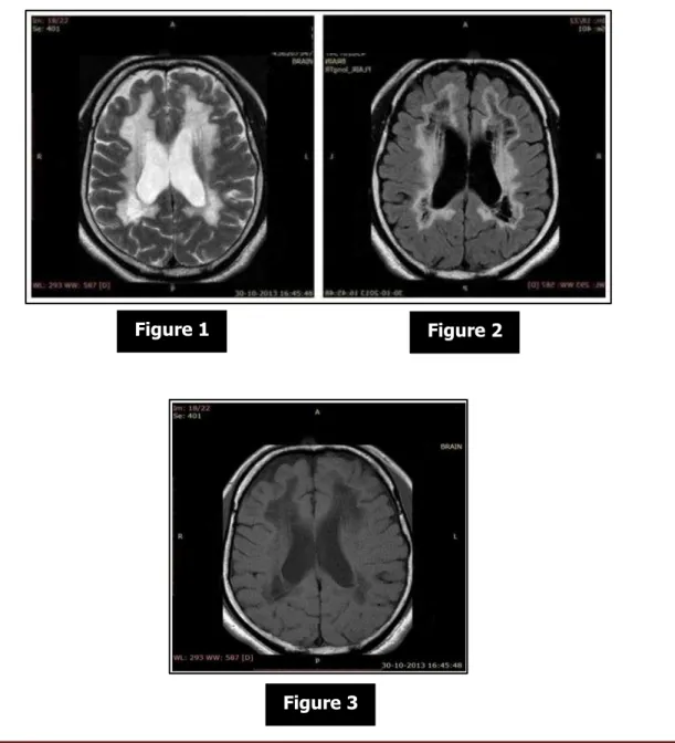
J of Evidence Based Med & Hlthcare, pISSN- 2349-2562, eISSN- 2349-2570/ Vol. 2/Issue 12/Mar 23, 2015 Page 1880 white matter showed severe decrease in NAA, choline and creatine and presence of lactate peak. Additional notable findings were diffuse extensive brain stem and thalamic atrophy. MRI revealed bilateral symmetrical diffuse extensive signal changes in white matter similar to that of CSF signal intensity in all lobes on T1W (hypointense), PD (hypointense), FLAIR (hypointense) and T2W (hyperintense) images with a very thin boundary between CSF and white matter. There was diffuse involvement of deep and subcortical white matter, arcuate fibers, internal capsules, external capsule and corpus callosum. The cerebral cortex was thin with normal sulcal and gyral pattern. Thalamus on either side was atrophic. There was severe brain stem and cerebellar atrophy. Ventricles were dilated, likely due to white matter changes. A cavum septum pellucidum was noted. Basal ganglia were normal. In vivo localized multi-voxel proton spectrometry was done (Hybrid 2D chemical shift imaging; TE = 135 ms) from the white matter and cerebral cortex. The MRS of white matter showed severe decrease in NAA. Lactate peak was also noted. MRS of cerebral cortex showed normal spectral pattern.
Figure 1 Figure 2
CASE REPORT
J of Evidence Based Med & Hlthcare, pISSN- 2349-2562, eISSN- 2349-2570/ Vol. 2/Issue 12/Mar 23, 2015 Page 1881 Axial T2 weighted image shows diffuse white matter hyperintensity similar to CSF intensity extending from periventricular white matter to the subcortical arcuate fibres. Axial FLAIR image shows white matter vanished and replaced by near-CSF intensity fluid i.e., it attenuated. Axial T1 weighted image shows diffuse white matter hypointensity similar to
CSF intensity.
DISCUSSION: The first time this disease was documented in 1962 when Eickle studied a 36 year old woman.1 In 1993-94, Dr. Hanefeld and Dr. Schiffmann and their colleagues identified the
disease. It is characterized by chronic progressive neurological deterioration with cerebellar ataxia, usually less prominent spasticity and relatively mild mental decline.2,3 Epilepsy is common.
VWM disease has an autosomal recessive mode of inheritance. Characteristically, there are additional episodes of major and rapid deterioration following minor head trauma and especially febrile infections.4,5 The classical and most common variant of Vanishing white matter disease has
its onset in childhood, at age 2-6 years.6,7 though this disease may have an early infantile or
antenatal onset.8-10 that had happened in our patient. Optic atrophy with loss of vision may occur8
but that was not present in our patient. The baby had not a history of acute frightening that recently has been reported as another provoking factor.9 MRI of the brain is usually diagnostic in
VWM. It shows an abnormal signal of all or almost all cerebral white matter with relatively spared U-fibers in some cases and progressive rarefaction and cystic degeneration of the affected white matter that is replaced by fluid.9,10,11 There have been important advances in the diagnosis of
VWM because of genetics since 1996. VWM leukoencephalopathy is caused by mutations in the five genes (eIF2B1–5) encoding the five subunits of eukaryotic translation initiation factor eIF2B (eIF2Bα, , , , and ).
Pathological study had shown axonal loss, hypomyelination, demyelination and gliosis, primarily involving the white matter with cortical sparing. Abnormal foamy oligodendroglial cells are identified and are unique for vanishing white matter.12 Pontine tegmental white matter may
also be involved.13 Increased cerebrospinal glycine level is noted and may be secondary to
excitotoxic brain damage.13,14 Diagnostic criteria for diagnosing this disease include normal initial
psychomotor development, deterioration following infection or trauma, presence of ataxia and spasticity with MR features of diffuse white matter signal changes with signal intensity of CSF on all pulse sequences.15,16,17 Additional features are lesions in central tegmental tracts and basis
pontis. Subcortical white matter involvement is early and severe. Cerebellar or primary vermian atrophy has been documented with or without involvement of cerebellar white matter. Basal ganglia are typically spared.18 Internal and external capsules may also be spared. MRS might
reveal reduced NAA, choline and creatine with mildly increased lactate and glucose peaks. In advanced disease, the white matter shows almost complete disappearance of all normal signals and presence of glucose and lactate, compatible with presence of mainly CSF and little brain tissue. Spectra of the cortex are preserved; however, signals representing lactate and glucose are described.19,20 Differential diagnosis on MR imaging include megalencephalic leukoencephalopathy
with subcortical cysts, Canavan's disease and Alexander's disease.21 Age of onset and
temporofrontal cysts differentiate it from megalencephalic leukoencephalopathy;22,23 classical
CASE REPORT
J of Evidence Based Med & Hlthcare, pISSN- 2349-2562, eISSN- 2349-2570/ Vol. 2/Issue 12/Mar 23, 2015 Page 1882
CONCLUSION: Vanishing White Matter leukoencephalopathy is one of the most prevalent hereditary diseases of white matter in childhood. Clinical and neuroimaging findings are very typical, and they suggest this diagnosis. Molecular genetic studies confirm up to 90% of affected patients. Nevertheless, it is still unknown why this disease starts up at a particular moment and why it follows an acute or chronic form. To date, no curative treatment has been found. Avoidance of stress situations known to provoke deterioration in VWM patients is essential. Liberal use of antibiotics and antipyretics, vaccinations, and abstinence of contact sports are simple but important measures. However, they are not sufficient to prevent onset or progression of the disease. The most important consequence of research findings of the last 5 years probably is that prenatal diagnosis has become available for families as soon as the disease-causing mutations in the index patient have been identified.
REFERENCES:
1. Hanefeld F, Holzbach U, Kruse B, Wilichowski E, Christen HJ, Frahm J. Diffuse white matter disease in three children: An encephalopathy with unique features on MRI and MRS. Neuropediatrics 1993; 24: 244-8. [PUBMED]
2. Schiffmann R, Moller JR, Trapp BD, Shih HH, Farrer RG, Katz DA, et al. Childhood ataxia with diffuse central nervous system hypomyelination. Ann Neurol 1994; 35: 331-40.
3. Van der Knaap MS, Barth PG, Gabreels FJ, Franzoni E, Begeer JH, Stroink H, et al. A new unknown leukoencephalopathy with vanishing white matter. Neurology 1997; 48: 845-55. 4. Van der Knaap MS, Kamphorst W, Barth PG, Kraaijeveld CL, Gut E, Valk J. Phenotypic
variation in leukoencephalopathy with vanishing white matter. Neurology 1998; 51: 540-7. 5. Tedeschi G, Schiffmann R, Barton NW, Shih HH, Gospe SM Jr, Brady RO, et al.
6. Proton magnetic resonance spectroscopic imaging in childhood ataxia with diffuse central nervous system hypomyelination. Neurology 1995; 45: 1526-32.
7. Labauge P, Fogli A, Niel F, Rodriguez D. CACH/VWM syndrome and leucodystrophies related to EIF2B mutations. Rev Neurol. 2007; 163: 793-9.
8. Dastych M, Gottwaldova J, Pohludka M, Prikryl P, Benovoska M. Determination of A sialotransferrin in the cerebrospinal fluid with HPLC method. Scand J Clin Lab Invest 2010; 70 (2): 87-91.
9. Hanefeld F, Holzbach U, Kruse B, Wilichowski E, Christen HJ, Frahm J. Diffuse white matter disease in three children: an encephalopathy with unique features on magnetic resonance imaging and proton magnetic resonance spectroscopy. Neuropediatrics 1993; 24(5): 244-8. 10.Vander Knaap MS, Barth PG, Gabreels FJ, Franzoni E, Begeer JH, Valk J, et al. A new
leukoencephalopathy with vanishing white matter. Neurology 1997; 48: 845–55.
11.Vander Knaap MS, Kamphorst W, Barth PG, Gut E, Valk J. Phenotypic variation in leukoencephalopathy with vanishing white matter. Neurology. 1998; 51: 540–47.
CASE REPORT
J of Evidence Based Med & Hlthcare, pISSN- 2349-2562, eISSN- 2349-2570/ Vol. 2/Issue 12/Mar 23, 2015 Page 1883 13.Vander Knaap MS, Van Berkel CGM, Herms J, Coster RV, Baethmann M, Naidu S, et al. eIF2B related disorders: antenatal onset and involvement of multiple organs. Am J Hum Genet 2003; 73(5): 1199–07.
14.Vermeulen G, Seidl R, Mercimek-Mahmutoglu S, Vander Knaap MS, Gert CS, Jan JR et al. Fright is a provoking factor in vanishing white matter disease. Ann Neurol 2005; 57(4): 560–3.
15.Labauge P. Magnetic resonance findings in leucodystrophies and MS. Int MS J 2009; 16(2): 47-56.
16.Serafini G, Pompili M, Innamorati M, et al. White matter hyperintensities and self-reported depression in a sample of patients with chronic headache. J Headache Pain. 2012; 13: 661– 7.
17.Schiffmann R, Van der Knaap MS. Invited article: an MRI-based approach to the diagnosis of white matter disorders. Neurology. 2009; 72: 750–9.
18.Hanefeld F, Holzbach U, Kruse B, Wilichowski E, Christen HJ, Frahm J. Diffuse white matter disease in three children: An encephalopathy with unique features on MRI and MRS. Neuropediatrics 1993; 24: 244-8. [PUBMED]
19.Schiffmann R, Moller JR, Trapp BD, Shih HH, Farrer RG, Katz DA, et al. Childhood ataxia with diffuse central nervous system hypomyelination. Ann Neurol 1994; 35: 331-40.
20.Van der Knaap MS, Barth PG, Gabreels FJ, Franzoni E, Begeer JH, Stroink H, et al. A new unknown leukoencephalopathy with vanishing white matter. Neurology 1997; 48: 845- 21.Van der Knaap MS, Kamphorst W, Barth PG, Kraaijeveld CL, Gut E, Valk J. Phenotypic
variation in leukoencephalopathy with vanishing white matter. Neurology 1998; 51: 540-7. 22.Tedeschi G, Schiffmann R, Barton NW, Shih HH, Gospe SM Jr, Brady RO, et al.
23.Proton magnetic resonance spectroscopic imaging in childhood ataxia with diffuse central nervous system hypomyelination. Neurology 1995; 45: 1526-32.
24.Senol U, Haspolat S, Karaali K, Luleci E. MR imaging of vanishing white matter. AJR Am J Roentgenol 2000; 175: 826-8. [PUBMED]
25.Chandrashekar HS, Guruprasad AS, Jayakumar PN, Srikanth SG, Taly AB. Megalencephalic leukoencephalopathy with subcortical cysts: MRI and proton spectroscopic features. Neurol India 2003; 51: 525-7.
CASE REPORT
J of Evidence Based Med & Hlthcare, pISSN- 2349-2562, eISSN- 2349-2570/ Vol. 2/Issue 12/Mar 23, 2015 Page 1884
4. Resident, Department of Radiology, J. J. M. Medical College, Davangere. 5. Resident, Department of Radiology, J. J. M. Medical College, Davangere.
NAME ADDRESS EMAIL ID OF THE CORRESPONDING AUTHOR:
Dr. Sindu P. Gowdar, # 3087, 9th Main,
3rdCross, ε.C.C. ‘B’ Block,
Davangere-577004.
E-mail: [email protected]
Date of Submission: 06/03/2015. Date of Peer Review: 07/03/2015. Date of Acceptance: 11/03/2015. Date of Publishing: 20/03/2015.
AUTHORS:
1. Sindu P. Gowdar 2. Naveen S. Maralihalli 3. Pramod Setty J. 4. Rajesh Venunath 5. Mithila P. V.
PARTICULARS OF CONTRIBUTORS:
1. Resident, Department of Radiology, J. J. M. Medical College, Davangere. 2. Associate Professor, Department of
Radiology, J. J. M. Medical College, Davangere.