Arq Neuropsiquiatr 2001;59(1):125-127
MENKES DISEASE
Case report of an uncommon presentation
with white matter lesions
Lúcia Maria Guimarães Santos
1, Carla da Silva Teixeira
1, Luiz Celso Pereira Vilanova
2,
Cecilia Micheletti
3, Carmem Silvia Curiati Mendes
1, Maria Lucia Borri
4, Ana Maria Martins
5ABSTRACT - Menkes disease is a rare X-linked disorder related to a defect in the copper metabolism. According to the current literature, the most frequent neuroimaging findings are cortical atrophy, chronic subdural effusion or hygroma, and vascular abnormalities. White matter lesions may be present before other features of the disease and may evolve into atrophy. We hereby report a case of Menkes disease with typical history and progression, and an early phase imaging study with important white matter abnormalities, which could have lead to diagnostic difficulties.
KEY WORDS: Menkes disease, neuroimaging.
Doença de Menkes: relato de caso com comprometimento da substância branca cerebral
RESUMO - A doença de Menkes é patologia ligada ao X relacionada a defeito no metabolismo de cobre. De acordo com literatura atual, os achados mais frequentes em exames de neuroimagem são atrofia cortical, efusão de subdural crônica ou higroma, e alterações vasculares. Lesões em substância branca podem estar presentes antes das alterações clinicas da doença e podem evoluir para atrofia. Apresentamos relato de caso da doença de Menkes com história e evolução típica, descrevendo os achados de neuroimagem na fase inicial da doença que revelaram sinais importantes de comprometimento da substância branca que poderiam conduzir a dúvidas diagnósticas.
PALAVRAS-CHAVE: doença de Menkes, neuroimagem.
Menkes disease is a neurological progressive disorder characterized by psychomotor deterioration, drug-treatment refractory epilepsy, and phenotypic characteristics such as thin and brittle hair, hypo-pigmented, and scattered eyebrows. The hair micros-cope study shows fragile and brittle filaments curled up in their own axis, which are called pili torti1-4. The
disease is caused by a defect in intracellular copper transport. It is a disease related to the X located in the Xg 13.3 area .The responsible gene is organized into 23 exons and the mRNA transcript encodes a copper-transporting P-type ATPase and is expressed in all tissues. The clinical picture is due to a deficiency of copper, resulting in defects of key copper depen-dent enzymes, including lysyl oxydase, cytochrome c oxidase, dopamine β-hydroxylase, tyrosinase, and superoxide dismutase. The pathogenic mechanism is not entirely explained. Depigmentation of hair and
Universidade Federal de São Paulo (UNIFESP) Escola Paulista de Medicina (EPM): 1Médica Neuropediatra do Setor de Neurologia Infantil; 2Professor Adjunto do Setor de Neurologia Infantil; 3Médica do Setor de Doenças Metabólicas Hereditárias; 4Médica Radiologista do
Departamento de Diagnóstico por Imagem; 5Professora Adjunto do Setor de Doeças Metabólicas Hereditárias.
Received 26 june 2000, received in final form 20 Setpember 2000. Accepted 25 September 2000.
Dr. Luiz Celso P. Vilanova - Setor de Neurologia Infantil UNIFESP-EPM - R Botucatu 740 - 04023-900 São Paulo SP - Brasil.
skin pallor are due to tyrosinase deficiency, hypo-thermia is due cytochrome c oxidase deficiency and lysyl oxidase deficiency causes tortuous arteries in brain with irregular lumen and a frayed and split intimal lining2,5-7.
The clinical spectrum of Menkes disease encom-passes several distinct variants. Individuals with the mild variant are developmentally delayed with cere-bellar ataxia, dysarthria and pili torti, and no seizures. Jankov et al.8 described a neonatal form with multiple
fractures and extensive vascular disease with early death. The occipital horn syndrome is considered Menkes disease variant. Skeletal dysplasia, soft brui-sable skin, hyper-extensible joints, diarrhea, and occi-pital exostoses6,8-10 characterize it.
126 Arq Neuropsiquiatr 2001;59(1)
neuroimaging record shows extensive lesions in white matter that seem secondary to a progressive brain destruction and collapse of the myelination process1,2,12,13. We report a case of Menkes disease
with neuroimaging study first conducted at two and then at four months of life, with substantial white matter lesion and vascular alterations.
CASE
LMR, 8 months-old, white male with non-consangui-neous parents. Early delivered at 8 months, after preg-nancy with no intercurrences. At two months of life epi-leptic manifestation began with various types of crisis, and frequency and intensity progressive aggravation despite the drug-oriented treatment. After crisis onset, the child remained hypoactive, poorly responsive to stimuli, and recurrent hypothermia. During the exam the child presented rectified and hypopigmented eyebrows,
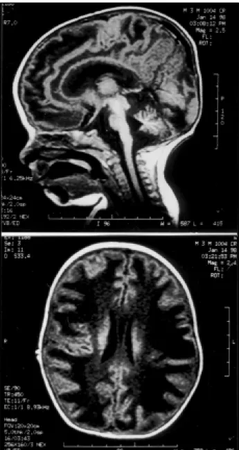
Fig 2 . Brain magnetic resonance at four months showed intense bilateral damaging of white matter and tortuosity of medium brain arteries.
Fig 1. Skull tomography at two months of age displayed hypoattenuation of front-parietal bilateral white matter, more evident to the right and minor in occipital areas.
Arq Neuropsiquiatr 2001;59(1) 127
micrognathia and scattered thin and brittle hair. Labo-ratory exams confirmed the Menkes disease diagnostic hypothesis, with copper dosage of 24mg/dl (normal values 70 to 140 mg/dl), and ceruloplasmin of 7 mg/dl (normal values between 20 and 40 mg/dl). The skull tomography at two months of age displayed hypoatte-nuation of front-parietal bilateral white matter, more evi-dent to the right, and minor damage, in occipital areas (Fig 1). The brain magnetic resonance at four months showed intense bilateral damaging of the white matter, with mild cortical atrophy and vascular alterations, and dilation and tortuosity of the medium brain arteries (Fig 2). The hair microscope analysis showed pili torti and monoletrix (Fig 3).
DISCUSSION
The pathology of the case at issue has a very suggestive clinical history, confirmed by the copper and ceruloplasmin serum dosage and hair analysis. The skull tomography displayed considerable white matter lesion at two months of life, and included a differential diagnosis regarding other leukodys-trophies. Brain magnetic resonance at four months showed intense bilateral damaging of white matter, very similar to diseases primarily affecting white matter. Usually radiological studies of patients with Menkes disease show cerebral atrophy, large sub-dural collections, and tortuosity of cerebral blood vessels. The neuroimaging study using brain magne-tic resonance shows local hypodensity areas, atrophy, necrosis, and gliosis of white matter that may be identified before the phenotypic alterations1,11,12. In
cases with diffuse and intense damaging of white matter, the arteriopathy with vascular insufficiency probably leads to a deficient myelination process, mainly in semi-oval centers and long tracts2. The
angiographic alterations are better visualized after three months of age.
Although the pathogenic mechanism of the Men-kes disease has not yet been established, we know there is a flaw in the copper absorption by the gastro-intestinal tract, and that this element ends up being poorly distributed in the tissues and very reduced in the brain, liver and white cells. Copper histidine is naturally present in normal human serum, histidine was shown to enhance the uptake of copper in hu-man trophoblast cells in the presence of serum, and
this is considered to be due the release of copper bound to albumin. Treatment with copper histidine had been described. In the mild variations copper histidine treatment led to normalization of plasma copper and ceruloplasmin with marked reduction of epileptic discharges, improved muscular tone and increased motor activity. The classical Menkes disease therapy is not effective and the parenteral reposition during the first weeks of life raises the copper serum level but not that of ceruloplasmin, thus not preven-ting neurological deterioration2,5,7,13.
The early diagnosis is mainly required for genetic counseling, X-Iinked inheritance with a recurrence risk of 50% for sons affected and 50% of daughters to be carriers. Less aggressive forms of the disease with compatible laboratory exams have also been described2,11. The intra-uterus diagnosis can be
performed through amniocentesis both using copper dosages in culture of amniotic liquid cells and chorionic villi cells; however, false negatives may occur as pregnancy age progresses11.
REFERENCES
1. Blaser SI, Berns DH, Ross JS, Lanska MJ, Weissman BM. Serial MR studies in Menkes disease. J Comput Assist Tomogr 1989;13:113-115. 2. Tumer Z, Horn N. Menkes disease: recent advances and new aspects. J
Med Genet 1997;34:265-274.
3. Reed UC, Rosemberg S, Diament AJ, Scaff M, Canelas HM, Lefevre AB. Menkes syndrome: review of the pathogenesis apropos of a clinico-pathological case. Arq Neuropsiquiatr 1984;42:262-273.
4. Menkes JH. Kinky hair disease. Pediatrics 1972;50:181-182.
5. Christodoulou J, Danks DM, Sarkar B, et al. Early treatment of Menkes disease with parenteral copper-histidine: long-term follow-up of four treated patients. Am J Med Genet 1998;76:154-164.
6. Proud VK, Mussel HG, Kaler SG, Young DW, Percy AK. Distinctive Menkes disease variant with occipital horns: delineation of natural history and clinical phenotype. Am J Med Genet 1996;65:44-51. 7. Menkes JH. Kinky hair disease: twenty five years later. Brain Dev
1988;10:77-79.
8. Jankov RP, Boerkoel CF, Hellmann J, et al. Lethal neonatal Menkes disease with severe vasculophaty and fractures. Acta Paediat 1998; 87:1297-1300.
9. Wakai S, Ishikawa Y, Nagaoka M, Okabe M, Minami R, Hayakawa T. Central nervous system involvement and generalized muscular atrophy in occipital horn syndrome Ehlers-Danlos type IX: a first Japanese case. J Neurol Sci 1993;116:1-5.
10. Haas RH, Robinson A, Evans K, Lascelles PT, Dubowitz V. An X-linked disease of the nervous system with disordered copper metabolism and features differing from Menkes disease. Neurology 1981;31:852-859. 11. Johnsen DE., Coleman L, Poe L. MR of progressive neurodegenerative
change in treated Menkes kinky hair disease. Neuroradiology 1991;33:181-182.
12. Ichihashi K, Yano S, Kobayashi S, Miyao M, Yanagisawa M. Serial imaging of Menkes disease. Neuroradiology 1990;32:56-59. 13. Geller TJ, Pan Y, Martin DS. Early neuroradiologic evidence of