Arq Neuropsiquiatr 2009;67(1):55-57
55
Prognostic Value of Proton Magnetic
resonance sPectroscoPy findings
in near drowning Patients
Reversibility of the early metabolite abnormalities
relates with a good outcome
Maria de Fátima Vasco Aragão
1,2,3, Meng Law
1, João Prola Netto
1,
Marcelo Moraes Valença
3, Thomas Naidich
1Abstract – In two children with near drowning hypoxic encephalopathy and normal-appearing structural MRI, acute proton magnetic resonance spectroscopy (1H MRS) showed biochemical alterations that correctly
indicated prognosis and helped to guide management decisions. Elevation of the lipid-lactate and glutamine-glutamate peaks, on the early (72 hour) 1H MRS, predicts a poor prognosis. Absence of lipid-lactate and
glutamine-glutamate peaks on the early 1H MRS and reversibility of early mild metabolite abnormalities on
follow up examination relates with good outcome.
KEy woRdS: hypoxia, near drowning, magnetic resonance spectroscopy.
Valor prognóstico da espectroscopia de prótons em vítimas de quase-afogamento: reversibilidade das anormalidades metabólicas precoces relacionou-se com bom prognóstico
Resumo – Em duas criancas vítimas de quase-afogamento com encefalopatia hipóxico-isquêmica, que apresentaram ressonância magnética por imagem normal, a espectroscopia de prótons por ressonância magnética (1H MRS) na fase aguda mostrou alterações bioquímicas que corretamente indicaram o prognóstico
e ajudaram a guiar o manejo terapêutico. Elevação dos picos de lipídeo-lactato e glutamina-glutamato na 1H
MRS precoce realizada com 72 horas previu um mau prognóstico. Relacionaram-se com bom prognóstico; a ausência dos picos de lipídeo-lactato e glutamina-glutamato na 1H MRS precoce, e a reversibilidade no exame
de controle (3 meses) das discretas anormalidades metabólicas encontradas no primeiro exame. PAlAvRAS-cHAvE: hipóxia, quase-afogamento, espectroscopia, ressonância magnética.
1department of Radiology, Mount Sinai School of Medicine, New york Ny, USA; 2Multimagem, Hospital Albert Sabin, Recife PE, Brazil; 3department of
Neuropsychiatry and Behavioral Studies, Federal University of Pernambuco, Recife PE, Brazil. Received 23 June 2008, received in inal form 22 october 2008. Accepted 10 december 2008.
Dra. Maria Fátima Viana Vasco Aragão – Estrada das Ubaias 332 / 1201 - 52061-080 Recife PE - Brasil. E-mail: [email protected].
The term “near drowning” signiies patient survival for more than 24-hours after cardiorespiratory arrest due to submersion. Early prognostic of good outcome versus sig-niicant neurological deicit or death is important for cor-rectly stratifying patient management1,2.
Magnetic resonance image (MRI) and computed to-mographic indings of acute hypoxia ischemic injury are often subtle1-3.we describe the indings of early proton
magnetic resonance spectroscopy (1H MRS) that helped
to predict the prognosis.
MetHod
In two children with hypoxic-ischemic insult due to fresh water near drowning and normal-appearing structural MRI are studied.
In each patient single-voxel 1.5 Tesla 1H MRS was performed
in the median biparietal-occipital gray matter using a stimulated echo acquisition mode (STEAM) technique with repetition time: 1,500 ms, echo time: 30 ms, eight excitations, volume of inter-est: 6 cm3, and acquisition time: 7 minutes. The 1H MRS data were
compared with the standard MRI study obtained at the same session using non contrast sagittal T1-weighted images (wI), cor-onal and axial T2-wI, axial T2*-wI, axial T2-FlAIR and axial diffu-sion-wI, followed by gadolinium-enhanced axial T1-wI.
results
Case 1
Arq Neuropsiquiatr 2009;67(1)
56
MRI and MRS: drowning patients Aragão et al.
resuscitated at poolside immediately after the accident, and arrived at hospital agitated but breathing spontane-ously. MRI and 1H MRS performed 72 hours after the near
drowning (Fig 1A-d) showed normal anatomic structure (Fig 1A,B,d) and decrease (Fig 1c) of 20% in the N-acetylas-partate/creatine (NAA/cr=1.04) ratio, an increase of 26% in the choline/creatine ratio (cho/cr=0.76) and 22% in the myoinositol/creatine (Myo/cr=0.74) ratios, but no lipid-lactate (lip-lac) peak. This patient had a good outcome and was discharged after 5 days with a normal neurologi-cal examination. Follow-up MRI and 1H MRS 3 months
af-ter the near drowning (Fig 1E-F) showed normal anatom-ic structure (Fig 1F)and normal metabolites ratios (Fig 1E). (NAA/cr=1.21, cho/cr=0.56 and Myo/cr=0.62).
Case 2
This 6-year-old boy also suffered near drowning in a swimming pool. He arrived at the hospital in cardiorespi-ratory arrest, but was resuscitated and stabilized. MRI and
1H MRS performed 72 hours after the near drowning (Fig
2A-c) showed apparently normal anatomic structure (Fig 2A-B) but signiicant metabolic abnormalities (Fig 2c), es-pecially increased lip-lac and glutamine-glutamate (Glx) peaks. There was also decrease of 8% of the NAA/cr (1.2) ratio and increase of 50% of cho/cr (0.9) ratio and 9% of Myo/cr (0.65) ratio. The metabolic indings were inter-preted as evidence of severe hypoxic injury with a poor prognosis. The patient remained comatose for 3 weeks, and progressed to non-progressive spastic/dystonic state, indicating chronic encephalopathy. The follow-up MRI and 1H MRS was done 3 months after the accident (Fig 2d
and E) and demonstrated severe brain atrophy (Fig 2d) and progression of metabolic changes (Fig 2E), with decrease of 54% in the NAA/cr (0.6) ratio, an increase of 75% in the Myo/cr (1.05) ratio, persistent of the lip-lac. However there was decrease in the cho/cr (0.6) ratio and Glx.
discussion
MRI and 1H MRS may demonstrate abnormalities the
irst day after a hypoxic-ischemic near drowning injury, but have greater prognostic value after the third day1.
Edema and T2-hyperintensity in the cerebral cortex or basal ganglia are highly sensitive (100%) and speciic (86%) predictors of poor prognosis (vegetative state and death)1.
The two children presented here did not exhibit these features.
The transitory decrease in NAA/cr ratio seen in our case 1 with good outcome appears to correspond to re-versible dysfunction of the neurons and glia after acute brain injury, not cell death, since the metabolites and the patient both recovered with no alteration in brain anat-omy MRI on follow-up study. Transitory reduced NAA/ cr ratio has not been reported yet in a patient with good outcome after hypoxic-ischemic insult from near drown-ing, although had been described in other pathologic conditions4,5 and represents cellular dysfunction rather
than cell death.
In case 2 with poor outcome, the Glx levels were el-evated on the 72-hour-1H MRS, then decreased on follow
up examination. This most likely relects early initiation of the excitotoxic cascade in a patient with severe brain injury6. The Myo/cr was increased at 72 hours in both
patients. The Myo/cr returned to normal in case 1 with
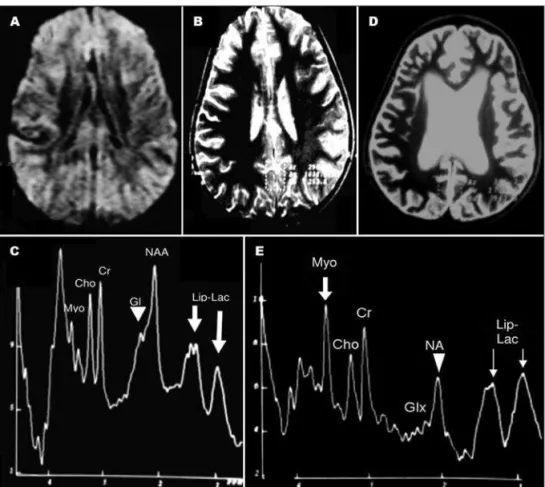
Fig 1. Case 1 – First exam, three days after near drowning. Axial FLAIR
(A) and axial DWI (B) are unremarkable. 1H MRS (C) VOI in the
medi-an biparietal gray matter, demonstrate on axial T2 weighted imag-ing (D), shows a mild decrease of the NAA/Cr ratio (arrow), and mild increase of the Cho/Cr and Myo/Cr ratios (arrowhead). On visual inspection, the Glx levels are normal and no Lip-Lac peak is identi-ied. On follow up exam (3-months later), axial T2 remains unremark-able (F). The follow up 1H MRS (E) in the median biparietal-occipital gray matter VOI demonstrated on the axial T2 (F), shows a normal spectrum (E) and reversibility and recovery of the metabolite
chang-es (arrow and arrowhead) seen on the initial 1H MRS (C), in this
Arq Neuropsiquiatr 2009;67(1)
57
MRI and MRS: drowning patients Aragão et al.
good outcome, but continued to increase in case 2 with poor outcome. Myoinositol is found primarily in astro-cytes and has a role in brain osmoregulation7,8. The early
but transient increase in Myo/cr in case 1 most likely re-lects its role in osmoregulation7. The progressive increase
in Myo/cr in case 2 most likely relects astrogliosis within the damaged brain6.
lactate is associated with anaerobic glycolysis, so the presence of lip-lac can relect brain ischemia7. case 1 with
no elevation of lip-lac on 1H MRS had a favorable
come. case 2 with elevated lip-lac levels had a poor out-come. These indings accord well with literature reports that elevated lip-lac peaks are strongly associated with poor outcome in brain injury, especially hypoxic-ischemic brain injury1,2.
According to dubowitz et al.1 the combination of MRI
and 1H MRS together is superior to either one alone,
de-creasing the false-negative potential. 1H MRS
comple-ments conventional MRI and clinical findings. 1H MRS
helps to predict the prognosis of near drowning victims with hypoxic encephalopathy and aids in triage, being par-ticularly important in the hyperacute and acute phases, when conventional imaging indings are not prognostic.
In patients with near drowning hypoxic encephalopa-thy, 1H MRS can demonstrate biochemical alterations that
aid prognosis and guide management decisions.
Eleva-tion of the lip-lac and Glx peaks, on the early (72 hour)
1H MRS, predicts a poor prognosis. Absence of
lipid-lac-tate and glutamine-glutamate peaks on the early 1H MRS
and reversibility of the early metabolite abnormalities on follow up examination correlates with a good outcome.
Acknowledgement – we would like to acknowledge the
assis-tance of leonardo da Fonte, claudia Fontana, Jose Ronaldo de Me-nezes, and Adelia Henriques in the preparation of this manuscript.
references
1. Dubowitz DJ, Bluml S, Arcinue E, Dietrich RB. MR of hypoxic enceph-alopathy in children after near drowning: correlation with quantitative proton MR spectroscopy and clinical outcome. AJNR Am J Neuroradiol 1998;19:1617-1627.
2. Kreis R, Arcinue E, Ernst T, et al. Hypoxic encephalopathy after near-drowning studied by quantitative 1H-magnetic resonance spectrosco-py. J Clin Invest 99;97:1142-1154.
3. Taylor SB, Quencer RM, Holzman BH, Naidich TP. Central nervous sys-tem anoxic-ischemic insult in children due to near-drowning. Radiolo-gy 1985;156:641-646.
4. Tiberio M, Chard DT, Altmann DR, et al. Metabolite changes in early relapsing-remitting multiple sclerosis. A two year follow-up study. J Neurol 2006;253:224-230.
5. Holshouser BA, Tong KA, Ashwal S, et al. Prospective longitudinal pro-ton magnetic resonance spectroscopic imaging in adult traumatic brain injury. J Magn Reson Imaging 2006;24:33-40.
6. Shutter L, Tong KA, Holshouser BA. Proton MRS in acute traumatic brain injury: role for glutamate/glutamine and choline for outcome prediction. J Neurotrauma 2004;21:1693-1705.
7. Danielsen E, Ross B. Magnetic resonance spectroscopy diagnosis of sur-gical diseases. Marcel Dekker, Inc USA, 1999.
8. Ashwal S, Holshouser B, Tong K, et al. Proton spectroscopy detect-ed myoinositol in children with traumatic brain injury. Pdetect-ediatr Res 2004;56:630-638.
Fig 2. Case 2 – First MRI examina-tion (A and B), three days after near drowning, shows unremarkable axi-al DWI (A) and axiaxi-al T2 (B). Howev-er the 1H MRS (C) with median bipari-etal-occiptal gray matter VOI, shows decrease of the NAA/Cr ratio and in-crease of Cho/Cr ratio and of Myo/
Cr ratio. Importantly the 1H MRS (C)