Dejan Petrovi}
1CLINICAL IMPORTANCE OF COLOR DOPPLER
ULTRASONOGRAPHY IN PREOPERATIVE
ASSESMENT OF HEMODIALYSIS VASCULAR
ACCESS CREATION
Primljen/Received 05. 08. 2012. god. Prihva}en/Accepted18. 09. 2012. god.
Summary:Chronic kidney failure is characterized with progressive and ireversible diminishing of glomer-ular filtration rate. Arterio-venous fistula (AVF) for he-modialysis should be created in patients with endogeno-us creatinine clearance£20 mL/min/1,73m2. Inner dia-meter of a.radialis³2.0 mm, inner diameter of v.cepha-lica³ 2.5 mm, flow velocity through the a.radialis — VmaxS³ 50 cm/s and flow through the a.radialis — Qa.radialis³40 mL/m intenable adequate maturing of distal radio-cephalic AVF. Diameter of v.cephalica³4.0 mm and blood flow QAV³500 mL/min, four weeks after the AVF creation, indicate adequately matured AVF and possibility of puncturing it. Maximal blood flow veloc-ity through AVF of 100–350 cm/s and blood flow of 500–1000 mL/min, are signs of good function of AVF and adequate hemodialysis. Color Doppler ultrasono-graphy enables preoperative planning of AVF, early complication detection, choice of appropriate therapeu-tical procedure for complication treatment, estimation of maturation, prime time for puncture and AVF func-tion, which all contribute to a significant morbidity dec-rease and better life quality in hemodialysis patients.
Key words:arterio venous fistula, hemodialysis, color doppler.
INTRODUCTION
Chronic kidney failure is characterized with pro-gressive and ireversible diminishing of glomerular fil-tration rate (GFR) (1–4). When glomerular filfil-tration
rate falls bellow 20 mL/min/1.73 m2, and serum creati-nine concentration rises above 220 mmol/L, chronic kidney failure progressively advances, leading to ire-versible renal parenchymal disorder and finally, to an end stage renal disease (1–4). In patients with diagno-sed chronic kidney failure cephalic veins should be preserved for arterio-venous fistula (AVF) creation (5–7). If hemodialysis planned, vascular access should be created when endogenous creatinine clearance — Ccr£ 20 mL/min/1.73 m2, and regular hemodialysis treatment should be started when Ccr£10 mL/min/1.73 m2. Dialysis should start earlier if there is hypervole-mia (Ccr = 15–20 mL/min/1.73m2) (5–7).
Native distal radio-cephalic AVF is anastomosys be-tween radial artery and cephalic vein (Brescia-Cimino fi-stula). Upon arterial flow venous limb of AVF becomes dilated and thickened (6–10 mm) (“maturation of AVF”). Maturation of native AVF should take at least a month, ideally 3–4 months before puncture (5–8). Planning of pri-me tipri-me for creation of vascular access for hemodialysis enables creation of good vascular access, adequate hemo-dialysis, cardiovascular morbidity and mortality decrease and better life quality in hemodialysis patients (5–9).
PREOPERATIVE ASSESSMENT
OF VASCULAR ACCESS
Before vascular access creation it is mandatory to take anamnestic data (data about previous central ve-nous dialysis catheter, diabetes mellitus, peripheral ar-terial and venous diseases, trauma and surgical inter-ventions at upper limbs, anticoagulant therapy and co-agulation disorders and previous vascular access),
form physical examination of upper limb vessels, Al-len test, upper limb vessels Color Doppler evaluation and echocardiography (6, 7, 8, 9). Preoperative evalua-tion of arteries and veins should be performed first in patient’s non-dominant arm (6, 7, 8, 9).
EVALUATION OF ARTERIES
FOR A-V FISTULA
The first step in the arterial system evaluation in-cludes palpation and measurement of arterial blood pressure in the upper limbs (a. brachialis). Difference of more than 20 mm Hg between both hands raises sus-picion of proximal stenosis on the side of the lower pressure value (poststenotic dilatation) (6, 7, 8, 9). Seco-nd step is Allen test. Allen test serves in the estimation of presence of anastomoses between a.radialis and a.ul-naris, basically the presence and functionality of palmar arches (superficial and deep arch) between the afore-mentioned arteries (6, 7, 8, 9). The third step in the arte-rial system evaluation is Color Doppler ultrasonography (CDU). Color Doppler ultrasonography is used for the measurement of arterial diameter, velocity and flow through the arteries of non-dominant arm (10, 11, 12).
EVALUATION OF VEINS
FOR A-V FISTULA
The first step in the venous system evaluation (“venous outflow”) includes inspection of the superfi-cial veins of the non-dominant arm (arm planned for access creation), after „outflow“ occlusion (cuff infla-ted in the upper arm above diastolic value for 5 minu-tes). During the inspection of the superficial veins one should pay attention to continuity and direction as well as their diameter (there should generally be > 5 mm to allow adequate development of A-V fistula), compres-sibility and extencompres-sibility of the veins (6, 7, 8, 9). Sec-ond step is Color Doppler ultrasonography (transducer of 10–12 MHz). It is used for the measurement of the cephalic and basilic vein diameter, and flow through the subclavian vein (10, 11, 12). The inner diameter of the cephalic vein³2.5 mm enables adequate matura-tion of a distal radio-cephalic A-V fistula. For better vi-sualisation of the venous system and accessory veins assessment the digital subtraction angiography (DSA) with 20–40 mL of contrast is employed (6, 7, 8, 9).
CLINICAL IMPORTANCE OF COLOR
DOPPLER ULTRASONOGRAPHY
Ultrasound equipment and technique of examination
Color Doppler ultrasonography reqires ultrasound machine equiped with B-mode and Color and Doppler
mode with linear transducer od 5–7.5 MHz as well. Upper limb arteries are traced longitudinally by Color Doppler ultrasonography from distal subclavian artery (a.subclavia) till the radial (a.radialis) and ulnar arteri-es (a.ulnaris). Arterial segments in which disturbance in color appearance is detected require additional asse-sment by B-mode and spectral doppler, for detection of stenosis or occlusion. Stenosis is considered hemodi-namically significant if more 50%, and stenosis disco-vered by ultrasonography before vascular access crea-tion, requires angiography (golden standard in blood vessels imaging) (13, 14).
Clinical importance
In evaluation of a.radialis, for preoperative assess-ment of arterio-venous fistula creation, one should exa-mine: inner diameter of a.radialis, morphology of the wall of a.radialis (measuring of thickness of intima-me-dia, atherosclerotic plaques, presence of calcifications), velocity and flow through a.radialis, as well as response of a.radialis to reactive hyperemia (10, 11, 12, 13, 14). Distal a.radialis diameter measurement (intima to intima distance) and measurement of intima-media thickness (IMT) is performed in longitudinal scan, in distal fore-arm, just above the wrist. Inner diameter of a.radialis — Id ³ 2.0 mm, flow velocity through the a.radialis — VmaxS³50 cm/s and blood flow through the a.radialis — Qa.radialis³40 mL/min maintain adequate maturing of distal radio-cephalic AVF (10, 11, 12, 13, 14). Measu-rement of diameter of a.radialis in systole and diastole enable assessment of its pulsatility (14).
Preoperative ultrasound examination of upper limb blood vessels for assessment of AVF outcome is indicated in patients with increased risk of arterio-ve-nous fistula insufficiency (elderly, female gender, obe-sity, diabetes mellitus, cardiovascular diseases) (14). It is specially indicated in patients with difficult physical examination blood vessel assessment (pulseless, previ-ous numerprevi-ous surgical attempts of vascular access cre-ation), those with any arterial disease (arteritis, athero-sclerosis) and patients with venous disease/disorder (previous punctures, previously placed central venous hemodialysis catheters) (14). In conclusion, pre-opera-tive ultrasonographic examination of upper limbs blood vessels should be performed in high risk patients for better outcome of AVF (14).
FAILURE OF AVF
FOR HEMODIALYSIS
Significant number of AVF doesn’t develop ade-quately (28–53%) (15, 16). Failure of the A-V fistula can be early and late. Early failure of the vascular ac-cess for hemodialysis is accounted for the period bet-ween creation and the beginning of the work of the fis-tula, or the first three months of its use (15, 16). Rea-sons for early haemodialysis access failure can be divi-ded into two groups. First group consists of inflow pro-blems, whilst second one consists of outflow problems ( table 1) (15, 16). Late failure of the A-V fistula on the other hand, is considered if it happens after three months of its use for hemodialysis (16).
Adequate maturation of the A-V fistula demands adequate inflow of the arterial blood (15, 16). One of the reasons of early failure of the vascular access is ste-notic process of the vein, nearby the anastomotic site, the so-called juxta-anastomotic stenosis (15, 16). Ma-nipulation with this segment of the vein during surgical access creation can be the cause of the vein damage. In the absence of stenosis, pulse on the spot of anastomo-sis is weak and compressible, continuous thrill (systo-lic-diastolic) is palpable and also auscultatory audible (15, 16). In the case of stenosis of the vein segment qui-te close to anastomosis the pulse is harder, inqui-tense, thrill is palpable only during systole, and auscultatory
only systolic component is present (15, 16). This type of stenosis requires either percutaneous transluminal angioplasty (PTA) or surgical revision of the vascular access (17, 18).
Good blood outflow is also very important for the adequate development of the vascular access for hae-modialysis. The most frequent problems of the outflow tract are small inner diameter of the vein, presence of the accessory veins and stenosis of the proximal part of the outflow vein due to previous trauma or veinpunctu-re (table 1) (15, 16).
ASSESSMENT OF PRIME TIME
FOR PUNCTURE OF AVF
In clinical practise, for nephrologists, it is very important to maintain good outcome of vascular access for hemodialysis (preopreative planning of vascular access creation, adequate maturing and prime time for puncture of vascular access for hemodialysis) (17, 18). Along the way to a good vascular access, which ena-bles adequate hemodialysis, there are numerous obsta-cles: late presentation of the patient to nephrologyst, preoperative vascular mapping of upper limb blood vessels, AVF creation, adequate maturation of AVF, prime time for puncture of AVF (17, 18). Every patient who begins hemodialysis treatment should have ade-quately matured AVF, ready for puncture. Practically, it means that AVF should be created well-timed, in pre-dialysis period of terminal chronic kidney insuffici-ency, along with previous preoperative mapping, which should give precise information to a surgeon about the diameter of the arteries and veins, presence of venous thrombosis or stenosis. Based on these, sur-geon makes decision about localisation and type of vascular access creation (17, 18). Three most frequent reasons for inadequate maturing of AVF are: local ste-nosis, presence of great accessory vein branches and very deep AVF (assessment of need for efferent vein superficialisation) (17, 18).
Procedures for improvement of native AVF outco-me are shown in table 2. Postoperative (four weeks af-ter the AVF creation) ultrasonography examination helps in assessment of AVF maturation. Diameter of efferent vein ³ 4.0 mm and blood flow QAV ³ 500 mL/min indicate adequately matured A-V fistula and possibility of its puncture, table 2 (17, 18).
ASSESSMENT OF AVF FUNCTION
In order to assess vascular access function Color Doppler ultrasonography of blood flow through the na-tive AVF is used. Blood flow through the vascular ac-cess is characterized by pulsatility, low resistance and Table 1. Causes of early fistula failure
INFLOW problems OUTFLOW problems Preexisting arterial anomalies Preexisting venous anomalies
• Anatomically small • Atherosclerosis disease
• Anatomically small • Accessory veins
Acquired Acquired
• Juxta-anastomotic stenosis • Fibrotic vein (stenotic)
high amplitude (high peak systolic and end dyastolic velocity) (19, 20). Peak systolic velocity through the vascular access is normaly 100–350 cm/s (19, 20). As a parameterfor assessment of vascular access function blood flow through the access is used — QAV. Flow through the vascular access is counted according to ap-propriate equation (19, 20):
QAV= r 2
p/4 x Vmean x 60 (mL/min)
where: r — inner radius of vascular access (cm), and Vmean — mean velocity through the access, calcula-ted from equation:
Vmean = (PSV – EDV)/PI,
where: PSV — peak systolic velocity (cm/s), EDV — end diastolic velocity (cm/s), and PI — pulsatility index.
Blood flow through the vascular access is normaly 500–1000 mL/min, for an AVF, and for A Vgraft as well. The lowest blood flow through the A Vfistula
in-dispensable for adequate hemodialysis is 300 mL/min. Flow below 300 mL/min leads to a sub dialysis and cessation of AVF, while flow above 1000 mL/min, le-ads to progressive left ventricle dilatation and heart fai-lure. Flow through the PTEF A V graft < 650 mL/min is followed by increased risk for thrombosis (19, 20).
Color Doppler ultrasonography of AVF should be prformed before dialysis or when dialysis is not plan-ned, in order to avoid infection (as a result of an exa-mintion) or bleeding from AVF puncture site (19, 20). Before ultrasonography examination of AVF anamne-stic data about vascular access should be collected (number of AVF created, actual fistula age, previous infections, previous diseases that might have damaged blood vessels) and data about the nature of the problem (low arterial blood flow, increased venous dialysis pressure, difficulties at needle puncture, collateral de-velopment, puncture site bleeding from AVF after he-modialysis, pain and swelling in the limb with vascular access, progressive heart failure as a consequence of increased flow through the vascular access, suspicious infection of vascular access) (19, 20).
CLINICAL IMPORTANCE OF GOOD
AVF HEMODIALYSIS OUTCOME
Adequate hemodialysis depends on quality and degree of functionality of vascular access. Complicati-ons of vascular access for hemodialysis are among ma-jor causes of morbidity and mortality increase in pati-ents with end stage renal disease, table 3 (21, 22, 23, 24, 25, 26).Blood flow through the AVF for hemodialysis in-fluences remodelling of cardiovascular system and he-art function. After creation, hemodialysis vascular ac-cess increases cardiac minute volume for 10–20% (25). Complications related to AVF creation contribute to a MEASURES
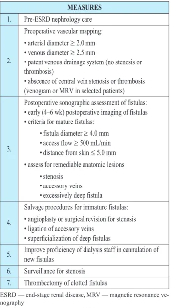
1. Pre-ESRD nephrology care
2.
Preoperative vascular mapping:
• arterial diameter³2.0 mm • venous diameter³2.5 mm
• patent venous drainage system (no stenosis or thrombosis)
• abscence of central vein stenosis or thrombosis (venogram or MRV in selected patients)
3.
Postoperative sonographic assessment of fistulas: • early (4–6 wk) postoperative imaging of fistulas • criteria for mature fistulas:
• fistula diameter³4.0 mm • access flow³500 mL/min • distance from skin£5.0 mm • assess for remediable anatomic lesions
• stenosis • accessory veins • excessively deep fistula
4.
Salvage procedures for immature fistulas:
• angioplasty or surgical revision for stenosis • ligation of accessory veins
• superficialization of deep fistulas
5. Improve proficiency of dialysis staff in cannulation of new fistulas
6. Surveillance for stenosis 7. Thrombectomy of clotted fistulas
ESRD — end-stage renal disease, MRV — magnetic resonance ve-nography
Modified according to reference Š17¹.
Table 2. Measures to increase fistula prevalence
Table 3. Risk factors for the development of cardiovascular complications in association
with arteriovenous shunt for hemodialysis Risk factors
1.
Increased flow through the AV shunt — QAV > 1000 mL/min
a) congestive heart failure b) distal steal phenomenon
2.
Decreased flow through the AV shunt — QAV < 300 mL/min
a) inadequate hemodialysis — Kt/V index < 1.2 b) malnutrition-hypoalbuminemia (albumin < 35 g/L)
3.
Infection of arteriovenuos shunt a) infective endocarditis
b) chronic microinflammation — CRP > 10 mg/L
left ventricle hypertrophy, worsening of coronary ischemia and development of congestive heart failure (25, 26, 27, 28, 29). Left ventricle hypertrophy is adap-tive response to increased cardiac work due to a left ventricle volume overload and risk factor for bad out-come in hemodialysis patients (25, 26, 27, 28, 29, 30). Patients with high-flow AVF have high risk for conges-tive heart failure development and express significant end-diastolic volume of left ventricle (25, 26). Most clinicians believe that AVF shouldn’t be created in pa-tients with diminished heart function (ejection fraction less than 30%) (25).
Prevalence of infective endocarditis in dialysis population is high up to 20% (31). In hemo-dialysis patients vascular access is mostly primary site of infection causing bacteriemia and infective endocar-ditis. Bacteriemia connected with vascular access pun-cture is frequently present in patients treated with he-modialysis (one episode in 100 patients per month).
Risk of bacteriemia depends on type of vascular access (native AVF, synthetic A-V graft, central venous cathe-ter for hemodialysis), and minimal risk is with punctu-re of native vascular access (31).
CONCLUSION
Color Doppler ultrasonography enables preopera-tive planning of AVF creation, assessment of prime ti-me for puncture, early detection of complications and choice of appropriate therapeutic procedure for their treatment, which contribute to a significant decrease of morbidity and better life quality in hemodialysis pati-ents (32, 33, 34).
Acknowledgments: Authors would like to express their deepest gratitude to the Serbian Ministry of Sci-ence and Technological Development for their Grant N°175014, which was used as one of the sources to fi-nancially support the study.
Sa`etak
KLINI^KI ZNA^AJ COLOR DOPPLER ULTRASONOGRAFIJE
U PREOPERATIVNOM PLANIRANJU IZRADE VASKULARNOG PRISTUPA
ZA HEMODIJALIZU
Dejan Petrovi}1
1 — Klinika za urologiju i nefrologiju, Centar za nefrologiju i dijalizu, Klini~ki centar „Kragujevac“, Kragujevac, Srbija
Hroni~nu slabost bubrega odlikuje progresivno i nepovratno smanjenje ja~ine glomerulske filtracije. Ar-terio-vensku fistulu (AVF) za hemodijalizu treba uraditi kod bolesnika kod kojih je klirens endogenog kreatinina £20 mL/min/1,73 m2. Unutra{nji dijametar a.radialis³ 2.0 mm, unutra{nji dijametar v.cephalica³2.5 mm, br-zina protoka krvi kroz a.radialis — VmaxS³50 cm/s i protok krvi kroz a.radialis — Qa.radialis³ 40 mL/m obezbe|uju adekvatno sazrevanje distalne radio-cefa-li~ne AVF. Dijametar v.cephalica³4.0 mm i protok krvi QAV³500 mL/min, ~etiri nedelje posle izrade AVF,
uka-zuju na adekvatno sazrelu AVF i na mogu}nost njene punkcije. Maksimalna brzina protoka krvi kroz AVF od 100 do 350 cm/s i protok krvi od 500 do 1000 mL/min, ukazuju na dobru funkciju AVF i adekvatnu hemodijali-zu. Color Doppler ultrasonografija omogu}ava preope-rativno planiranje izrade AVF, rano otkrivanje kompli-kacija, procenu razvoja, pravovremene punkcije i rada AVF, {to doprinosi zna~ajnom smanjenju morbiditeta i pobolj{anju kvaliteta `ivota bolesnika na hemodijalizi.
Klju~ne re~i:arterio-venska fistula, hemodijaliza, color doppler.
REFERENCES
1. Schmitz PG. Progressive renal insufficiency: office stra-tegies to prevent or slow progression of kidney disease. Postgrad Med. 2000; 108(1): 145–54.
2. National Kidney Foundation K/DOQI. Clinical practice guidelines for chronic kidney disease: evaluation, classification, and stratification. Am J Kidney Dis. 2002; 39(2 Suppl 1): S17–246.
3. Stojimirovi} B, Petrovi} D. Clinical significance of risk factors control in prevention of chronic renal failure progres-sion. Vojnosanit Pregl. 2006; 63(6): 585–91.
4. Petrovi} D, Stojimirovi} B. Proteinuria as a risk factor for progression of chronic renal disease. Vojnosanit Pregl. 2008; 65(7): 552–8.
5. Rossert JA, Wauters J-P. Recommendations for the scre-ening and management of patients with chronic kidney disease. Nephrol Dial Transplant. 2002; 17(Suppl 1): S19–28.
6. Besarab A, Brouwer D. Aligning Hemodialysis Treat-ment Practices with the National Kidney Foundation, s K/DOQI Vascular Access Guidelines. Semin Dial. 2004; 33(11): 694–711.
Correspondence to/Autor za korespodenciju
Doc. Dr Dejan Petrovi}
Clinic for Urology and Nephrology, Center for Nephrology and Dialysis, Clinical Center „Kragujevac“, Kragujevac, Zmaj Jovina 30, 34000 Kragujevac,
Tel.: +381 34 370302, Fax: +381 34 300380
8. Konner K, Nonnast Daniel B, Ritz E. The Arteriove-nous Fistula. J Am Soc Nephrol. 2003; 14(6): 1669–80.
9. Asif A, Merrill D, Pennell P. Vascular Access Educa-tion, Planning and Percutaneus Interventions by Nephrologists. In: Ronco C, Brendolan A, Levin NW, editors. Cardiovascular Disorders in Hemodialysis. Basel: Karger; 2005. p. 138–49.
10. Brimble KS, Rabbat CG, Schiff D, Ingram AJ. The Cli-nical Utility of Doppler Ultrasound Prior to Arteriovenous Fis-tula Creation. Semin Dial. 2001; 14(5): 314–7.
11. Malovrh M. Native arteriovenous fistula: Preoperative evaluation. Am J Kidney Dis. 2002; 39(6): 1218–25.
12. Malovrh M. The Role of Sonography in the Planning of Arteriovenous Fistulas for Hemodialysis. Semin Dial 2003; 16(4): 299–303.
13. Petrovi} D, Novakovi} B. Clinical importance Color Doppler ultrasonography in evaluation of maturation and work vascular access for hemodialysis. In: Ne{i} V, ed. Hypotension in dialysis and vascular access. Lazarevac: Elvod-print; 2005. p. 103–6.
14. Ferring M, Henderson J, Wilmink A, Smith S. Vascular ultrasound for the pre-operative evaluation prior to arteriovenu-os fistula formation for haemodialysis: review of the evidence. Nephrol Dial Transplant. 2008; 23(6): 1809–15.
15. Beathard GA. An Algorithm for the Physical Examina-tion of Early Fistula Failure. Semin Dial. 2005; 18(4): 331–5.
16. Jagi} N, Petrovi} D, Miloradovi} V, Novakovi} B. Cli-nical importance of early detection of vascular access failure in haemodialysis patients. Medicus. 2006; 7(3): 103–6.
17. Allon M. Current Management of Vascular Access. Clin J Am Soc Nephrol. 2007; 2(4): 786–800.
18. Asif A, Roy-Chaudhury P, Beathard GA. Early Arteri-ovenous Fistula Failure: A Logical Proposal for When and How to Intervene. C J Am Soc Nephrol. 2006; 1(2): 332–339.
19. Sands JJ, Ferrell LM, Perry MA. The role of color flow Doppler ultrasound in dialysis access. Semin Nephrol. 2002; 22(3): 195–201.
20. Wiese P, Nonnast Daniel B. Colour Doppler ultraso-und in dialysis access. Nephrol Dial Transplant. 2004; 19(8): 1956–63.
21. Dikow R, Schwenger V, Zeier M, Ritz E. Do AV Fistu-als Contribute to Cardiac Mortality in Hemodialysis Patients? Semin Dial. 2002; 15(1): 14–7.
22. London GM, Guerin AP, Marchais SJ. Hemodynamic Overload in End-Stage Renal Disease Patients. Semin Dial. 1999; 12(2): 77–83.
23. Rigatto C, Parfrey PS. Uraemic Cardiomyopathy: an Overload Cardiomyopathy. J Clin Basic Cardiol. 2001; 4(2): 93–5.
24. Sood MM, Pauly RP, Rigatto C, Komenda P. Left ven-tricular dysfunction in the haemodialysis population. NDT Plus. 2008; 1(4): 199–205.
25. MacRae JM, Levin A, Belenkie I. The Cardiovascular Effects of Arteriovenous Fistulas in Chronic Kidney Disease: A Cause for Concern? Semin Dial. 2006; 19(5): 349–52.
26. Petrovi} D, Stojimirovi} B. Vascular access blood flow for hemodialysis — a risk factor for development of cardiovas-cular complications in hemodialysis patients. Med Rew. 2007; 60(3–4): 183–6.
27. Petrovi} D, Stojimirovi} B. Left ventricular hypertro-phy in hemodialysis patients. Med Rew. 2008; 61(7–8): 369–74. 28. Petrovi} D, Miloradovi} V, Poskurica M, Stojimirovi} B. Diagnostics and treatment of ishemic heart disease in hemo-dialysis patients. Vojnosanit Pregl. 2009; 66(11): 897–903.
29. Petrovi} D, Jagi} N, Miloradovi} V, Stojimirovi} B. Non-tradicional risk factors for development of cardiovascular complications in haemodialysis patients. Ser J Exp Clin Res. 2009; 10(3): 95–102.
30. Petrovi} D, Jagi} N, Miloradovi} V, Stojimirovi} B. Left ventricular hypertrophy — risk factor for poor outcome in hemodialysis patients. Ser J Exp Clin Res. 2008; 9(4): 129–35. 30. Nucifora G, Badano LP, Viale P, Gianfagna P, Allocca G, Montanaro D, et al. Infective endocarditis in chronic haemo-dialysis patients: an increasing clinical challenge. Eur Heart J. 2007; 28(19): 2307–12.
31. Beathard GA, Litchfield T (Physician Operators Fo-rum of RMS Lifeline). Effectiveness and safety of dialysis vas-cular access procedures performed by interventional nephrolo-gists. Kidney Int. 2004; 66(4): 1622–32.
32. Petrovi} D, Stojimirovi} B. Cardiovascular morbidity and mortality in hemodialysis patients — epidemiological anal-ysis. Vojnosanit Pregl. 2008; 65(12): 893–900.