*Corresponding author: Faeze Zeinali, Emergency Medicine Department, Shahid Sadoughi University of Medical Sciences, Yazd, Iran. Tel: 03538284089; Fax: 03538223514; Email: [email protected]
© 2016 mums.ac.ir All rights reserved.
This is an Open Access article distributed under the terms of the Creative Commons Attribution License (http://creativecommons.org/licenses/by/3.0), which permits unrestricted use, distribution, and reproduction in any medium, provided the original work is properly cited.
Aortic
Dissection
of
Unknown Origin in a Young Patient: A Case Report
Majid Hajimaghsoudi
1, Faeze Zeinali
2*, Mehdi Bagherabadi
1, Morteza Saeedi
31. Emergency Medicine Physician, Trauma Research Center, Emergency Department, Shahid Rahnemoon Hospital, Shahid Sadoughi University of Medical Sciences, Yazd, Iran
2. Resident of Emergency Medicine, Department of Emergency Medicine, Shahid Sadoughi University of Medical Sciences, Yazd, Iran 3. Emergency Medicine Physician, Department of Emergency Medicine, Shariati Hospital, Tehran University of Medical Sciences, Tehran, Iran
A R T I C L E I N F O A B S T R A C T
Article type:
Case Report
Aortic dissection occurs when a tear develops in the wall of the aorta, which is rare in the young population. This fatal disorder is hard to diagnose, especially in young patients. We present the case of aortic dissection in a 15-year-old boy referred to the Emergency Department of Yazd University of Medical Sciences in November 2015. The patient presented to our department with sudden acute chest pain. Emergent computed tomography (CT) scanning of the brain, chest, and abdomen reflected bilateral pleural effusion, biluminal aorta, arterial flap in the upper part of the abdominal aorta, and dilated small bowl loop. The patient did not have any aortic dissection risk factors such as history of connective tissue disease, congenital heart disease, coarctation of the aorta,
and hypertension. The only noticeable point in the patient’s history was
swimming two hours before the onset of the chest pain. Aortic dissection is a rare differential diagnosis in children with acute sudden chest pain.
Article history:
Received: 27 Dec 2015 Revised: 05 Apr 2016 Accepted: 06 Apr 2016
Keywords:
Aortic Dissection Risk Factors Young Adults
►Please cite this paper as:
Hajimaghsoudi M, Zeinali F, Bagherabadi M, Saeedi M. Aortic Dissection of Unknown Origin in a Young Patient: A Case Report. J Cardiothorac Med. 2016; 4(2):461-463.
Introduction
Aortic dissection is secondary to damage or tear in the wall of the aorta (1). Aortic dissection is not relatively common, but it most frequently occurs in 60 to 70-year-old males. Incidence of the thoracic aortic dissection is 3–4 cases per 100,000 persons per year (2). Sometimes, this fatal disorder is misdiagnosed, especially in young patients; therefore, true incidence rate of this disorder is underestimated (3).
Major risk factors for aortic dissection are uncontrolled hypertension, atherosclerosis, weakened and bulging artery, aortic valve defect, and coarctation of the aorta. Aortic dissection is rare in the pediatric population (4); in this study, we present the case of a 15-year-old boy presenting to the Emergency Department of Yazd University of Medical Sciences, Iran.
Case Report
The patient was a 15-year-old boy with acute and sharp chest pain initiated about two hours before hospital admission, while swimming in a public pool. He described a sudden retrosternal
and epigastric pain with radiation to the back. After a few seconds, his pain migrated to periumbilical region. The patient’s past medical, familial, and drug history was unremarkable.
At admission, the patient looked ill, with a heart rate of 130 bpm, initial blood pressure of 125/80 mmHg in both hands, respiratory rate of 30 bpm, and . C body temperature. Laboratory test results for this patient showed hemoglobin level of 12.9 g/dL, white blood cell count of 17,800/µl, and platelet count of 241,000/µl; serum
electrolytes, including Na⁺, K⁺, and Ca⁺⁺ were
normal. Liver function test was minimally higher than the normal range, indicating a normal amylase level. Electrocardiogram demonstrated a normal sinus rate without any abnormal changes.
Hajimaghsoudi M et al Aortic dissection of unknown origin in a young patient
462 J Cardiothorac Med. 2016; 4(2):461-463.
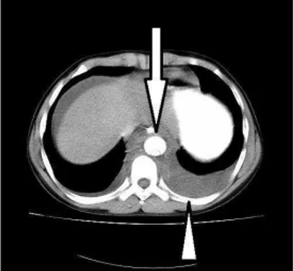
Figure 1. The arrow shows double
lumens of descending aorta and arrow-head shows the effusion of thorax mainly on the left side
Figure 2. The arrows show significant
landmarks of bowel obstruction
Figure 3. The next cut of computed
tomography scan showing aortic wall flap
abdominal pain and dilated small bowl loop in the CT scan, diagnostic laparotomy was carried out; during surgery, ischemic blackish small-bowel loop was noticed. The patient died due to cardiac arrest.
Discussion
The incidence of aortic dissection is rare in young patients. In 2009, Fikar et al. reviewed more than 1000 aortic dissection patients 3.5% of whom were aged under 19 years old (5). Major risk factors for aortic dissection are hypertension, connective tissue disease, congenital heart disease, severe chest trauma, and genetic defect in fibrin production (4).
Through studying the medical records of our patient, we found no personal or familial history of connective tissue disease such as Ehlers-Danlos, Marfan, or Turner syndromes, or consumption of any drugs causing connective tissue disturbance. In addition, there were no symptoms or history of congenital heart disease such as bicuspid aortic valve, coarctation of the aorta, and hypertension. The only point in the
patient’s history was diving in a pool about two
hours before the onset of the chest pain.
In cases of aortic dissection, the main finding is abdominal pain immediately after the onset of severe chest pain. In the previous studies, similar to our patient, the first symptom was acute and severe knife-like chest pain with changing position over the time (3, 6, 7). Ayrik et al. and Nadour et al. reported cases of painless aortic dissection (8, 9). Ngan et al. reported a case of aortic dissection in a 17-year-old male patient, without any known risk factors. Their patient had acute abdominal pain and retroperitoneal hematoma, and died two days after admission (10).
In aortic dissection, extension of hematoma or flap commonly results in obstruction of the renal (8%), mesenteric (8-13%), and cerebral arteries. Based on the involved vessel, the signs and symptoms (e.g., azotemia and oliguria in the
involved renal artery, abdominal pain and hematochezia in the involved mesenteric artery, and neurological defect in the involved cerebral artery) can be different (7).
Intravenous contrast CT scan is the gold standard for diagnosis in emergency patients, although magnetic resonance imaging (MRI) and Transesophageal Echocardiography (TEE) can be useful in stable patients, as well (7). In our case, abdominal pain was one of the major complaints; therefore, abdominal CT scan was performed.
Generally, in aortic dissection, treatment is based on reduction of hydrodynamic pressure and heart beat energy through administering beta and calcium channel blockers, pain relief, and surgical methods for tissue regeneration such as endovascular stent grafting (11). The extension of vascular involvement and the interval between dissection and the onset of treatment can affect the rate of mortality. In the early hours, the rate of mortality increases 1-3%/hour from symptom to treatment onset, which increases to 25%/hour after 24 hours.
In our patient, the extension of artery involvement was not justified by trauma, and another unknown etiology such as fibrinogen deficiency might be involved. In management of complicated dissections with inadequate perfusion in aortic branches, emergent endovascular stent grafting is recommended (7).
Conclusion
According to this case, aortic dissection is an important differential diagnosis in children with acute chest pain, which is hard to diagnose. We should always keep in mind that this fatal disorder is a possible diagnosis even in the young peoples without known risk factors. The early recognition and rapid treatment is lifesaving.
Running title Hajimaghsoudi M et al
J Cardiothorac Med. 2016; 4(2):461-463. 463
Aortic dissection of unknown origin in a young patient
References
1. Kentaro H, Nobutaka H, Kazuhiko S, Izumi N. Aortic dissection complicated with fatal cerebral infarction: case report and review of literatures. Open J Modern Neurosurg. 2012; 2:21-4.
2. LeMaire SA, Russell L. Epidemiology of thoracic aortic dissection. Nat Rev Cardiol. 2011; 8:103-13. 3. Pineault J, Ouimet D, Pichette V, Vallee M. A case of
an aortic dissection in a young adult: a refresher of the literature of this "great masquerader". Int J Gen Med. 2011; 4:889-93.
4. Howard DP, Sideso E, Handa A, Rothwell PM. Incidence, risk factors, outcome and projected future burden of acute aortic dissection. Ann Cardiothorac Surg. 2014; 3:278-84.
5. Fikar CR, Fikar R. Aortic dissection in childhood and adolescence: an analysis of occurrences over a
0‐year interval in New York State. Clin Cardiol.
2009; 32:E23-6.
6. Ngan KW, Hsueh C, Hsieh HC, Ueng SH. Aortic dissection in a young patient without any predisposing factors. Chang Gung Med J. 2006; 29:419-23.
7. Nienaber CA, Eagle KA. Aortic dissection: new frontiers in diagnosis and management part II: therapeutic management and follow-up. Circulation. 2003; 108:772-8.
8. Ayrik C, Cece H, Aslan O, Karcioglu O, Yilmaz E. Seeing the invisible: painless aortic dissection in the emergency setting. Emerg Med J. 2006; 23:e24-7. 9. Nadour W, Goldwasser B, Biederman RW, Taffe K.
Silent aortic dissection presenting as transient locked-in syndrome. Tex Heart Inst J. 2008; 35:359-61.
10.Ngan K, Hsueh C, Hsieh HC, Ueng S. Aortic dissection in a young patient without any predisposing factors. Chang Gung Med J. 2006; 29:419-23.