Original Research
Marginal Assessment of Crowns by the Aid of Parallel Radiography
Farnaz Fattahi
1, Rashin Giti
1, Kianoosh Torabi
11
Department of Fixed Prosthodontics, School of Dentistry, Shiraz University of Medical Sciences,
Shiraz, Iran
Received 18 September 2014 and Accepted 13 December 2014
Abstract
Introduction: Marginal adaptation is the most critical item in long-term prognosis of single crowns. This study aimed to assess the marginal quality as well asthe discrepancies in marginal integrity of some PFM single crowns of posterior teeth by employing parallel radiography in Shiraz Dental School, Shiraz, Iran. Methods: In this descriptive study, parallel radiographies were taken from 200 fabricated PFM single crowns of posterior teeth after cementation and before discharging the patient. To calculate the magnification of the images, a metallic sphere with the thickness of 4 mm was placed in the direction of the crown margin on the occlusal surface. Thereafter, the horizontal and vertical space between the crown margins, the margin of preparations and also the vertical space between the crown margin and the bone crest were measured by using digital radiological software. Results: Analysis of data by descriptive statistics revealed that 75.5% and 60% of the cases had more than the acceptable space (50µm) in the vertical (130±20µm) and horizontal (90±15µm) dimensions, respectively. Moreover, 85% of patients were found to have either horizontal or vertical gap. In 77% of cases, the margins of crowns invaded the biologic width in the mesial and 70% in distal surfaces. Conclusion: Parallel radiography can be expedient in the stage of framework try-in to yield some important information that cannot be obtained by routine clinical evaluations and may improve the treatment prognosis.
Key words: Marginal adaptation, metal-ceramic crowns, radiography.
--- Fattahi F, Giti R, Torabi K. Marginal Assessment of Crowns by the Aid of Parallel Radiography. J Dent Mater Tech 2015; 4(1): 29-36.
Introduction
Regardless of the place of margins, proper marginal fit of dental fillings and crowns is essential to prevent periodontal diseases and recurrent caries (1-3). Several researchers have announced that clinical methods (visual inspection and/or an explorer) can be used to evaluate the gingival margin of filling and crown. They have also considered it to be a difficult diagnostic task, particularly when the restoration margin is interproximal and subgingival (1,2). Some other researchers have taken the use of explorer into question, as it tends to stick, whether or not the tissue adjacent, to the filling is carious; thus, radiographic methods are suggested to be used for diagnosis of the lesions adjacent to restorations (1,4). Despite the fact that all defective restorations do not necessarily bring about disease, marginal misfits are crucial to be appropriately diagnosed so that the tooth and the surrounding tissues can be maintained as much as possible; so it should be included as a part of the overall evaluation of the quality of the restoration. The margin of a single unit extracoronal restoration is the most critical item in long-term prognosis and the most susceptible part to distort (5). Therefore, success and failure of the fixed restoration are completely related to adaptation and location of the crown margin (5). Some other items that affect the marginal adaptation are marginal beveling, venting, type and thickness of the cement, type of the impression material (dimensional accuracy), and the design of the margin preparation (6,7).
The best place for the crown margin is where the best access is provided both for the dentist to make impression, and for the patient to clean the restoration (5). The fourdeterminingfactors in choosing the marginal location are periodontal consideration, esthetic consideration, adequate retention, and finally extending the preparation to the sound tooth structure (2,8).
nonrestored ones (9). The extent of inflammation around the restored teeth depends on particular elements such as emergence profile, adequate access to polish the subgingival margins and biologic width invasion (3). Biological width is defined as the distance between the alveolar crest and the junctional epithelium and is estimated to be approximately 2.04mm (10). Migration of the junctional epithelium and bone resorptionare the consequences of marginal invasion of the crown to biologic width; which occurs to create the former biological distance (10).
Evaluation of the marginal adaptation can be performed either qualitatively or quantitatively. Qualitative assessment is done by direct visualization and sense of touch(by use of an explorer) (11), by using impression materials, or through radiological assessment (12). For quantitative assessment, employing a microscope in high magnification would be the best choice to measure the gap space. Graded explorers and parallel radiography can also be used for this purpose (13,14). Radiography is routinely used for quantitative evaluation in Nance and Hixon-Old father’s methods which are specialized for space measurement in mixed dentition (15).
In the case of rehabilitation with dental implants, most implant systems consist of two components – the implant screw and a connecting transmucosal structure, the abutment. The prosthetic crown can be either connected to the abutment or be an independent element (16), consequently, a gap can exist between the implant and the abutment.Also between the crown and the abutment, there might be a gap or an overextension of the luting agent. Since the presence of excess cement may result in peri-implant inflammation, radiographic evaluation has been proposed by some studies to ensure the appropriate seating and debridement of subgingival restorations (17, 18). Marginal misfit in cement-retained implant single crowns can also be accompanied by changes in crestal bone (17). Researches on radiographic assessment have reported the radiopacity of the restorative material and the technique to have effects on the assessment of marginal misfits (1). A number of in-vitro studies have used conventional and digital radiography to evaluate the diagnosis of gaps, and adopted marginal discrepancies from 0.01 to 0.5 mm between the restoration and the tooth (12, 19). Also Bjorn used radiography to measure the size of overhang and the marginal gap, as well as the distance between the crown margin and the bone crest (20). Radiography is particularly essential to estimate and calculate the bone dimensions before dental implant surgery(21). Hence, various radiographic techniques such as digital radiography, periapical, and computed tomography have been devised to evaluate the proximal surfaces; however, within the literature there exists little
consensus on their individual use. Thus, in an attempt to search for a rationale on the use of parallel radiography and to suggest the best protocol, the current study was enrolled to evaluate the marginal adaptation and location of the crowns (the most critical item in fixed restoration) at the delivery point by employing radiographic assessment in Shiraz Dental School, Shiraz, Iran.
Materials and Method
In this descriptive study, 200 fabricated single-unit crowns were evaluated for the location and adaptation of margins by parallel radiography using Kodak E speed dental x-ray film (Eastman Kodak, Rochester NY, USA). The study was performed in the Department of Fixed Prosthodontics and OMF Radiology of Shiraz Dental School, Shiraz, Iran in 2012. The research protocol was submitted for consideration, comment, guidance and was approval by the research ethics committee of Shiraz School of Dentistry (ID: EC-2013-166). Considering the Declaration of Helsinki as the ethical principles, allpatients were informed in details about the nature of the trial, and individual voluntary informed consent was signed.
All the crowns were related to the posterior teeth and all of them were porcelain fused to metal (PFM). The type of margin preparation for all of them was shoulder bevel, and all of them were cemented by zinc phosphate cement.
Figure 1. Method of measuring horizontal and vertical marginal discrepancy
The final values were recorded regarding the coefficient of magnification. The values for marginal discrepancies were evaluated by descriptive statistical analysis of the registered data.
Generally the biologic width around different teeth is different and can just be measured by histological evaluation. In the current study, however, the lowest biological width (2mm) was considered as standard and the extents less than thatwere considered as invasion to the biological width. Since a space of at least 0.5mm should be available between the preparation finishing line and the junctional epithelium crest (10), the spaces less than 2500µm between the crown margin and the bone crest were considered as biologic width violation;besides, the open margin was attributed to the marginal gap of more than 50µm (5).
Results
The largest recorded value around the tooth is considered as the marginal discrepancy value; however, the buccal and lingual surface of the teeth could not be
detected in radiographic evaluation. Hence, only mesial and distal surfaces were evaluatedin this survey. The acceptable marginal gap in this study was 50µm. Accordingly, the crowns with horizontal and/or vertical gap of more than 50µm in mesial or distal surfaces were considered as having open margin. As represented in table 1, 75.5% of the crowns had vertical and 60% of the crowns had horizontal discrepancy. Likewise, 85% of all crowns had a marginal gap more than the acceptable extent in either horizontal or vertical dimensions.As demonstrated in table 2, 77% and 70%of the crowns invaded the biological width in the mesial and distal surfaces, respectively.
Out of 200 evaluated teeth in this study, 70 teeth were detected to have some remaining cement around the teeth in the gingival sulcus, indicating that they had not been completely cleaned.
The margins of 15 crowns (7.5%) were on amalgam restoration with overhang. A total of 8 (4%) did not have the effective emergence profile, although they did not have marginal gap either. In 40 cases restored by a castable post, 8 core and 10 crowns had space between the post and Gutta-percha in the root canal and in 3 cases, the prepared post was deviated from the root canal direction.
Table1. Number and percentage of crowns with or without horizontal or vertical gap
Table 2. Number and percentage of crowns have more or less space than 2500µ between the crown margin and the crestal bone
Total Either horizontal or vertical
Horizontal dimension Vertical dimension
Without gap With gap
Without gap With gap
Without gap With gap
200 30
170 80
120 49
151 Number
100 15%
85% 40%
60% 24.5%
75.5% Percentage
Percentage Number
Marginal space to the crestal bone(µm)
23% 46
More than 2500 Mesial
77% 154
Less than 2500
24% 48
More than 2500 Distal
70% 140
Discussion
The current study was carried out to find a rationale on using radiography as an adjunct method for diagnosing the misfit in dental prostheses and restorations. When a filling or a crown is placed, the restoration surface should be aligned with the tooth margin (4, 22). Misfit is usually defined as the lack of adaptation between the restoration and the prepared tooth. They must be prevented because they are prone to cause accumulation of biofilm and consequently lead to development of carious lesions (1,2,23).It might be disadvantageous to the gingival and marginal bone tissues, as well (23,24). Likewise, healthy periodontal conditions are maintained through providing proper fitting of abutments on implants (17,25). There are a number of studies in which Radiographic examination has been recommended for the assessment of dental prosthesis or restorations (19,26) as well as the abutment adaptation (18). Two recent publications have suggested the presence of digital radiographic artifacts beside the metal restorations, which could impede the interpretation of such images (27,28). Another study compared the digital and conventional radiographs to assess the diagnostic accuracy of metal restoration misfit, and found no significant difference between conventional and digital original images (29). Based on the controversial results of this study, the presence of vertical marginal gap in 75.5% and horizontal gap in 60% of crowns can endorse the fact that evaluation of the marginal adaptation by using only explorer (30), rouge and chloroform, or even by an impression material would not be adequate. The results from clinical and radiographic examination were brought to comparison; the study that had assessed marginal gaps adjoining to implant components hadreported higher accuracy from the radiographic recording (31). In the case of assessing Class II restorations, the number of false positive diagnosis was reduced due to making use of both clinical and radiograph evaluation together (32). On the other hand, the number of correct cases diagnosed by the two methods were found to have no difference in an in-vivo study (33), whereas another study reported radiographs to have helped diagnosing more correct cases (34).Some studies were conducted concerning the impact of x-ray beam angulation (35,36) and they all agreed that the most accurate angulation for marginal misfit diagnosis in dental restorations is the orthogonal projection. Regarding image acquisition system, conventional film radiography was used in majority of the studies, while some others had used digital radiography, both CCD-based sensors and PSP systems (12,21). However, the use of computed tomography in diagnosing the misfits was assessed by none of them.The vertical marginal gap could probably be due to incomplete seating of the crowns (37), and
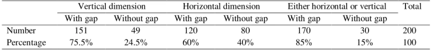
incomplete seating by itself can be caused by tight proximal contact or presence of pressure spot.Vertical marginal gap can also be the result of short ditching in the laboratory by the technician and inaccuracy in making the impression of marginal preparation by the clinician (Fig. 2) (37). Practically, the subgingival margins, especially in the posterior regions, cannot be registered accurately by the impression material due to the poor access and the presence of blood (37). Some authors have proposed that radiography can assess the restored proximal surfaces better (1,2). In a study the proximal margins of Class II amalgam restorations were evaluated by employing radiographic examination solely and in association with clinical method. The resultrevealedbetter quality of diagnosis with the combined method (32). The horizontal flaws can be in two forms of ledge or overhang. Ledge is the result of wrong preparation (37); for instance, the chamfer margin should never be prepared wider than half of the thickness of a diamond bur tip, otherwise an unsupported lip of enamel may create marginal ledge (Fig. 3a) (37).
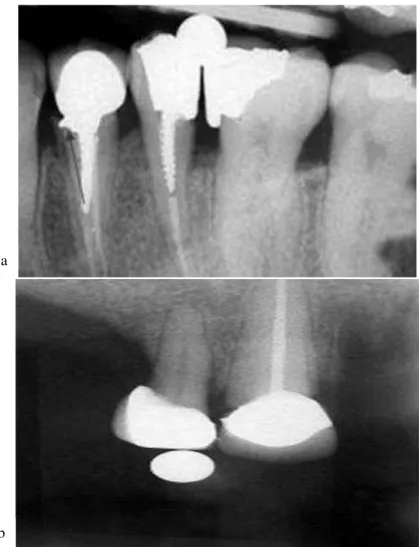
Presence of positive horizontal space (an overhang) can be allied to the technician or the operative dentist. In some instances, the technician may ditchthe die beyond the finishing line of the preparation and subsequently this flaw would lead to an overhang margin that can invade the gingival tissue and develop inflammation (Fig. 3b) (23). Sometimes also the undercut which is created in the preparation of the crown by the operative dentist can cause the wax pattern distortion during removal from the die and would produce marginal gap and overhang (5). Invasion of the biologic width can reversely be associated with the operative errors of the dentist(23). It must be concerned that the preparation should not be extended more than 0.5mm into the gingival sulcus (Fig. 4) (23).
Figure 2. Radiographic image showing vertical marginal gap
Figure 3.a. Radiographic projection illustrating horizontal marginal discrepancy in the form of ledge formation. b. Radiographic picture showing horizontal marginal discrepancy in the form of overhang
Figure 4. Radiographic picture showing invasion of the restoration margin to the biologic width (arrow)
Figure 5. Radiographic projection displaying the Figure 3a. Radiographic projection illustrating horizontal marginal discrepancy in the form of ledge formation3b Radiographic picture showing horizontal marginal discrepancy in the
form of overhange residual cement that has not been completely cleaned
Conclusion
In general, dental radiography is used to shows the proximal surfaces of the teeth butpracticalinformation can be obtained from it about adaption of the crown margins, its location and its relation to the bone crest. Accordingly, dental radiographic evaluation can be used as an adjunct to the clinical examinations to yield a better treatment prognosis.
References
1. Mjor IA. Clinical diagnosis of recurrent caries. J
Am Dent Assoc 2005;136:1426-33.
2. Larson TD. The clinical significance of marginal
fit. Northwest Dent 2012;91:22-9.
3. Papageorgiou SN, Papadelli AP, Koidis PT,
Petridis HP. The effect of prosthetic margin
location on caries susceptibility. A systematic
review and meta-analysis. British Dent J
2013;214:617-24.
4. Kidd EA. Caries diagnosis within restored teeth.
Adv Dent Res 1990;4:10-3.
5. Shilling burg HT, Sather DA, Wilson EL, Cain JR,
Mitchell DL, Blanco LJ. Fundamentals of fixed
prosthodontics. Chicago: Quintessence publishing
Co, Lnc, 2012.
6. Eames WB, O'Neal SJ, Monteiro J, Miller C, Roan
JD, Jr., Cohen KS. Techniques to improve the
seating of castings. J Am Dent Assoc 1978;96:
7. Henry PJ, Harnist DJ. Dimensional stability and
accuracy of rubber impression materials. Aust Dent
J 1974;19:162-6.
8. Gardner FM. Margins of complete
crowns--literature review. J Prosth Dent 1982;48:396-400.
9. Reeves J. Periodontal health--challenges in
restorative dentistry. Primary Dent J 2014;3:73-6.
10. Newman MG, Takei HH, Klokkevold PR, Crranza
FA. Clinical periodontology. Tothed. ST. Louis:
Elsevier/Saun Ders, 2006.
11. Akin A, Toksavul S, Toman M. Clinical Marginal
and Internal Adaptation of Maxillary Anterior
Single All-Ceramic Crowns and 2-year
Randomized Controlled Clinical Trial. J Prosth
2014 (In Press).
12. Haak R, Wicht MJ, Hellmich M, Noack MJ.
Detection of marginal defects of composite
restorations with conventional and digital
radiographs. Eur J Oral Sci 2002;110:282-6.
13. Varol S, Kulak-Ozkan Y. In Vitro Comparison of
Marginal and Internal Fit of Press-on-Metal
Ceramic (PoM) Restorations with
Zirconium-Supported and Conventional Metal Ceramic Fixed
Partial Dentures Before and After Veneering. J
Prosth 2014 (In Press).
14. Alshiddi IF, Habib SR, Al-Mazrou FY, Aly AM,
Al-Zaid AM. Comparing Government (School) vs.
Private (non-school) Dental Laboratories in
Marginal Adaptation of Single
Porcelain-Fused-to-Metal Crowns. Oral Health Dent Manage
2014;13:707-11.
15. Dean JA, Avery DR, Mcdonalal RE. Dentistry for
the child and adolescent. Mary land Heights:
Elsevier Mosby, 2011.
16. Schwarz MS. Mechanical complications of dental
implants. Clin Oral Implants Res 2000;11:156-8.
17. Wadhwani C, Rapoport D, La Rosa S, Hess T,
Kretschmar S. Radiographic detection and
characteristic patterns of residual excess cement
associated with cement-retained implant
restorations: a clinical report. J Prosth Dent
2012;107:151-7.
18. Pette GA, Ganeles J, Norkin FJ. Radiographic
appearance of commonly used cements in implant
dentistry. Int J Periodont Res Dent 2013;33:61-8.
19. Weyns W, De Boever J. Radiographic assessment
of the marginal fit of cast restorations. J Prosth
Dent 1984;51:485-9.
20. Bjorn AL, Bjorn H, Grkovic B. Marginal fit of
restorations and its relation to periodontal bone
level. II. Crowns. Odontologisk Revy 1970;21:
337-46.
21. Sharkey S, Kelly A, Houston F, O'Sullivan M,
Quinn F, O'Connell B. A radiographic analysis of
implant component misfit. Int J Oral Maxillofac
Implants 2011;26:807-15.
22. Holmes JR, Bayne SC, Holland GA, Sulik WD.
Considerations in measurement of marginal fit. J
Prosth Dent 1989;62:405-8.
23. Giollo MD, Valle PM, Gomes SC, Rosing CK. A
retrospective clinical, radiographic and
microbiological study of periodontal conditions of
teeth with and without crowns. Braz Oral Res
2007;21:348-54.
24. Sorensen SE, Larsen IB, Jorgensen KD. Gingival
and alveolar bone reaction to marginal fit of
subgingival crown margins. Scand J Dent Res
1986;94:109-14.
25. Chen CJ, Papaspyridakos P, Guze K SM, Weber
HP, Gallucci GO. Effect of misfit of cement
-retained implant single crowns on crestal bone
changes. Int J Prosthodont 2013;26:135-7.
26. Croll TP, Epstein DW, Castaldi CR. Marginal
adaptation of stainless steel crowns. Pediatric
dentistry. 2003;25(3):249-52.
27. Schweitzer DM, Berg RW. A digital radiographic
artifact: A clinical report. J Prosth Dent
2010;103:326-9.
28. Brettle D, Carmichael F. The impact of digital
image processing artefacts mimicking pathological
features associated with restorations. Br Dent J
2011;211:167-70.
29. Liedke GS, Spin-Neto R, Vizzotto MB, Da Silveira
conventional and digital radiography for detecting
misfit between the tooth and restoration in
metal-restored teeth. J Prosth Dent 2014 (In Press).
30. Spedding RH. Two principles for improving the
adaptation of stainless steel crowns to primary
molars. Dent Clin North Am 1984;28:157-75.
31. Konermann AC, Zoellner A, Chang BM, Wright
RF. In vitro study of the correlation between the
simulated clinical and radiographic examination of
microgaps at the implant-abutment interface.
Quintessence Int 2010;41:681-7.
32. Espelid I, Tveit AB. Diagnosis of secondary caries
and crevices adjacent to amalgam. Int Dent J
1991;41:359-64.
33. van Amerongen WE, Eggink CO. The cervical
margin of amalgam restorations: a radiographic and
clinical assessment. ASDC J Dent Child
1986;53:177-83.
34. Kroeze J, Ruiken R, van 't Hof M. Evaluation of an
indirect method for assessing the quality of
amalgam restorations in epidemiological studies.
Commun Dent OrAL epidemiol 1988;16:208-11.
35. Begona Ormaechea M, Millstein P, Hirayama H.
Tube angulation effect on radiographic analysis of
the implant-abutment interface. Int J Oral
Maxillofac Implants 1999;14:77-85.
36. Papavassiliou H, Kourtis S, Katerelou J,
Chronopoulos V. Radiographical evaluation of the
gap at the implant-abutment interface. J Esthet
Restor Dent 2010;22:235-50.
37. Rosental SF, Lond MF, Fuyimoto J. Contemporary
fixed prostothodontics. St. Louis: Elsevier/Mosby;
2006.
38. Antonijevic D, Obradovic-Djuricic K, Rakocevic Z,
Medigovic I. In vitro radiographic detection of
cement overhangs on cement-retained implant
restorations. Int J Oral Maxillofac Implants
2013;28:1068-75.
Corresponding Author: Kianoosh Torabi
School of Dentistry
Shiraz University of Medical Sciences, Shiraz, Iran Tel: 00989171085682