American Journal of Animal and Veterinary Sciences 6 (3): 121-124, 2011 ISSN 1557-4555
© 2011 Science Publications
Corresponding Author: Garedaghi Yagoob, Department of Pathobiology, Tabriz Branch, Islamic Azad University, Tabriz, Iran Tel: 0098 914 311 0499
121
Prevalence of bovine Cysticercosis of
Slaughtered Cattle in Meshkinshahr Abattoir
1
Y. Garedaghi,
2A.P. Rezaii Saber and
3M. Saberie Khosroshahi
1
Department of Pathobiology,
2
Department of Clinical Sciences,
3
Department of Veterinary Medicine,
Tabriz Branch, Islamic Azad University, Tabriz, Iran
Abstract: Problem statement: The occurrence of the larvae of Taenia saginata (T. saginata) in cattle musculature causes T. saginatacysticercosis or bovine cysticercosis while the adult worms in human small intestines cause taeniasis. Approach: In this study, the prevalence of Taenia saginata cysticercosis in cattle slaughtered for meat in Meshkinshahr Abattoir, Iran between September 2010 and August 2011 was reported. Results: The examination of various organs of 500 cattle in Meshkinshahr Abattoir showed that 15(3%) were infected with T. saginatacysticercosis. The tongue, masseter muscles, cardiac muscles, triceps muscles and thigh muscles were the main predilection sites of the cysts. The cysts of bovine cysticercosis were also identified on the spleen, intercostal muscles, diaphragm and liver. Out of 460 male cattle, examined, 14 (3%) had cysts of bovine cysticercosis while 1 (2.5%) of the 40 female animals investigated were infected. Conclusion: The animals slaughtered were all adults. No significant difference in prevalence rates was recorded between the sexes. The prevalence of bovine cysticercosis was higher in local sarabi cattle breeds than Holstein-Frisian cattle.
Key words: Prevalence, bovine cysticercosis, meshkinshahr abattoir
INTRODUCTION
The occurrence of the larvae of Taenia saginata (T. saginata) in cattle musculature causes T. saginata cysticercosis or bovine cysticercosis while the adult worms in human small intestines cause taeniasis (Solusby, 1982; Neva and Brown, 1994; Carpio, 2002). In humans, T. saginata infestation is accompanied with mild symptoms ranging from nausea, abdominal discomfort, epigastric pain, diarrhea, vitamin deficiency, excessive appetite or loss of appetite, weakness and loss of weight to digestive disturbances and intestinal blockage (Neva and Brown, 1994). However, in cattle, heavy infestation by T. saginatacysticercosis may cause myocarditis or heart failure (Gracey et al., 1999). The life cycle and transmission of the parasite occur most commonly in environments characterized by poor sanitation, primitive livestock husbandry practices, and inadequate meat inspection, management and control policies (Smith and Corn, 2003; Phiri et al., 2003). The life cycle of the parasite, Taenia saginata, involves humans and cattle the final and intermediate hosts respectively (Lees et al., 2002). T. saginata
cysticercosis is found almost all over the world, albeit at very low prevalence in developed countries. Moderate prevalence levels are seen in southern Asia. High prevalence rates occur in Sub- Saharan Africa, especially in Eastern Africa where it causes an important economic loss due to condemnation of meat (Cabaret et al., 2002). In Ethiopia, reported prevalence rates of bovine cysticercosis in cattle ranged from 2.2-3.2%. Bovine cysticercosis is one of those animal health problems that reduce export earnings of the country EARO (Edem, 2009). It is, therefore, important that due attention be given to this disease to improve the quality and quantity of beef so as to satisfy the domestic consumption and to increase the foreign export revenue. The aim of this study was to know the current prevalence of bovine
cysticercosis in cattle using the conventional
parasitological methods at Meshkinshahr Abattoir, North-west of Iran.
MATERIALS AND METHODS
American J. Animal & Vet. Sci., 6 (3): 121-124, 2011
122 cattle, sheep, goats and camels are slaughtered and animals for slaughter come from different regions of the city.
Study animals: Postmortem inspection was conducted on 500 cattle slaughtered at Meshkinshahr Abattoir, which originate from neighboring localities and different regions. Of the animals slaughtered 125 were Holstein-Frisian cattle breeds culled due to various problems from dairy farms where as the remaining 375 were local sarabi cattle breeds. Particular attention was given to factors such as sex, age, and origin of the animals.
Postmortem inspection: Postmortem examinations were carried out in Meshkinshahr Abattoir on randomly selected 500 slaughtered cattle, between Septembers 2010 and August 2011. Carcasses of these animals were thoroughly inspected; incisions and inspection were done following the methods earlier described Ministry of Agriculture (Munyeme et al., 2010; Belino, 1975). In the abattoir both exotic and local sarabi cattle breeds were slaughtered. The abattoir was visited three days in a week and was such that the butchers were met in the afternoon while bringing their animals to the abattoir for slaughter. This was done to determine the age and sex of each animal slaughtered. The diaphragm, triceps, thigh muscles, masseter muscle, heart muscle, intercostal muscles, lungs, liver, tongue, kidney, intestinal mucosa and spleen of 500 cattle slaughtered during the study period were examined for bovine cysticercosis. During the routine meat inspection of these organs one or more incisions were made. The cysts observed were categorized into live and calcified cysts. The cysts were carefully dissected from the tissues and the number in each organ recorded for each animal. At the end of the examination, the cysts collected were taken to the laboratory for further diagnosis following the procedures already reported (Opara et al., 2006). The cysts were released using bovine bile or pepsin, usually occurred within 30-60 min. The cysts were then identified as T. saginata cysticercosis if there were no hooks on the evaginated scolex (Munyeme et al., 2010; Opara et al., 2006).
Statistical analysis: The occurrence of bovine cysticercosis was calculated by dividing the number of animals harboring a cyst by the total number of animals examined. Percentages (%) to measure occurrence and chi-square (χ2) to measure association were the statistical tools applied.
RESULTS
All the cattle slaughtered in the Meshkinshahr Abattoir were adults. Of the total 500 animals inspected,
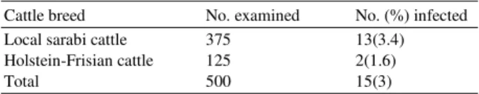
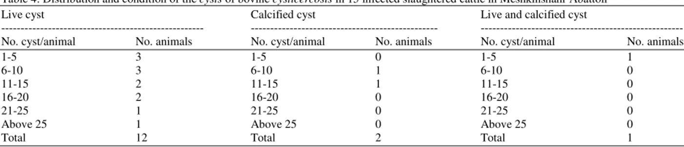
15 animals were positive for bovine cysticercosis. The prevalence of bovine cysticercosis was 3%. The prevalence between Holstein-Frisian cattle breeds and local sarabi cattle breeds were described (Table 1). The sex distribution of T. saginata cysticercosis infection of cattle was described (Table 2). Of the 500 cattle examined, 92% were adult males. Although more males than females were examined, the prevalence of infection did not show any significant difference (p>0.05). On the predilection sites of the cysts 36.6, 35.6, 34.9 and 33.4%, of the cysts were collected from the masseter muscles, tongue, heart muscles and triceps muscles respectively. Infection was also recorded from the liver (2.7%), intercostal musceles (1.4%), spleen (0.9%) and intestinal mucosa (0.1%). Bovine cysticercosis was not found in the lungs and kidney (Table 3). The distribution and condition of bovine cysticercosis in slaughtered cattle in Meshkinshahr Abattoir, North-west of Iran was described (Table 4). The cysts of bovine cysticercosis found in the spleen, intercostal muscles and intestinal mucosa were all live while those in the liver 4 were live and 19 were dead. Both live and dead cysts of bovine cysticercosis were found also in other organs inspected.
Table 1: The prevalence of T. saginata cysticercosis infection of cattle in Meshkinshahr Abattoir
Cattle breed No. examined No. (%) infected
Local sarabi cattle 375 13(3.4)
Holstein-Frisian cattle 125 2(1.6)
Total 500 15(3)
Table 2: Sex distribution of bovine cysticercosis infection of cattle in Meshkinshahr Abattoir
Sex No. examined No. (%) infected
Male 460 14(3)
Female 40 1(2.5)
Total 500 15(3)
p>0.05
Table 3: Predilection sites of bovine cysticercosis cysts in slaughtered cattle in Meshkinshahr Abattoir
Predilection sites No (%) infected
Masseter muscles 102(36.6)
Tongue 92(35.6)
Heart muscles 83(34.9)
Triceps 79(33.4)
Thigh muscles 61(11.9)
Diaphragm 54(11.2)
Liver 23(2.7)
Intercostals muscles 12(1.4)
Spleen 8(0.9)
American J. Animal & Vet. Sci., 6 (3): 121-124, 2011
123
Table 4: Distribution and condition of the cysts of bovine cysticercosis in 15 infected slaughtered cattle in Meshkinshahr Abattoir
Live cyst Calcified cyst Live and calcified cyst
--- --- ---
No. cyst/animal No. animals No. cyst/animal No. animals No. cyst/animal No. animals
1-5 3 1-5 0 1-5 1
6-10 3 6-10 1 6-10 0
11-15 2 11-15 1 11-15 0
16-20 2 16-20 0 16-20 0
21-25 1 21-25 0 21-25 0
Above 25 1 Above 25 0 Above 25 0
Total 12 Total 2 Total 1
DISCUSSION
During the present study period, we found that 3% of cattle presented for slaughter at Meshkinshahr Abattoir, North-west of Iran harbored bovine cysticercosis. Cysticerci are easily missed, as they may not be present on routine cuts considering that most cases of cysticercosis are light infections. Moreover, observations indicated that except for the dead, degenerate or calcified cysticerci that usually form white and fibrotic lesions a careless meat inspector could most likely miss out quite a number of viable cysticerci, which blend the pinkish-red colour of the meat and be passed on for human consumption. Differences in the skills and motivation of meat inspectors, the speed of the slaughter activity, and the meat inspection facilities, are among the many other contributory factors. Our observations showed that the masseter muscles, tongue, heart muscles, triceps muscles and thigh muscles among others were the preferred organs (predilection sites) for the cysts of bovine cysticercosis similar to earlier reports in various endemic areas (Zahedi, 2009; Munyeme et al., 2010; Pawlowski and Schultz, 1972; Opara et al., 2006) DOA. It appears that several factors, such as activity of the muscles, age, and the geographical area concerned determine largely the predilection sites in slaughtered cattle (Minozzo et al., 2002; Opara et al., 2006; Opara et al. 2006). It was observed that some live cysts were isolated from the intercostal muscles, spleen, liver, intestinal mucosa but not from the kidneys and lungs. Sexrelated distribution of bovine cysticercosis infection of the slaughtered cattle in this study showed that sex of the animals and infection were independent. Though more males than females were infected, it could be related to the sample size and not sex. Since the animals slaughtered were all adults it was not possible to compare the relationship of prevalence with the ages of the animals. Breed related distribution of bovine cysticercosis indicated higher prevalence (3.4%) in local sarabi cattle breeds than Holstein-Frisian cattle (1.6%). This is may be associated with management system where Holstein-Frisian cattle breeds were from
dairy farms having less exposure to contaminated pasture with human excreta while the local sarabi cattle breeds were from fattening or extensive management system. In Meshkinshahr, the habit of eating raw beef dishes and backyard slaughter might have contributed for the high prevalence of bovine cysticercosis in cattle. Therefore, to reduce the transmission of taeniasis/ bovine cysticercosis, public education to avoid consumption of raw meat, and use of latrines and improved standards of human hygiene were recommended.
CONCLUSION
The animals slaughtered were all adults. No significant difference in prevalence rates was recorded between the sexes. The prevalence of bovine cysticercosis was higher in local sarabi cattle breeds than Holstein-Frisian cattle.
ACKNOWLEDGMENT
The researchers wish to thanks the Islamic Azad University, Tabriz Branch and Tabriz, Iran for the financial supports, and all laboratory technicians for technical aids in this project.
REFERENCES
Belino, E.D., 1975. Some observations on Taenia saginatacysticercosis in slaughter cattle in Nigeria. Int. J. Zoonoses, 2: 92-99. PMID: 1223011 Cabaret, J., S. Geerts, M. Madeline, C. Ballandonne and
D. Barbier, 2002. The use of urban sewage sludge on pastures: The cysticercosis threat. Vet. Res., 33: 575-597 DOI: 10.1051/vetres:2002040
Carpio, A., 2002. Neurocysticercosis: An update. Lancet Infect Dis., 2: 751-762. DOI: 10.1016/S1473-3099(02)00454-1
American J. Animal & Vet. Sci., 6 (3): 121-124, 2011
124 Gracey, F.J., D.S. Collins and R.J. Huey, 1999. Meat
Hygiene. 10th Edn., Elsevier Health Sciences, London, ISBN: 0702022586, pp: 758.
Lees, W., J. Nightingale, D. Brown, B. Scandrett and A. Gajadhar, 2002. Outbreak of Cysticercus bovis (Taenia saginata) in feedlot cattle in Alberta. Can. Vet. J., 43: 227-228. PMID: 11901599
Minozzo, J.C., R.L.F. Gusso, E.A. De Castro, O. Lago and V.T. Soccoi, 2002. Experimental bovine infection with Taenia saginata Eggs: Recovery rates and cysticerci location. Braz. Arch. Biol. Technol, 45. DOI: 10.1590/S1516-89132002000600008
Munyeme, M., H.M. Munangandu, J.B. Muma, A.M. Nambota and D. Biffa et al., 2010. Investigating effects of parasite infection on body condition of the Kafue lechwe (Kobus leche kafuensis) in the Kafue basin. BMC Res. Notes, 3: 346-346. DOI: 10.1186/1756-0500-3-346
Neva, F.A. and W.H. Brown, 1994. Basic Clinical Parasitology. 6th Edn., Appleton and Lange, Norwalk, Conn., ISBN: 9780838506240, pp: 356. Opara, M.N., U.M. Ukpong, I.C. Okoli and J.C.
Anosike, 2006. Cysticercosis of Slaughter Cattle in Southeastern Nigeria. Ann. N.Y. Acad. Sci., 1081: 339-346. DOI: 10.1196/ annals.1373.048
Pawlowski, Z. and M.G. Schultz, 1972. Taeniasis and cysticercosis (Taenia saginata). Adv. Parasitol., 10: 269-273. DOI: 10.1016/S0065-308X(08)60176-1
Phiri, I.K., H. Ngowi, S. Afonso, E. Matenga and M. Boa et al., 2003. The emergence of Taenia solium cysticercosis in Eastern and Southern Africa as a serious agricultural problem and public health risk. Acta. Trop., 87: 13-23. DOI: 10.1016/S0001-706X(03)00051-2
Smith, E.A. and R.M. Corn, 2003. Surface plasmon resonance imaging as a tool to monitor biomolecular interactions in an array based format. Applied Spectrosc., 57: 320A-332A. PMID: 14658142
Solusby, E.J.W., 1982. Helmiths, Arthropods and Protozoa of Domestic Animals. 7th Edn., Lea and Febiger, London, Philadelphia, ISBN: 9780812107807, pp: 809.
Zahedi, M.J., S.D. Moghadam, M.A. Mosavi, T. Mirshekari and M. Hayatbakhsh, 2009. Helicobacter pylori colonization in biopsies of the adenotonsillectomy specimens. Am. J. Applied
Sci., 6: 2050-2053. DOI: