___________________________
Corresponding author: Miljkovi Danijela, Department of Evolutionary Biology,Institute for Biological Research, “S. Stankovi “University of Belgrade,Despota Stefana 142,11000 Belgrade, Serbia,phone: +381 11 207 83 76, Fax: +381 11 761 433,Mobtel.:+381 64 24 17 228,e-mail: [email protected]
UDC 575 DOI: 10.2298/GENSR1302297M
Original scientific paper
BETWEEN-CLONE, BETWEEN-LEAF AND WITHIN-LEAF VARIATION IN LEAF EPIDERMIS TRAITS IN Iris pumila CLONES
Danijela MILJKOVI 1*, Stevan AVRAMOV 1, Vukica VUJI 2, Luka RUBINJONI 2, Nataša KLISARI BARISI 1, Uroš ŽIVKOVI 1, Aleksej TARASJEV 1
1Institute for Biological Research “S. Stankovi “,University of Belgrade, Belgrade, Serbia 2 Faculty of Biology, University of Belgrade, Belgrade, Serbia
Miljkovi D., S. Avramov, V. Vuji , L. Rubinjoni, N. Klisari Barisi , U. Živkovi and A. Tarasjev (2013): Between-clone, between-leaf and within-leaf variation in leaf epidermis traits in Iris pumila clones.Genetika, Vol 45, No. 2, 297-308.
The goal of this study was to analyze variation and covariation in epidermal characteristics (epidermal cell density ECD, stomata density SD, and stomata index -SI) on Iris pumila clones on between-clone, between-leaf and within-leaf levels. ECD (similar to the pattern previously observed for SD) increased from the base to the top of leaf, while SI remained constant. Results of profile analyses indicated that clones, individual plants whitin clones (ramets), and three successive leaves on the same plant were not significantly different for examined characteristics, but genetic variation for position effect was detected (significant Zone x clone interaction). Results of the contrast analysis confirmed differences between the base and middle leaf positions for ECD (similar to those for SD) as well as between clone variation for those differences. Observed differences between leaf zones and correlations between analyzed traits were mostly consistent with the expansion hypothesis of stomata differentiation.
Key words:epidermal traits, stomatal density, epidermal cell density, stomata index, Iris pumila.
INTRODUCTION
and CO2 levels). Stomata are the most important control system of CO2 influx and water vapor
efflux, and as such are the principal leaf epidermal structure for the survival of land plants (NADEAU and SACK, 2002). Investigation of the pattern of stomata distribution, from a whole plant perspective, is very useful because both the epidermal cell number and stomata number present a response to the plant/environment interaction (GALMÉS et al.2007; AVRAMOV et al. 2007; XU and ZHOU; 2008. LORANGER and SHIPLEY, 2010; TARASJEVet al. 2012).
The pattern of stomata density (of the aerial organs including leaves, stems, flowers, fruits and seeds) is organ and species specific, and depends on the phase of organ’s development (young organs have fewer total stomata than mature organs). The frequency and positioning of stomata is also affected by environmental factors (VATÉN and BERGMANN, 2011). Stomata patterns within and between leaves influence plant productivity and water use efficiency (WEYERS and LAWSON 1997). As the plant grows, the epidermis expands by cell division and cell elongation. Stoma pattering presents a result of interaction between plant and the environment, that modified and regulated the stomata density during leaf development (GEISLER and SACK, 2002; POMPELLIet al.2010). Specific epidermal cells differentiate into stomata guard cells, and their correct distribution on leaf surface is important for leaf physiological function in environmental conditions (BROWNLEE, 2000). SD often decreases as neighboring epidermal cells expand during maturation (VATÉN and BERGMANN, 2011).
Large differences in stomata characteristics were found among species and cultivars (FERRISet al. 2002; LORANGER and SHIPLEY, 2010), as well as for other factors such as light intensity (TUCI et al. 1999; CAO and BOOTH, 2001; AVRAMOVet al. 2007), nutrient availability (CROXDALE, 2000), or interpopulation variability (BATOSet al. 2010).
Monocots and dicots take different paths of stomata development. Numerous studies were published in recent years, with intent to define development proces of stomata, their ontogeny and structure in monocots (ABUBAKAR et al. 2011 and references therein). In monocotyledon leaves with polarized growth, stomata are arranged in rows along the main leaf axis and they are characterized by a temporal and spatial gradient. The linear proces of growth (from the "division and expansion zone" in the base, to the middle and top of leaf as the "elongation zone") is characterized for monocot leaves (VAN VOLKENBURGH et al.1998; PEMAC and AVRAMOV, 2001). The dissipated distribution of stomata is characterized for dicot leaf type (CROXDALE, 2000; GEISLER and SACK 2002).
Stomata patterning has been studied in monocotyledons (CHINet al. 1995; CROXDALE, 2000) and, in most cases, the formation of stomata on any mature leaf reflects the contribution of cell divisions and expansion to leaf growth (KOUWENBERG et al. 2004). The parallel rows of stomata and epidermal cells that are located between the veins are typical for the Iris pumila monocotyledon type leaf.
The ratio between stomata and epidermal cell density, stomata index (SI) is a sensitive parameter that quantifies the effects of epidermal cell expansion due to contrasting light conditions, leaf age, and other environmental conditions (POOLE and KURSCHNER, 1999). Possible source of variation of stomata pattern between individual plants and individual leaves is the different time and mode of leaf growth of monocotyledonous and dicotyledonous leaf types (CROXDALE, 2000). The complexity of this problem indicates that a large number of stratified samples is required to obtain a truly representative value (WEYERS and LAWSON,1997).
et al 2011). The factors that are responsible for stomata differentiation are not completely investigated, but observed patterns can be result of differences in differentiation itself, result of irregular epidermal cell expansion, or result of both (the differentiation, expansion and mixed hypotheses (BEERLING and CHALONER ,1993).
In this work we studied variability and covariation of epidermal traits on Iris pumila, monocot clonal plant that was previously extensively used in genetic, ecological and evolutionary studies (TARASJEVet al. 2012 and references therein). The goal of this preliminary study was to:
- Examine is there significant between-leaf variation (between three successive leaves) , and within-leaf variation (between the three sampling zones -base, middle and top of the leaf) for analyzed epidermal characteristics.
- Examine is there significant variation between clones in analyzed epidermal characteristics, in order to check for genetic variability of analyzed traits
- Estimate significance and sign of the correlations between stomata density, epidermal cell density and stomata index, in order to test the hypothesis of stomata differences development over the leaf surface of Iris pumila.
MATERIALS AND METHODS
Iris pumila is a rhizomatous perennial herb and is very abundant in the dune system at Deliblato Sands, a situated about 50-km northeast of Belgrade (44o 48' N, 20o 58' E). Three
successive last fully expanded leaves were examined, from three ramets of the same clone. The first leaf is the youngest and the third is the oldest (the period of new leaf appearance is about five to seven days). Stomata frequency could be biased due to the counting of fields overlapping vein areas (which had a low number or zero stoma) (LARKIN et al. 1997). Total leaf stomata census is inpractical to count, which is why an optimal sampling strategy must be established. The number of readings per area can be estimated using objective statistical criteria. In this experiment, samples were taken from three zones on every leaf: the base (first cm of the second quarter of leaf); the middle (first cm of the third quarter of leaf), and the top (first cm of fourth quarter of leaf). To obtain the impression of leaf surface replicas of harvested leaf material were made, using clear nail polish applied to the leaf surface. The clear nail polish was painted across the selected leaf part, with a 0.5cm-wide band.
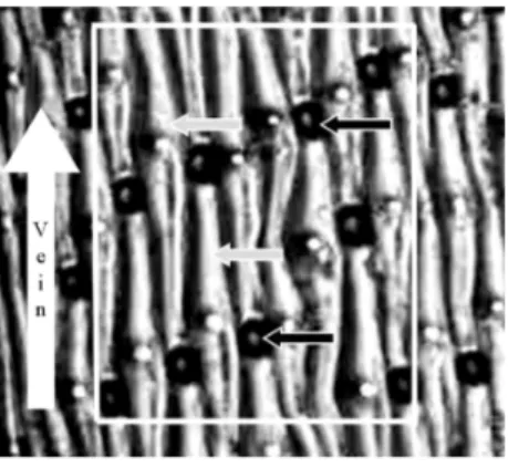
The dry film of polish was pulled from the leaf by a piece of adhesive tape and mounted on a microscope slide. With this replica technique stomata and epidermal cells were visible and easy for counting (Figure 1). Stomata density (SD) and epidermal cell density (ECD) were sampled in 20 randomly chosen microscope fields at each of three zones on individual leaf. Microscope measurements of these preparations were made in a band across the leaf in the middle of a segment, using the Olympus "Vanox" microscope (magnification of 6.7 x 10). Number of stomata and epidermal cells on a given zone of the leaf were counted by frames of dimension 700m x 436m (area of 0.327mm2) (software UTHSCSA Image Tool 3.00).
Methodology of cell count was previously given in PEMAC and AVRAMOV (2001).
Stomata index (SI) was calculated according to the formula of SALISBURY 1927 (after CHENet al. 2001; ABUBAKARet al. 2011)
SI= (SD/ECD+SD) x100
Figure 1. Images of the abaxial surface of Iris pumila leaf showing epidermal cells (grey arrows) and stomata (dark grey arrows) and vein (white arrow). White square presents the sampling area for the counting of stomata.
Scheffe's test was used to assess differences between the analyzed epidermal traits for different zones on leaf. Traits from different zones could be treated as a same trait that was repeated thought the time of each leaf development. According to this assumption we analyzed data using multivariate repeated-measures analysis (REPEATED option in SAS procedure). The samples are referred to as the subjects and repeated observations (zones: base, middle, and top) on each leaf as the “within-subject” factor, by repeated terminology (VON ENDE, 1993), while the individual clone was designated as the “between-subject” factor. The pattern of response of the within-subject factor zones (base, middle, top) was analyzed using the profile analysis, which transforms the within-subject repeated observations to a set of differences (contrasts of first and second order), and then makes the univariate analysis on the contrasts.
Linear correlation coefficients between leaf characteristics were calculated for every zone on each of the three successive leaves (PROC CORR). The correlations between SD, ECD and SI may be used to test the hypotheses on the mechanisms of contributing to spatial variation in epidermal characteristics. Allstatistical analyses were performedusing SAS9.1 (SAS, Cary, NC).
RESULTS
Statistically significant differences in epidermal traits between three successive leaves in the three zones along the leaf have not been obtained (Table 1), and therefore the leaf mean values for mean stomata density, epidermal cell density and stomata index are presented (Figure 2). The stomata density (SD) had a range from 61.23 (#/mm2) for the base to the 94.57 (#/mm2)
to the top. ECD had a same pattern of gradients for all three successive leaves: 145.45 (#/mm2)
for the base to the 216.40 (#/mm2) to the top. Stomata index (SI) was not significantly different
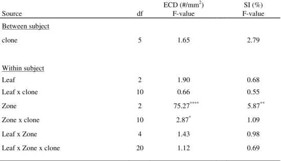
Table 1. Results of profile analysis (MANOVA) epidermal cell density (ECD) and stomatal index (SI) observed across three succesive leaves and three adjacent positions on each of Iris pumila leaves.
ECD (#/mm2) SI (%)
Source df F-value F-value
Between subject
clone 5 1.65 2.79
Within subject
Leaf 2 1.90 0.68
Leaf x clone 10 0.66 0.55
Zone 2 75.27**** 5.87**
Zone x clone 10 2.87* 1.09
Leaf x Zone 4 1.43 0.98
Leaf x Zone x clone 20 1.12 0.69
* P<0.05, ** P<0.01, *** P<0.001, **** P<0.0001
According the REPEATED profile analysis (Table 1), between-subject source variation was not significantly different among clones for all of the examined traits (SD - PEMAC and AVRAMOV 2001, ECD and SI (Table 1)). For within-subject factor, zones, statistically significant difference was observed for all three traits (p < 0.05). Different zones on the same leaf vary among clones, according the observed statistically significant zones and clone interaction for SD (PEMAC and AVRAMOV, 2001) and ECD (p < 0.05) (Table 1).
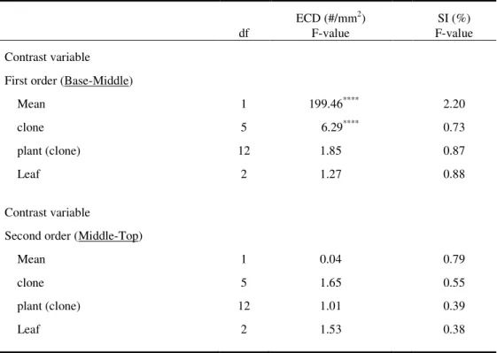
Table 2. Individual ANOVAs on each of the contrasts of within-subject factor between three adjacent zones on each leaf first order (Base-Middle) and second order (Middle-Top).
ECD (#/mm2) SI (%)
df F-value F-value
Contrast variable
First order (Base-Middle)
Mean 1 199.46**** 2.20
clone 5 6.29**** 0.73
plant (clone) 12 1.85 0.87
Leaf 2 1.27 0.88
Contrast variable
Second order (Middle-Top)
Mean 1 0.04 0.79
clone 5 1.65 0.55
plant (clone) 12 1.01 0.39
Leaf 2 1.53 0.38
* P<0.05, ** P<0.01, *** P<0.001, **** P<0.0001
Individual Repeated ANOVA was computed on each of the contrast variables: first order (base-middle) and second order (middle-top) contrast variable (Table 2). Mean effects were statistically significant for SD and ECD, indicating that these two traits display a gradient along the leaf for base-middle contrast. A significant clone effect was observed for the contrast variable of first order (Base-Middle) for SD and ECD. This result suggests that the zone patterns of SD and ECD were clone specific, at the leaf part between base and middle zone (SD – (PEMAC and AVRAMOV,2001), ECD, and SI (Table 2)).
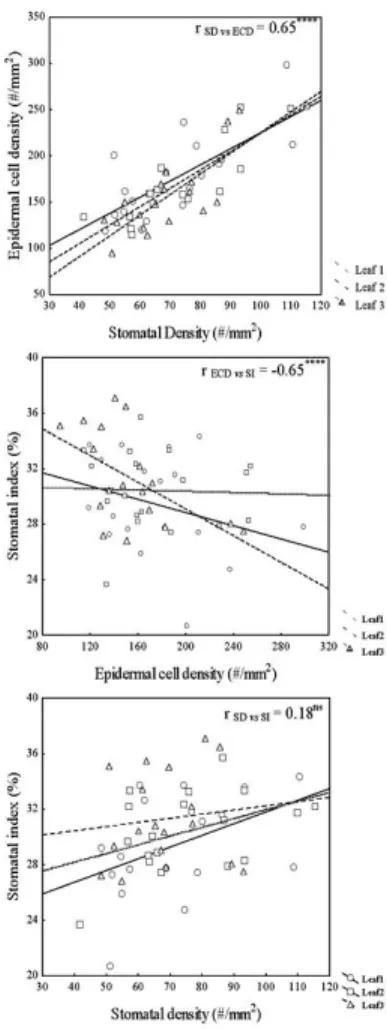
A significant positive linear relationship was found between SD and ED (r SD vs. ECD =
p<0.0001) (Figure 3). In both cases lines produced for different leaves were parallel, e.g relationship between traits was similar in successive leaves.
Figure 3. Correlation analysis among epidermal traits for three successive leaves of Iris pumila. Scatter
plots showing the relation between epidermal cells, stomatal density and stomatal index. The trandlines and values of Pearson’s coefficients for all three comparations of epidermal traits were also presented (nsP > 0.05, ****P < 0.0001). Individual values of epidermal traits circles present data for first leaf, squares for second and triangles for third leaf.
DISCUSSION
After stomata initiation in complete, leaf expansion could still be modified by a change in light level. The Stomata density is a trait that can change after exposure to new light conditions, and SD modifications were observed after the light regime change (GAY and HURD, 1975, TUCI et al. 2003). Differences between the three successive leaves were tested in this study because first leaf was the youngest (relatively to the second and third leaf), and we did not detect differences in epidermal traits patterns between three successively developed leaves (Table 1). In some species, differences between cultivars, clones and genotypes in stomata parameters were observed (WILLMER and FRICKER 1996; MISHRA, 1997, PEK EN et al. 2006). In our study there was no difference between clones for analyzed epidermal traits (no significant clone effect in Table 1) but we detected significant Zone x clone interactions for SD and ECD (Table 1. and PEMAC and AVRAMOV, 2001) that was consequence of genetic difference in differentiation between base and the middle part of I.pumila leaves (significant Clone effect - Table 2). Therefore, genetic aspects should be taken into account in further analysis of epidermal traits variation in I. pumila.
Stomata density is often not found to be constant over the whole leaf surface (GITZ and BAKER, 2009). Leaf elongation in monocots is a linear process and new cells were produced in a basal meristem so leaf size can vary with cell length, cell number, or both (VAN VOLKENBURGH et al. 1998). Cells are displaced away from the base, in parallel longitudinal cell files, by the continuous production and elongation of new cells in this zone. After leaving the 'division and expansion zone' these cells go through the 'elongation zone' (SCHNYDER et al. 1990). In Iris pumila leaves, stomata were denser on top of the leaf (PEMAC and AVRAMOV,2001) and this study showed the same pattern for epidermal cells, too (Figure 2, Tables 1 and 2).
The factors that are responsible for stomata differentiation are not completely investigated (BEERLING and CHALONER, 1993) but three possible explanations for stomata differences over leaf surfaces are given:
1. Stomata differentiation was uneven over the leaf surface, resulting in different densities of stomata, or epidermal cells, or both (the differentiation hypotheses).
2. The leaf undertakes irregular expansion after the cells had differentiated, resulting in uneven spacing of the stomata (the expansion hypothesis).
3. Spatial variation in both leaf expansion and stomata differentiation contributed to the uneven spacing of stomata (the mixed differentiation and expansion hypothesis).
comparisons by Scheffe’s test (Figure 2) indicate that there is possibility that differentiation also has some role in observed patterns.
Stomata end epidermal frequency is correlated while Because of the observed within– leaf variation in epidermal traits, for future examination of epidermal traits in Iris pumila (and especially for their analysis as means of plant adaptation to different environmental conditions) we suggest that it would better to take measurements on two different positions of the Iris pumila leaf (middle and top).
ACKNOWLEDGEMENT
This work was supported by Ministry of Education, Science and Tehnological Development, No. OI 173025, Government of the Republic of Serbia.
Received September 9 th 2012
Accepted April 05th, 2013
REFERENCES
ABUBAKAR, B. Y., O. T. MUSTAPHA, D. N. IORTSUUN (2011): Variability of epidermal structure and stomatal ontogeny in some species of Asparagaceae from Falgore Game Reserve. New Clues in Sciences 1: 80-83.
AVRAMOV, S., D. PEMAC, B. TUCI (2007): Phenotypic plasticity in response to an irradiance gradient in Iris pumila: adaptive value and evolutionary constraints. Plant Ecol. 190 (2): 275-290.
BATOS, B., D. VILOTI, S. ORLOVI , D. MILJKOVI (2010): Inter and intra-population variation of leaf stomatal traits of Quercus robur L. in Northern Serbia.Arch. Biol. Sci. 62(4): 1125-1136.
BEERLING, D. J.and W. G. CHALONER (1993): Stomatal density responses of Egyptian Olea europaea L. leaves to CO2 changes since 1327 BC. Ann. Bot. 71: 431-435.
BOCCALANDRO, H. E., M. L. RUGNONE, J. E. MORENO, E. L. PLOSCHUK, L. SERNA, J. M. YANOVSKY, J. JORGE (2009): Phytochrome B enhances photosynthesis at the expense of water-use efficiency in Arabidopsis. Plant. Physiol. 150: 1083–1092.
BROWNLEE, C. (2000): Plant development: Keeping your distance. Current Biology 10: R555–R557.
CAO, K. and E.W. BOOTH (2001): Leaf anatomical structure and photosynthetic induction for seedlings of five dipterocarp species under contrasting light conditions in a Bornean heath forest. J. Trop. Ecol. 17: 163-175.
CHEN, L. Q., C. S. LI, W. G. CHALONER, D. J. BEERLING, Q. G. SUN, M. E. COLLINSON, P. L. MITCHELL (2001): Assessing the potential for the stomatal characters of extant and fossil Ginko leaves to signal atmospheric CO2 change. Am. J. Bot. 88(7): 1309-1315.
CHIN, J., Y. WAN, J. SMITH, J. CROXDALE (1995): Linear aggregations of stomata and epidermal cells in Tradescantia leaves: evidence for their group patterning as a function of the cell cycle. Dev Biol. 168: 39–46.
CROXDALE, J. L. (2000): Stomatal patterning in angiosperms. Am. J. Bot. 87(8): 1069–1080.
FERRIS, R., L. LONG, S. M. BUNN, K. M. ROBINSON, H. D. BRADSHAW, A. M. RAE, G. TAYLOR (2002): Leaf stomatal and epidermal cell development: identification of putative quantitative trait loci in relation to elevated carbon dioxide concentration in poplar. Tree Physiol. 22: 633–640.
GALMÉS, J., J. FLEXAS, R. SAVÉ, H. MEDRANO (2007): Water relations and stomatal characteristics of Mediterranean plants with different growth forms and leaf habits: responses to water stress and recovery. Plant and Soil 290: 139– 155.
GAY, A. P. and R. G. HURD (1975): The influence of light on stomatal density in the tomato. New Phytol. 75: 37-46. GEISLER, M. J. and F. D. SACK (2002): Variable timing of developmental progression in the stomatal pathway in
GITZ, D. C. and J. T. BAKER (2009): Methods for creating stomatal impressions directly onto archivable slides. Agron. J. 101: 232-236.
KOUWENBERG, L. R., W. M. KURSCHNER, H. VISSCHER (2004): Changes in stomatal frequency and size during elongation of Tsuga heterophylla needles. Ann. Bot. 94: 561–569.
LARKIN, J. C., M. D. MARKS, J. NADEAU, F. SACK (1997): Epidermal Cell Fate and Patterning in Leaves. The Plant Cell 9: 1109-1 120.
LORANGER, J. and B. SHIPLEY (2010): Interspecific covariation between stomatal density and other functional leaf traits in a local flora. Botany 88: 30-38.
MISHRA, M. K. (1997): Stomatal characteristics in different ploidy levels in Coffea L. Ann. Bot. 80: 689-692.
NADEAU, J. A., and F. D. SACK (2002): Control of stomatal distribution on the Arabidopsis leaf surface. Science 296: 1697-1700.
PEKSEN, E., A. PEKSEN, C. ARTIK (2006): Comparison of leaf and stomatal characteristics in faba bean (Vicia faba L.). J. Biol. Sci. 6: 360-364.
PEMAC, D. and S. AVRAMOV (2001): Method for estimating stomatal density in grass-type leaves: an example using Iris
pumila. Arch. Biol. Sci. 53(1-2): 7P-8P.
POMPELLI, M. F., S. C. V. MARTINS, E. F. CELIN, M. C. VENTRELLA, F. M. DAMATTA (2010): What is the influence of ordinary epidermal cells and stomata on the leaf plasticity of coffee plants grown under full-sun and shady conditions? Braz. J. Biol. 70(4): 1083-1088.
POOLE, I., J. D. B. WEYERS, T. LAWSON, J. A. RAVEN (2000): Effect of elevated CO2 on the stomatal distribution and leaf physiology. New Phytol. 145(3): 511-52.
POOLE, I. and W. M. KÜRSCHNER (1999): Stomatal density and stomatal index: the praxis. In Jones T. P. & Rowe N. P. (eds.): Fossil plants and spores: modern techniques. Geological Society, Special Publications, London: 257-260.
ROYER, D. L. (2001): Stomatal density and stomatal index as indicators of paleoatmospheric CO2 concentration. Rev. Palaeobot. Palynol. 114: 1-28.
SALISBURY, E. J. (1927): On the causes and ecological significance of stomatal frequency with special reference to the woodland flora. Philosophical Transactions of the Royal Society B 216: 1-65.
SCHNYDER, H., S. SEO, I. F. RADEMACHER, W. KUHBAUCH (1990): Spatial distribution of growth rates and of epidermal cell lengths in the elongation zone during leaf development in Lolium perenne. L. Planta 181: 423-431.
SKIRYCZ, A., H. CLAEYS, S. DE BODT, A. OIKAWA, S. SHINODA, M. ANDRIANKAJA, K. MALEUX, N. B. ELOY, F. COPPENS, S. D. YOO, K. SAITO, and D. INZÉ (2011): Pause-and-stop: The effects of osmotic stress on cell proliferation during early leaf development in Arabidopsis and a role for ethylene signaling in cell cycle arrest. Plant Cell 23: 1876–1888.
TARASJEV, A., S. AVRAMOV, D. MILJKOVI (2012): Evolutionary biology studies on the Iris pumila clonal plant: Advantages of a good model system, main findings and directions for further research. Arch. Biol. Sci. 64: 159-174.
TAY, A. and A. FURUKAWA (2008): Variations in leaf stomatal density and distribution of 53 vine species in Japan. Plant Spec. Biol. 23: 2–8.
TICHA, I. (1982): Photosynthetic characteristics during ontogenesis of leaves. Stomata density and sizes. Photosynthetica
16(2): 375-471.
TUCI , B., D. PEMAC, B. STOJKOVI , S. AVRAMOV (1999). Coping with environmental changes in Iris pumila: a pilot experiment. Arch. Biol. Sci. 51: 137-148.
TUCI , B., D. PEMAC, S. AVRAMOV (2003): Plasticity to day length of Iris pumila leaf phenological traits. Popul. Ecol. 45: 31–39.
VON ENDE, C. N. (1993): Repeated measures analysis: growth and other time-dependent measures. In S. M. Scheiner and J. Gurevitch (eds.), design and analysis of ecological experiments. Chapman & Hall, New York: 113-137. WEYERS, J. D. B. and T. LAWSON (1997): Heterogeneity in stomata characteristics. Adv. Biol. Res. 26: 317-352. WILD, A. and G. WOLF (1979): The effect of different light intensities on the frequency and size of stomata, the size of
cells, the number, size and chlorophyll content of chloroplasts in the mesophyll and the guard cells during the ontogeny of primary leaves of Sinapis alba. Z. Pflanzenphysiol. 97: 325-342.
WILLMER, C. M.and M. FRICKER (1996): "Stomata", 2nd edn. Chapman and Hall, London.
V A R I R A N J E E P ID E R M A L N IH O S O B I N A L I S T A I r i s p um i l a
K L O N O V A ( I Z M E U K L O N O V A , I Z M E U L I S T O V A I U O K V IR U L IS T A )
Danijela MILJKOVI 1*, StevanAVRAMOV 1, Vukica VUJI 2, Luka RUBINJONI 2,
Nataša KLISARI BARISI 1, Uroš ŽIVKOVI 1, Aleksej TARASJEV1
1
Institut za biološka istraživanja “S. Stankovi “,Univerzitet u Beogradu, Beograd, Srbija, 2 Biološki fakultet, Univerzitet u Beogradu, Beograd, Srbija
Izvod
Cilj ovog istraživanja je analiza variranja i kovariranja epidermalnih osobina lista (gustina epidermalnih elija ECD, gustina stoma SD i stomatalni indeks SI) izme u i unutar lista kao i izme u klonova Iris pumila. Gustina epidermalnih elija (sli no obrascu gustina stoma, ranije publikovano) raste od baze ka vrhu lista, dok vrednost stomatalnog indeksa ostaje konstantna. Rezultati Profile analize (MANOVA) ukazuju da se klonovi, individualne biljke svakog klona (rameta), kao i tri sukcesivna lista na svakoj od biljaka ne razlikuju za ispitivane karakteristike epidermisa lista (epidermalna gustina lista i stomatalni indeks). Me utim, variranje unutar lista na razli itim pozicijama lista (baza, sredina i vrh lista), uslovljeno razlikama genotipa (klonovima) je potvr eno statisti ki zna ajnom interakcijom zona (pozicija na listu) x klon (P < 0.05). Razlike izme u baze i sredine lista za gustinu epidermalnih elija (F = 199.46, P < 0.0001), kao i zna ajna razlika izme u klonova izme u pomenutih pozicija je prema rezultatima analize kontrasta statisti ki zna ajna (F = 6.29, P < 0.001). Dobijene razlike izme u pozicija na listu i korelacija izme u analiziranih epidermalnih osobina lista I.pumila klonova su potvrda hipoteze širenja epidermalnih elija prilikom diferencijacije stoma unutar lista.
Primljeno 09. IX 2012.