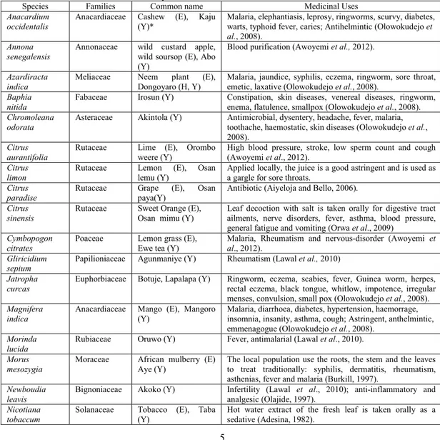
IDENTIFICATION AND AUTHENTICATION OF DRY SAMPLES OF SOME MEDICINAL PLANTS USING LEAF EPIDERMAL FEATURES AS MARKER
Texto
Imagem

Documentos relacionados
Apesar dos resultados não terem sido consistentes para todas as variáveis analisadas ocorreu uma tendência de me- lhora da saúde, principalmente no que diz respeito à melhoria
of time on culture medium and substrates during the acclimatization of fig tree plantlets produced in vitro , characterizing some leaf anatomy aspects of plantlets cultured in vitro
Pearson correlation between canopy temperature differences and leaf water potential, transpiration rate, and stomatal resistance of Phaseolus vulgaris plants submitted to
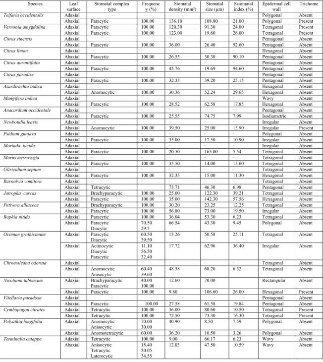
All leaves of these species are hypostomatic, and present: peltate trichomes on both surfaces; stomata sunk in epidermal depressions; small epidermal cells with thick walls
O OpenCV implementa uma func¸˜ao pronta para fazer a fragmentac¸˜ao do blob conforme Ahmadzadeh, por´em n˜ao possui func¸˜ao para determinac¸˜ao da ´area do blob
Rainfall (mm), soil water content (SWC) (%), and average soil temperature (°C) at the depth 0-15 cm in the Caatinga areas of Olho D’Água do Casado (Area I) (A) and Delmiro
Root length, leaf and stem dry weight, leaf weight ratio, stomatal index of the adaxial surface, adaxial epidermis thickness, and spongy parenchyma thickness of plants remained
“As famílias e a instituição de educação pré-escolar são dois contextos sociais que contribuem para a educação da mesma criança; importa, por isso, que haja uma relação
