mobile unit
Alex Murillo Baixas
Relatório do Projecto Final do MIEM Orientador na FEUP: Prof. Jorge Lino – MEC Prof. Carlos Aguiar – FEUP/Design Studio
Faculdade de Engenharia da Universidade do Porto Mestrado Integrado em Engenharia Mecânica
Julho 2009
I would like to dedicate this thesis to my friend at university Joan Llull, you will always be in our hearts.
SUMMARY
The objective of this project is to design a mobile unit for medical assistance around the country in the area of ophthalmology. The final goal is to open the way so that a mini-bus can be constructed.
This project is an application of a global project that is being done in the Design Studio at FEUP, with the goal to construct a new inter-universitary bus which would connect travellers coming from metro or bus to their universities.
The initial design work, including the design of the structural components, was made by Lúcia Fernandes and Carla Rocha from the Universidade de Aveiro.
The aim of this project is to apply the knowledge acquired during the engineering studies in a social way and the main objective of this project is to give more medical attention to the people who don’t have access to. There are a number of possible scenarios:
People who don’t have a hospital close to their home, and thus may only go in case of emergency. These people would not normally go periodically for health check-ups. People who would only go to the witch doctor as they may be the only ones who go regularly to their district. This may sometimes cause more problems instead of solving them due to a lack of hygiene, proper medicines, etc.
People who are unable to leave their home without help due to their health problems, such as disabled people or blind people.
Those who live in little villages and have to wait for more than one month until one ophthalmologist visits the village.
It is also important to mention that alongside checking their eyes and detecting visual disorders the unit should also give information about prevention and hygiene. This mobile unit is designed to check the population’s visual disorders, who may need surgery in hospitals. It could be also worthwhile having materials such as tents and disinfection
equipment so that some field hospitals could be installed, if not in the same day, the coming days so that needy cases could be operated on, in these specified areas.
The interior design of the bus should be designed so that it provides the most important necessities determined and required, flexible and aesthetically pleasing for its potential users. With regards to the outside design, the environment and the climatology of the probable areas where it might have to pass have to be also taken into consideration.
Adding to this, a reciprocal dialogue has to be maintained with doctors, nurses, patients, designers and others in order to have a continual improvement of the final design/product. With that input, the colours, forms, furniture, necessary equipment and other aspects must be analyzed carefully in the design stage of the product.
The next stage in this process is for the prototype to be scrutinised with regard to the main difficulties that such a mobile unit may experience in terms of space, dimensions, vibrations, lightening and others such problems.
PREFACE
Due to the lack of a proper medical coverage, an inefficient public health system combined with the global growing population, it makes sense to support hospitals with mobile units that can help the hospitals in order not to collapse, and give attention to under‐served patient
populations
Every country has its own necessities but the type of assistance needed in occidental countries is different from the necessities of non-occidental countries.
While the assistance programs, mainly in rich countries, can be to just give more comfort to those patients that live far away from the hospital or to comfort those who do not have access to ophthalmologists in their hospital, in poorest countries the type of assistance can be more including of educational programs or even dealing with diseases that hardly have some case in occidental countries.
There’s an increasing industry and business of mobile units trucks, bus etc. These are mainly used as a support unit in hospitals, as itinerant caravans through all over the country, having as stakeholders the state or even a private company.
The transportation of specific medical equipment needed to carry to places where often the roads are not paved and are full of dust, shocks, and mud might need careful study. Not only space but vibrations and electrical power sources are problems that need to be taken into consideration while the design of mobile units are in the process.
ABSTRACT
This project borne from a project that currently is performed for a company in the Design Studio of FEUP and that uses a standard metallic chassis. The goal is to use the same chassis/frame and conceive a mobile unit for the medical sector.
Considering that one of the advisors had already made contact with a doctor that expressed a real interest in the development of a small bus that is able to run around the country to give assistance to population that needs basic medical care in the field of ophthalmology, this project appears as a natural sequence of the current Design Studio work.
To design the global concept of this mobile unit it is fundamental to understand the limitations of the transportation of the medical basic equipment that is fundamental in ophthalmology, in order to conceive the necessary vibration dumping systems and others. It is necessary to design the whole mobile unit and all it interior to accommodate and
transport in safe conditions the adequate equipment, machines, objects and materials that are fundamental for this kind of assistance.
It is intended that the project of this mobile unit gives information and construction solutions that could be used in other mobile units for other types of applications. Concretely the main themes that are studied and analyzed are:
Mobile
Unit
Vibration Analysis Illumination study Interior & Exterior design Feasibility plan Photovoltaic Installation
GRATITUDE
I would like to express my sincere thanks to my advisers at FEUP: Jorge Lino, Carlos Aguiar, and Xavier Carvalho. In addttion I would like to thanks César Vasques, to my family and to my friends Borja Santoro, Fernando Castán and Vitor Ribeiro from Auto-Ribeiro.
1 TABLE OF CONTENTS ... 7 2 LIST OF FIGURES ... 11 3 LIST OF TABLES ... 14 4 INTRODUCTION... 16 4.1 FACTS ABOUT BLINDNESS AND VISUAL IMPAIRMENT ... 16 4.2 MAIN CAUSES OF VISUAL IMPAIRMENT ... 17 4.2.1 Cataract ... 19 4.2.2 Refractive errors ... 21 4.2.3 Childhood blindness ... 22 4.2.4 Low vision ... 23 4.2.5 Trachoma ... 24 4.2.6 Onchocerciasis ... 26 4.2.7 Glaucoma ... 26 4.2.8 Diabetic retinopathy ... 27 4.2.9 Age related macular degeneration ... 28 4.2.10 Corneal opacities ... 28 4.2.11 Genetic eye diseases ... 29
4.3 SUMMARY OF THE VISIT AT S. JOÃO HOSPITAL, OPHTHALMOLOGIC DEPARTMENT: ... 30
5 LITERATURE REVIEW: ... 31 5.1 STATE OF THE ART: ... 31 5.1.1 MMIC Mobile medical: ... 31 5.1.2 La boit Inc. ... 35 5.2 EXAMPLE CASE: ... 37 5.2.1 The Armenian Eye Care Project: ... 37 5.2.2 Aquila (Italy) earthquake: ... 38 5.3 EQUIPMENT AND DISPOSABLE MATERIAL: ... 39 5.3.1 Autorefractometer: ... 40 5.3.2 Tonometer: ... 40
5.3.4 Phoropter: ... 41 5.3.5 Optotype projector: ... 42 5.3.6 Refraction lens analyzer ... 42 5.3.7 Retinoscope: ... 42 5.3.8 Ophthalmoscope:... 43 5.3.9 Three mirror lens or superfield. ... 44 5.3.10 Disposable material: ... 44 5.4 VEHICLE VIBRATIONS ... 44 5.4.1 Introduction: ... 45 5.4.2 Excitation sources: ... 46 5.4.3 Road roughness: ... 47 5.4.4 Vehicle response properties: ... 51 5.4.5 The quarter‐car model: ... 51 5.4.6 Suspension Stiffness: ... 56 5.4.7 Suspension Damping ... 58 5.4.8 Pitch and Bounce motions ... 59 5.5 ILLUMINATION ... 60 5.5.1 Introduction ... 60 5.5.2 Definitions ... 60 5.5.3 Case Study Minibus ... 66 6 EXPERIMENTAL WORK ... 69 6.1 CONSULTATION DISTRIBUTION ... 69 6.2 NECESSITIES AND FUNCTION ANALYSIS ... 70 6.2.1 Necessities ... 70 6.2.2 Functions... 70
6.3 FUNCTIONAL ANALYSIS. FAST ... 71
6.4 RISK ANALYSIS ... 72
6.5.1 Common characteristics ... 76 6.6 DIFFERENT ALTERNATIVES ... 90 6.6.1 Premium: ... 90 6.6.2 Deluxe: ... 91 6.6.3 Avant: ... 91 6.7 RIDE ANALYSIS ... 93 6.7.1 Introduction ... 93 6.7.2 Computations: ... 94 6.8 DIALUX SIMULATION ... 98 6.9 FINANCIAL ANALYSIS ... 99 6.9.1 Expenses: ... 99 6.9.2 Incomes: ... 100 7 ANALYSIS OF THE RESULTS: ... 101 7.1 JUSTIFICATION OF THE SOLUTION: ... 101 7.2 RIDE ANALYSIS: ... 103 7.2.1 Design Parameters: ... 103 7.2.2 Weight distribution: ... 103 7.2.3 Ride and mass frequency analysis ... 105 7.3 SIZING THE PHOTOVOLTAIC SYSTEM: ... 107 7.4 ILLUMINATION ... 109 7.4.1 Equipment selected: ... 109 7.4.2 Results: ... 109 7.5 FINANCIAL ANALYSIS:... 112 7.5.1 Results: ... 112 8 CONCLUSIONS: ... 114 9 WORKS CITED ... 115 10 BIBLIOGRAPHY ... 116 APPENDIX A: ... 117
INVERTER ECOSFERA 1500W CHARACTERISTICS: ... 117
10.1 SOLAR PANELS SUNPOWER 315 CHARACTERISTICS: ... 118
AIR CONDITIONING CARRIER AC 420 I CHARACTERISTICS: ... 119
COMPUTER AND PRINTER CHARACTERISTICS ... 120 DIALUX OUTPUT FILES: ... 122 UGR results: ... 122 Brand and model of the lights: ... 122 Coordenates and ubication of the lights ... 124 APPENDIX B ... 126
MOBILE UNIT SEEN IN FEUP, OWNED BY A OPTICIAN’S SHOP: ... 126
APPENDIX C ... 127
DETAILED FEASIBILITY ANALYSIS: ... 127
“TAXAS MODERADORAS” DESCRIPTION: ... 129
APPENDIX D ... 131
TOYOTA COASTER CHARACTERISTICS: ... 131
APPENDIX E ... 133 OTHER RENDERS ... 134
2
LIST OF FIGURES
Figure 4-1: Global causes of blindness as a proportion of total blindness in 2002. ... 17
Figure 4-2: Blindness prevalence 2002. ... 18
Figure 4-3: The cataract cloude. ... 19
Figure 4-4: Distribution of global blindness by age group. ... 20
Figure 4-5: Ageing trend 1980-2002. Percentage of world population aged 60 years or older. ... 20
Figure 4-6: Forecast of the needed cataract surgeries. ... 21
Figure 4-7: 1.4 million of blind children in the world, three quarters of them situated in the poorest regions of Africa and Asia. ... 22
Figure 4-8:About 124 million people suffer low vision. ... 23
Figure 4-9: Global distribution of active trachoma. Year 2002. ... 24
Figure 4-10: Lack of donors make access to the corneal surgery difficult to reach. ... 28
Figure 5-1: MMIC commercial logotype. ... 31
Figure 5-2: MMIC 16 meter trailer. ... 32
Figure 5-4: 3-D designed MMIC mobile Laboratory/Pharmacy unit ... 33
Figure 5-3: Interior and exterior of an expandable MMIC mobile surgery unit ... 33
Figure 5-5: MMIC Mobile unit equipped with scan. ... 34
Figure 5-6: MMIC Ophthalmology Unit. ... 34
Figure 5-7: Another perspective of the MMIC CT Scan mobile unit. ... 35
Figure 5-8: La Boit is a US company that transforms buses into medical mobile units. ... 35
Figure 5-9 La boit Mobile AIDS Information and laboratory point. ... 36
Figure 5-10: Laboit Children’s information point. ... 36
Figure 5-11: The AECP trailer parked in Armenia. ... 37
Figure 5-12: The ophalmologist examinates one patient with the slit lamp. ... 38
Figure 5-13: The unit arrived in the firsts days. ... 39
Figure 5-14: Different views of the mobile unit. ... 39
Figure 5-15: One of the new autorefractometers models. ... 40
Figure 5-17: The slit lamp is one of the most important required equipment. ... 41
Figure 5-18: New generation phoropter. ... 41
Figure 5-19: The optptype projector is used in combination with the phoropter. ... 42
Figure 5-20: A simply lens analyzer. ... 42
Figure 5-21: Portable Retinoscope. ... 43
Figure 5-22: Direct ophtalmoscope. ... 43
Figure 5-23: Three mirror lens. ... 44
Figure 5-24: The ride dynamic system (3) ... 45
Figure 5-25: Examples of simulated road profiles with superimposed w values: (a) w=1.5 ; (b) w=2 ; (c) w =2.5; (d) w=3; (e) w=3.5. (4) ... 47
Figure 5-26: Examples of longitudinal profiles of in-situ roads. (2) ... 48
Figure 5-27: Obstacles are considered in the cosine form with their random alternation and position on the track. (2) ... 48
Figure 5-28: Comparison of PSDs profile with obstacles (black one) vs. profile without obstacles (grey) for v=90Km/h. (2) ... 49
Figure 5-29: Example of measured random profile with exaggerated obstacle shapes. (2) ... 50
Figure 5-30:PSD of the measured random profile shown in Figure 4-29, with two single and two straight-line approximations. (2) ... 50
Figure 5-31: Undamped natural frequency versus static deflection of a suspension. (1) ... 53
Figure 5-32: The quarter-car model. ... 54
Figure 5-33: Quarter-car response to road, tire/wheel, and body inputs. (1) ... 55
Figure 5-34: Isolation of raod acceleration by the quarter-car model. (1) ... 57
Figure 5-35: On-road acceleration spectra with different spung mass natural frequencies. (1) ... 57
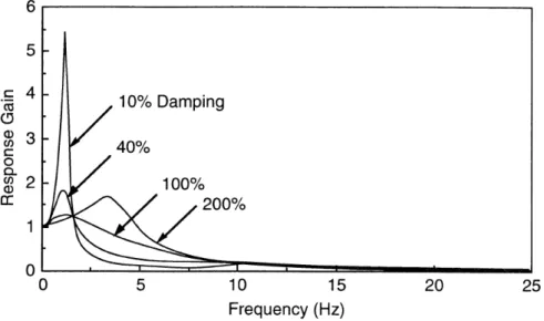
Figure 5-36: The effect of damping on suspension isolation behaviour. (1) ... 58
Figure 5-37: Effect of wheelbase filtering on vertical and longitudinal response gain of a truck. ... 59
Figure 5-38: Spectral colors. ... 61
Figure 5-39: Luminous intensity concept. ... 62
Figure 5-40: The differences between the real and the apparent surface. ... 64
Figure 6-1: Distribution of one box at the department of ophthalmology at Hospital Sao Joao, Porto, Portugal.. 69
Figure 6-2: Diagram of the Functional Analysis System Techinique (FAST). ... 72
Figure 6-4: Cause-effect diagram. ... 74
Figure 6-5: Cause-effect diagram. ... 74
Figure 6-6: Cause-effect diagram. ... 75
Figure 6-7: Cause-effect diagram. ... 75
Figure 6-8: The Carrier AC420 I Air conditioning... 77
Figure 6-9: Table for the ophtalmological exploration. ... 78
Figure 6-10: Bosch KTR14V21. ... 78
Figure 6-11: Genergy Generator... 81
Figure 6-12: Average daily solar radiation (6) ... 82
Figure 6-13: Portugal and Spain map of yearly sum of global irradiation on a horizontal surface. (7) ... 82
Figure 6-14: Project description. ... 83
Figure 6-15: Climate data of Porto,Pedras Rubras, Portugal. ... 84
Figure 6-16: Base case diesel power system (the diesel generator). ... 84
Figure 6-17:Load characteristics, daily electricity demand. ... 85
Figure 6-18: Maximum depth of discharge depending on the battery type. (8) ... 86
Figure 6-19: Inverter and Battery characteristics. ... 86
Figure 6-20: Daily and annual solar radiation horizontal and titled. ... 87
Figure 6-21: Efficency depending on the cell type. (8) ... 88
Figure 6-22: Characteristics of the photovoltaic panels. ... 89
Figure 6-23: Top view of the solar panels. ... 89
Figure 6-24: Green House Gas (GHG) emissions. ... 90
Figure 6-25: Top view of the interior of the Premium model. ... 90
Figure 6-26: Deluxe model distribution, The wardrobes are situated on the right. ... 91
Figure 6-27: Different views of the Avant model. ... 92
Figure 6-28. Oscillations of a vehicle passing over a road bump. (10) ... 93
Figure 6-29: Vehicle Coordiantes axes.(1) ... 96
Figure 7-1: 3D view of the exterior of the mobile unit. ... 101
Figure 7-2: Left side viex of the exterior. ... 102
Figure 7-4: ... 107
Figure 7-5: From left to right, solar panels, battery and the inverter selected for this study. ... 107
Figure 7-6: Solar panels orientation. ... 108
Figure 7-7: Render of the interior with all the lights switched on. ... 110
Figure 7-8: Top view of the consultory illumination effect. ... 111
Figure 7-9: False colors representation of the illuminance levels. ... 111
Figure 7-10: Waiting room false colors representation of illuminace levels and the correspondent values, this values are also valid for Figure 6-9. ... 112
3 LIST OF TABLES
Table 4-1: Estimation of the number or prevalent cases of children with severe visual impairment and blindness due to avoidable causes, by level of socioeconomic development. ... 23Table 5-1: European recomendations for the illuminance of working places and surroundings EN 12464-1. ... 67
Table 5-2: EN-12464-1 Color appearance. ... 67
Table 5-3: EN-12464-1 Levels of Em, UGRL and Ra that ophtalmological office must accomplish. ... 68
Table 6-1: Analysis of Necessities matrix. ... 70
Table 6-2: Description of the functions. ... 71
Table 6-3:Correlation matrix of functions. ... 71
Table 6-4: Total active power calculations. ... 79
Table 6-5: Computations of the active, reactive and apparent power that will be installed in the mobile unit. .... 80
Table 6-6: Masses and gravity centers of all the components of the mobile unit. ... 97
Table 6-7: List of all the weights considered for the computations. (12) ... 98
Table 6-8: Annual costs for the disposable material. ... 100
Table 7-1: Center of gravity of all the components, its distribution between front and rear axle initially (without equipment) and the distribution considering all the components. ... 103
Table 7-3: Results obtained in different study surfaces of the mobile unit. ... 109 Table 7-4: Feasibility analysis for the first five years. ... 112 Table 7-5: : Feasibility analysis for the last five years. ... 113
4 INTRODUCTION
With the purpose of giving the best description and approach at present situation of the visual impairment and blindness all over the world, some data and graphics have been reported so that people with no extended previous knowledge in medicine can have a clearly idea about the most common disease.
The description will explain: what exactly they are, how can be treated, wich are the regions or areas more affected all over the world and finally if they can be treated or prevented with the help of the mobile unit.
Please note that in chapters 1.1 and 1.2, all text and data, pictures and figures are taken from the World Health Organization (1) and (2).
4.1 FACTS ABOUT BLINDNESS AND VISUAL IMPAIRMENT
Worldwide, more than 161 million people are visually impaired; among them, 124 million have low vision and 37 million are blind.
Another 153 million people suffer from visual impairment due to uncorrected
refractive errors (near-sightedness, far-sightedness or astigmatism). Virtually all these people could restore normal vision with eyeglasses or contact lenses.
More than 90% of the world’s visually impaired people live in low- and middle-income countries.
Except in the most developed countries, cataract remains the leading cause of blindness.
Cataract surgery is one of the most cost-effective treatments that can be offered in developing countries. It can allow people to increase their economic productivity by up to 1500% of the cost of the surgery during the first post-operative year.
Age-related causes of visual impairment and blindness are increasing, as is blindness due to uncontrolled diabetes.
The good news is that up to 75% of all blindness in adults is avoidable through prevention or treatment. Worldwide, corneal scarring is the single most important cause of avoidable blindness, followed by cataract and retinopathy of prematurity (ROP).
Infectious causes of blindness are decreasing globally as a result of public health action. The number of people affected by blinding trachoma has decreased from 360 million people in 1985 to approximately 80 million people today.
An estimated 1.4 million children under age 15 are blind. Yet approximately half of all childhood blindness can be avoided by early treatment of disease and correcting abnormalities at birth such as cataract and glaucoma.
4.2 MAIN CAUSES OF VISUAL IMPAIRMENT
Except for the most developed countries, cataract remains the leading cause of blindness in all regions of the world. Associated with ageing, it is even more significant as a cause of low vision.
Glaucoma is the second leading cause of blindness globally as well as in most regions, with age-related macular degeneration (AMD) ranking third on the global scale. However, in developed countries, AMD is the leading cause of blindness, due to the growing number of people over 70 years of age.
Other major causes are trachoma, other corneal opacities, diabetic retinopathy, and eye conditions in children (e.g. cataract, retinopathy of prematurity and vitamin A deficiency).
Cataract, glaucoma, corneal opacity, diabetic retinopathy, onchocerciasis, childhood blindness, trachoma, and some other causes of blindness can potentially all be prevented and/or treated. WHO estimates that, globally, up to 75% of all blindness is avoidable.
However, the proportion of the specific causes of blindness varies considerably from region to region, depending on local circumstance. Only about half the cases of childhood blindness are avoidable.
Some of these diseases, such as trachoma and river blindness, are prevalent primarily in less developed areas of the world where there are also specific environmental hazards.
In many middle income and industrialized countries, three other eye conditions have emerged as potential threats to the status of sight of their populations. The increase of diabetes among many population groups has caused diabetic retinopathy to be added to the priority list, while glaucoma, an eye disease known for centuries, remains on the public health agenda due to difficulties in its early diagnosis and frequent necessity of lifelong treatment. Age-related macular degeneration (AMD) ranks third among the global causes of visual impairment with a blindness prevalence of 8,7%. It is the primary cause of visual deficiency in industrialized countries. An emerging important cause of visual impairment is uncorrected refractive errors.
4.2.1 CATARACT
Definition:
Cataract is clouding of the lens of the eye which impedes the passage of light. Although most cases of cataract are related to the ageing process, occasionally children can be born with the condition, or a cataract may develop after eye injuries, inflammation, and some other eye diseases. Magnitude:
According to the latest assessment, age related cataract is responsible for 48% of world blindness, which represents about 18 million people.
Although cataracts can be surgically removed, in many countries surgical services are inadequate, and cataract remains the leading cause of blindness. As people in the world live longer, the number of people with cataract is growing. Cataract is also an important cause of low vision in both developed and developing countries. Even where surgical services are available, low vision associated with cataract may still be prevalent, as a result of the long period spent waiting for operations and barriers to surgical uptake, such as cost, lack of information, and transportation problems.
Prevention and treatment:
Comprehensive prevention of cataract development is not known yet. Reduction of cigarette smoking, ultraviolet light exposure, and alcohol consumption may prevent or rather delay the development of cataract. Diabetes mellitus, hypertension and high body mass index are identified as additional risk factors.
The treatment of cataract is an operation, which is very successful in restoring sight. The opaque lens is removed and replaced by an artificial intraocular lens. In many remote parts of the developing world, people remain blind from cataract, due to a lack of access to quality eye care at an affordable cost. Among the first 5 most cost-effective health intervention.
Why is cataract increasing so rapidly?
Approximately 85% of all cataract is age-related, the cause of which is unknown. The other 15% are the result of a variety of known causes. By definition, the prevalence of age-related cataract increases with age and, ultimately, everyone in their nineties will be affected. (Figure 9) Cataract also increases with age in developing countries - but often earlier in life and to a greater extent.
Figure 4‐3: The cataract cloude.
Figure 4‐4: Distribution of global blindness by age group.
Between 2000 and 2020, the world’s population is estimated to increase from 6.08 to 7.52 billion. This growth will occur mainly in developing countries.
During the same period, the number of people aged 65 years and older is estimated to increase from 425 million to 677 million globally. In the World Bank regions of China, India, Latin America and the Caribbean, the Middle Eastern Crescent and other Asia and islands, the population aged 65 years and older will double during this period. (Figure 10) While the number of cataract operations is increasing in most countries, this is not enough to compensate for the rise in incidence due to the ‘ageing’ of the population.
Figure 4‐5: Ageing trend 1980‐2002. Percentage of world population aged 60 years or older.
Due to modern surgical techniques with IOL implantation, cataract patients currently undergo operations at a much earlier stage than before. This has led to a threefold to fourfold increase in cataract surgery in a number of industrialized countries. As extra capsular cataract
extraction with IOL implantation also becomes available in more developing countries, this will have huge implications for the number of needed operations.
Figure 4‐6: Forecast of the needed cataract surgeries.
4.2.2 REFRACTIVE ERRORS
Refractive errors can be divided into three major groups:
People over 40 years of age with presbyopia (difficulty in near vision and reading) Myopia
Uncorrected aphakia (eyes with lenses removed and not replaced by IOL or spectacles).
The global magnitude of refractive errors is not reliably known, as there is great variation in groupings according to age, definitions of blindness, and examination methods. Reports suggest that 5-25% of blindness in some countries is caused by refractive errors and as much as 4% of the population sees less than 6/18 (20/60, 0.33) because of this condition. Correction of significant refractive errors requires a well-trained refractionist and access to affordable and good-quality spectacles.
Presbyopia may be easiest to solve through bulk purchase of standard spherical reading spectacles. The costs of such spectacles are low and in most cases full refraction is not required.
Myopia usually develops at the age of 10-15 years. Intervention should focus on screening children in this age group using a simple test for refractive errors. Those who fail the test should be referred for refraction and provided with spectacles. Large-scale screening may increase awareness of refractive errors and motivate parents and grandparents to come forward for testing as well. Increasing demand for spectacles may also promote the development of more optical services.
Uncorrected aphakia is unfortunately still a frequent cause of blindness or low vision. Provision of aphakic glasses is an essential component of cataract intervention.
Training of refractionists and the development of affordable and good quality optical services should be an essential component in most VISION 2020 action plans.
4.2.3 CHILDHOOD BLINDNESS
Definition:
Childhood blindness refers to a group of diseases and conditions occurring in childhood or early adolescence, which, if left untreated, result in blindness or severe visual impairment that are likely to be untreatable later in life. The major causes of blindness in children vary widely from region to region, being largely determined by socioeconomic development, and the availability of primary health care and eye care services. In high-income countries, lesions of the optic nerve and higher visual pathways predominate as the cause of blindness, while corneal scarring from measles,
vitamin A deficiency, use of harmful traditional eye remedies, ophthalmic neonatorum, and rubella cataract are the major causes in low-income countries. Retinopathy of prematurity is an important cause in middle-income countries. Other significant causes in all countries are congenital abnormalities, such as cataract, glaucoma, and hereditary retinal dystrophies Magnitude:
According to Gilbert and Foster, the prevalence of blindness in children varies according to socioeconomic development and under-5 mortality rates. In low-income countries with high under-5 mortality rates, the prevalence may be as high as 1.5 per 1000 children, while in high-income countries with low under-5 mortality rates, the prevalence is around 0.3 per 1000 children. Using this correlation to estimate the prevalence of blindness in children, the number
Figure 4‐7: 1.4 million of blind children in the world, three quarters of them situated in the poorest regions of Africa and Asia.
of blind children in the world is approximately 1.4 million. Approximately three-quarters of the world’s blind children live in the poorest regions of Africa and Asia.
Prevention and treatment:
Prevention and treatment of childhood blindness is disease specific. For Vitamin A
deficiency, at a cost of only 5 US cents a dose, vitamin A supplements reduce child mortality by up to 34% in areas where Vitamin A deficiency is a public health problem. As vitamin A deficiency manifests often during an outbreak of measles, properly planned and implemented national vaccination programs against measles has reduced the prevalence of eye
complications. In middle income countries, retinopathy of prematurity (ROP) is among the leading causes of blindness, the incidence of which can be reduced through availability and affordability of screening and curative services. Early treatment of cataract and glaucoma can be beneficial, while low vision devices are helpful in children with residual vision.
Table 4‐1: Estimation of the number or prevalent cases of children with severe visual impairment and blindness due to avoidable causes, by level of socioeconomic development.
4.2.4 LOW VISION
Definition:
Refractive errors include myopia (short-sightedness) and hyperopia (long-sightedness) with or without astigmatism (when the eye can sharply image a straight line lying only in one meridian).
For low vision, the following two definitions are in use: • (WHO) Low vision is visual acuity less than 6/18 and equal to or better than 3/60 in the better eye with best correction.
• (Low Vision Services or Care) a person with low vision is one who has impairment of visual functioning even after treatment and/or standard refractive correction, and has a visual acuity of less than 6/18 to light perception, or a visual field less than 10 degrees from the point of
Figure 4‐8:About 124 million people suffer low vision.
fixation, but who uses, or is potentially able to use, vision for the planning and/or execution of a task for which vision is essential.
Magnitude:
Recent studies have confirmed the existence of a large burden of uncorrected refractive errors, although the interventions required are significantly cost effective, and have an important impact on economic development and quality of life. Severe refractive errors have been estimated to account for about 5 million blind people. According to the most recent data available to WHO, there are an estimated 124 million people in the world with low vision. About a fourth of these would benefit from low vision services.
Prevention and treatment:
Refractive errors can be rectified with appropriate optical correction while people with low vision may be helped with low vision devices.
4.2.5 TRACHOMA
The disease and how it affects people:
Trachoma is an infection of the eyes that may result in blindness after repeated re-infections. It is the world’s leading cause of preventable blindness and occurs where people live in
overcrowded conditions with limited access to water and health care. Trachoma spreads easily from person to person and is frequently passed from child to child and from child to mother within the family. Infection usually first occurs in childhood but people do not became blind until adulthood. The disease progresses over years as repeated infections cause scarring on the inside of the eyelid, earning it the name of the “quiet disease” The eyelashes eventually turn in. This causes rubbing on the cornea at the front of the eye. The cornea becomes scarred leading to severe vision loss and eventually blindness.
The cause:
Trachoma is caused by an organism called Chlamydia trachomatis. Through the discharge from an infected child’s eyes, trachoma is passed on by hands, on clothing, or by flies that land on the face of the infected child.
Distribution
Trachoma occurs worldwide and most often in poor rural communities in developing
countries. Blinding trachoma is widespread in the Middle East, North and Sub-Sahara Africa, parts of the Indian subcontinent, Southern Asia and China. Pockets of blinding trachoma occur in Latin America, Australia (among native Australians) and the Pacific Islands. Scope of the Problem:
The World Health Organization (WHO) estimates that six million worldwide are blind due to trachoma and more than 150 million people are in need of treatment.
Interventions:
Primary interventions advocated for preventing trachoma infection include improved sanitation, reduction of fly breeding sites and increased facial cleanliness (with clean water) among children at risk of disease. The scaring and visual change for trachoma can be reversed by a simple surgical procedure performed at village level which reverses the inturned
eyelashes.
Good personal and environmental hygiene has been proven to be successful in combating trachoma. Encouraging the washing of children’s faces, improved access to water, and proper disposal of human and animal waste has been shown to decrease the number of trachoma infections in communities.
4.2.6 ONCHOCERCIASIS
Onchocerciasis, or river blindness, is endemic in 30 countries in Africa and also occurs in small foci in six Latin American countries and in Yemen. Currently, there are an estimated 18 million people who are infected with onchocerciasis. Among these, approximately 0.3 million people are already blind from this disease. The disease is expected to be brought under control by the year 2010 if present efforts in endemic countries are successfully completed.
An approach has recently been developed and introduced, whereby community-directed treatment with annual doses of ivermectin would make it possible to eliminate this blinding disease burden from the affected countries in Africa and Latin America. However, the increased numbers of patients needing treatment (many of whom are living in areas of conflict), and the duration of treatment (at least 20 years), are raising concern about whether the problem will be brought under control by 2010.
Over the last 25 years, considerable progress has been made by the Onchocerciasis Control Program (OCP) in 11 countries in West Africa through both vector control and ivermectin distribution. This success, when expressed in health, economic and developmental terms, was the reason behind the launch in December 1995 of a new program, the African Program for Onchocerciasis (APOC) in 19 other African countries.
In Latin America, the Onchocerciasis Elimination Program in the Americas (OEPA) is successfully using ivermectin distribution in six countries in Central and South America. A coordination group of NGOs is working closely with all three onchocerciasis control programs and with national counterparts in virtually all endemic countries.
4.2.7 GLAUCOMA
Definition:
Glaucoma can be regarded as a group of diseases that have as a common end point a
characteristic optic neuropathy which is determined by both structural change and functional deficit. The medical understanding of the nature of glaucoma has changed profoundly in the past few years and a precise comprehensive definition and diagnostic criteria are yet to be finalized. There are several types of glaucoma, however, the two most common are primary open angle glaucoma (POAG), having a slow and insidious onset, and angle closure glaucoma (ACG), which is less common and tends to be more acute.
Magnitude:
The number of persons estimated to be blind as a result of primary glaucoma is 4.5 million, accounting for slightly more than twelve per cent of all global blindness. Risk factors are those limited to the onset of disease and those associated with progressive worsening in already established disease. The primary risk factors that are linked to the individual and the
onset of the disease are age and genetic predisposition. The incidence of POAG rises with age and its progression is more frequent in people of African origin. ACG is the common form of glaucoma in people of Asian origin.
Prevention and treatment:
There is little known about primary prevention of glaucoma; however, there are effective methods of medical and surgical treatment if the disease is diagnosed in its early stage. Through appropriate treatment, sight may be maintained; otherwise the progression of the condition leads eventually to severe restriction of the visual field and irreversible blindness.
4.2.8 DIABETIC RETINOPATHY
Definition
Diabetic retinopathy is composed of a characteristic group of lesions found in the retina of individuals having had diabetes mellitus for several years. The abnormalities that characterize diabetic retinopathy occur in predictable progression with minor variations in the order of their appearance. Diabetic retinopathy is considered to be the result of vascular changes in the retinal circulation. In the early stages vascular occlusion and dilations occur. It progresses into a proliferative retinopathy with the growth of new blood vessels. Macular edema (the
thickening of the central part of the retina) can significantly decrease visual acuity. Magnitude:
There are important differences over the past few decades in diagnosis, medical care, socioeconomic factors and other risk factors that influence the prevalence and geographic distribution of diabetes and retinopathy as well. It is estimated that in 2002 diabetic
retinopathy accounted for about 5% of world blindness, representing almost 5 million blind. As the incidence of diabetes gradually increases, there is the possibility that more individuals will suffer from eye complications which, if not properly managed, may lead to permanent eye damage.
Prevention and treatment:
Risk factors for diabetic retinopathy include duration of diabetes, level of glycemia, presence of high blood pressure, dependence on insulin, pregnancy, levels of selected serum lipids, nutritional and genetic factors. Medical interventions can decrease some of the risk to vision caused by diabetic retinopathy. The control of glycemia decreases the risk of the incidence and the progression of the retinopathy. If sight threatening retinopathy is present, timely laser photocoagulation of the retina decreases the risk of a subsequent severe visual lesion.
4.2.9 AGE RELATED MACULAR DEGENERATION
Definition:
Age-related macular degeneration (AMD) is a condition affecting people over the age of 50 and involves the loss of the person’s central field of vision. It occurs when the macular (or central) retina develops degenerative lesions. It is thought that circulatory insufficiency, with reduction in the blood flow to the macular area, also plays a part. Several forms of AMD exist.
Magnitude:
Globally, AMD ranks third as a cause of visual impairment with a blindness prevalence of 8.7%. It is the primary cause of visual impairment in industrialized countries. The main risk factor is ageing. Other risk factors may include the use of tobacco, genetic tendencies, the degree of pigmentation (with light colored eyes being at higher risk), arterial hypertension, the ultraviolet rays, and consumption of a non-balanced diet.
Prevention and treatment
At present, there is neither prevention nor a cure. Palliative treatments which seem to be able to retard the progress somewhat include the use of lasers, dynamic phototherapy and
sometimes surgery. Rehabilitative training of those with impaired vision includes the availability of bright lighting in the living and work spaces and the use of special aids for viewing and computer use.
4.2.10 CORNEAL OPACITIES
Definition
Corneal visual impairment encompasses a wide variety of infectious and inflammatory eye diseases that cause scarring of the cornea, the clear membrane that covers the outside of the eye. Significant scarring ultimately leads to functional vision loss.
Magnitude
The 4th cause of blindness globally (5.1%), corneal blindness is one of
the major causes of visual deficiency after cataract, glaucoma and age related macular degeneration (AMD). Trachoma is responsible for nearly 4.9 million blind, mainly as a result of corneal scarring and vascularization. Ocular trauma and corneal ulcerations are significant causes of corneal blindness. They are often underreported but they are estimated at 1.5 to 2.0 million new cases of unilateral blindness every
year. Among the causes of childhood blindness (approximately 1.5 million cases in the world and 5 million children with visual impairment) appear xerophthalmia (350,000 cases per
Figure 4‐10: Lack of donors make access to the corneal surgery difficult to reach.
year), new-born conjunctivitis, and rarer ocular infections like herpes and keratoconjunctivitis.
Even though the control of onchocerciasis and leprosy are public health success stories, these diseases are still significant causes of blindness, affecting approximately 250,000 individuals each. Traditional eye medicines have also been implicated as a major risk factor in the current epidemic of corneal ulceration in developing countries.
Corneal visual impairment is encompasses a wide variety of infectious and inflammatory eye diseases that cause corneal scarring, which ultimately leads to functional vision loss.
Prevention and treatment
Public health prevention programs are the most cost-effective means of decreasing the global burden of corneal blindness Indeed, the only currently available curative treatment is the surgery, by graft of cornea. But the access to this surgery is very difficult, even in the developed countries, for lack of donors.
4.2.11 GENETIC EYE DISEASES
Definition
Genetic eye diseases include a large number of ocular pathologies which have in common the transmission from parents to children by their genetic inheritance. All do not cause visual impairment.
Magnitude
Knowledge about genetic eye diseases has increased dramatically during the last twenty years. Although there are no global statistics which let us know the extent of the burden of visual impairment from genetic causes, it does seem that genetic eye pathology represents a significant percentage of the causes of blindness in industrialized countries.
Prevention and treatment
The only current means of prevention of genetic eye pathology is genetic counseling.
Treatment of genetic eye disorders is largely experimental, with the exception of surgeries on the cornea, lens and vitreous, which are well-documented in certain cases. The best hopes for treatment, however, lie in the use of gene therapy, growth promotion therapies for
Treatment at minibus:
From all of these 11 most common diseases, there are some that can be analyzed and treated with no surgery so it can be solved in the mini-bus. There are others that might need surgery, so they should be practiced in a Hospital or even in disinfected camp tends. And finally there’s a third type of disease that in some cases it might need surgery and some can be solved without surgery just with some medication.
Diseases that should be treated in a Hospital or disinfected camp tends: Genetic eye diseases.
Corneal opacities.
Age related macular degeneration (AMD) and Cataract Diseases that can be treated in the minibus
Diabetic retinopathy: Some extra laser equipment would be needed.
Onchocerciasis: The treatment is for almost 20 years, but it can be solved with pills. Trachoma: Some antibiotic are needed.
Low Vision
Refractive errors Diseases that in some cases can be treated and others not: Glaucoma: Some cases can be treated with eye drops.
Childhood Blindness: Vaccination programs against measles and vitamin A supplements.
4.3 SUMMARY OF THE VISIT AT S. JOÃO HOSPITAL, OPHTHALMOLOGIC DEPARTMENT:
Attended by:
Luis Torrão: Chief Doctor Sofia Fonseca: Doctor Rodolfo: Optic technician.
In the visit to the hospital one can see how they organize their work and which equipment they use in a normal visit. They only treat adult patients, as with children they need to do the “lang” and also the binocular sight test”. The role of the optic technician is basic so he helps to reduce the time that one normal consultancy lasts. He does an initial reading with the
autorefractometer this machine gives a first approximation to the eye graduation. Later, this is further analysed with the projector and the lenses box.
He also uses the tonometer to check if the pressure of the eye is in its normal range. Other diseases could be treated in the consultancy, including:
Trachoma: The Bill Gates foundation is already working in an antibiotic to finish this disease. Onchocerciasis: Could be treated with pills.
Diabetic retinopathy: This one can be treated with laser equipment. Glaucoma: Some kind of this can be treated with eye drops.
Low vision: Refractive errors can be rectified with appropriate optical correction while people with poor vision may be helped with low vision devices
Time:
A normal visit takes about 15 minutes, in one afternoon of 5 hours of work they can visit between 15 and 20 patients.
The Dra. Sofia usually classified the visits in two phases. People without any visual
impairment just need the first one and then they can go at home. The second phase is only necessary for patients that have some visual impairment or risk like diabetics.
The first phase is about the revision of the retina: They use the projector, the retinoscopy, and the lamp, they also use the phoropter, and some disposable material as silicone gloves, and napkins.
Before the second phase begins, patients need to have some eyedrops, and let them wait 30 minutes. After this time, they are ready to have a more exhaustive treatment.
5 LITERATURE REVIEW:
5.1 STATE OF THE ART:
5.1.1 MMIC MOBILE MEDICAL:
It’s the most experienced company in medical mobile units with more than 10 years. They also built ophthalmological units where doctors can also operate because their units are trailers (16 meters long).
Main Characteristics:
Set-up can be done in 1 hour by 1 person.
When configured as hydraulically expandable trailer provides almost three times of space than other mobile units. It can also be non-expandable trailer.
Figure 5‐1: MMIC commercial logotype.
They provide to each unit lighting, integrated power, heating, air conditioning equipped with HVAC/HEPA filtration, plumbing, communications, fire suppression and alarm.
In order to assure sealing from weather and thermal protection it’s furnished with two part floor.
They offer multiple transportation options such as train, air, or ship.
Figure 5‐2: MMIC 16 meter trailer.
Optional Equipment:
Power, communications, water and vacuum have redundant systems in case one fails. Monitoring systems, nurse call can be integrated.
Tanks for storage of fresh, black and grey water. Diesel generator
Differenced and separated pre and post-operative rooms.
Wheel chair lift accompanied of a spacious entrance and transition areas.
Telemedicine
Telemedicine employs monitors and fiber optic or copper cable in order to provide streaming connection to anywhere in the world or also to local networks.
As it is designed to be easy to use, and it provides exceptional mobility, it is changing the future of healthcare. Surgeries, doubts, data transfer and much more can be seen and control all over the world.
Mobile Surgery Unit
The operation room was designed to convene or surpass the U.S healthcare standards. Depending on configuration, pre or post operation rooms have the capacity of two or
three patients.
Clean area has been designed separate from the mucky room. Sterilization if instruments are assured in the clean utility room.
Mobile Diagnostic Unit
This unit is equipped with a multi-function lab premeditated for microbiological, biochemical, immune and blood lab work.
Mobile Breast Care Center:
It includes ultrasound equipment, digital mammography, digital imaging and stereotactic breast biopsy table.
Mobile Laboratory/Pharmacy Unit
Different rooms and zones for phlebotomy, pharmacy, lab, microbiology, blood testing & storage, patient waiting and processing, are installed in this unit model.
Figure 5‐4: 3‐D designed MMIC mobile Laboratory/Pharmacy unit Figure 5‐3: Interior and exterior of an
Mobile CT Scan-Ophthalmology-Dental Unit
It it’s designed with CT scan room and two general exam stations which can be configured for specific applications.
Figure 5‐5: MMIC Mobile unit equipped with scan.
Mobile Intensive Care Unit
Equipped with 6 beds but it can be designed to achieve customer desires. It can be used in large dimension disasters as surgery room.
Multiple uses of two rooms can be easily adapted. Mobile Ophthalmology Unit
It’s a non-expandable unit, divided in exam and laser room, and ultrasound room. Different areas are divided by curtains.
Each exam room if full equipped with the necessary equipment.
Mobile Dialysis Unit
It can be equipped with six dialysis stations and also can have one in reserve. Licensing & State Regulations:
MMIC received CMS authorization in October 1997. In March 1998, California officially certified the first Mobile Surgery Unit™ as a self-supporting ambulatory surgery center (ASC); the first time in the history of U.S. healthcare that a Mobile Surgery Unit™ was licensed as a self-supporting surgery center.
Since then, Florida has passed legislation allowing Mobile Surgery Units™ to be licensed for use in their Department of Corrections and Virginia now has two operational units. Other states have either reviewed or are in the process of reviewing applications for their specific requirements.
Figure 5‐7: Another perspective of the MMIC CT Scan mobile unit.
5.1.2 LA BOIT INC.
La Boit's are made in all-aluminum cage in order to give the user all the strengh need to anchor equipment and cabinetry . Main Characteristics:
- Two meter high interiors - Proven standard designs
-Custom made cabinetry with slam-shut latches - All non-pourous materials for easy cleaning Optional Equipment: Dental Chairs Chair Lighting Figure 5‐8: La Boit is a US company that transforms buses into medical mobile units.
Compressor Dry Vacuum Delivery Systems X-ray Capabilities Auto Clave Ultrasonic Cleaner Handicap Accessible
Flat Screen TVs (for patient education) Figure 5‐9 La boit Mobile AIDS Information and laboratory point. Figure 5‐10: Laboit Children’s information point.
5.2 EXAMPLE CASE:
5.2.1 THE ARMENIAN EYE CARE PROJECT:
The 16 meter tractor-trailer equipped with entire sterile and operating rooms, examination rooms was established at Malayan, Arshtarak (Armenia). This mobile eye hospital is owned by the AECP (Armenian Eye Care Protect). It operates during three weeks, seven days a week. American ophthalmologists and its Armenian colleagues made 200 surgeries and almost 90% made by local doctors. (and laser treatments), observed 5000 patients and referred 500 to the hospital.
The cost of this vehicle, with the equipment included is about $1 million, and is supposed to be the first of its kind in the world.
The initial purpose of the AECP was to come to Armenia and assist in operations that local doctors were not as experienced as American one’s. They trained Armenian doctors and some of them travelled to U.S to take supplementary training.
The project chose Ashtarak to initiate its program, since the town is close to Yerevan and doctors could go back and forth from the Malayan Center. In the end, however, the mobile unit will be spending numerous weeks a year in each of the 11 regions of Armenia, plus Karabakh.
Figure 5‐11: The AECP trailer parked in Armenia.
The initial phase of treatment is a series of screenings, in which doctors gather information and determine what treatment patients will require when the truck arrives in a certain place. Another task of the screening is to find out if the patient social status corresponds with the “socially vulnerable” criteria established by the Armenian government. If they fit it, they are given free treatment, including operation.
The Mobile Eye Hospital first found that the majority of the patients are elderly; some of them have never had any treatment for its diseases either because they could not pay it or they don’t trust in the Armenian health care system. This skepticism come from soviet times and also some of them are afraid of what doctors may say about the health.
Some examples of patients are:
75 years-old woman who never went before to check her eyes, and being examined by the AECP staff they discover that she has a cataract. Although she would have free treatment, because her family was deported to Siberia under Stalin’s regime, she state that her destiny is in God’s hands only. Even thought this case is not only one, is rare.
Another example would be the case of a 72 year old woman, who appointed for a surgery because she knew that she was suffering from cataracts for more than four years. She never went to a doctor because the total cost of the examination and surgery was between $120 from $200 and her pension was of $10 a month.
Figure 5‐12: The ophalmologist examinates one patient with the slit lamp.
It’s also a great experience for residency students to work side by side with experienced physicians who might not have this chance in a simple hospital.
The mobile unit will also provide data taken from all over the places through the
questionnaires that the AECP made; this data will be used by the government of Armenia who said that they don’t have the means to do a nationwide canvassing.
5.2.2 AQUILA (ITALY) EARTHQUAKE:
During the spring of year 2009, there was an earthquake where hundreds of people died and much more were injured, and buildings and roads were also severely damaged.
Figure 5‐13: The unit arrived in the firsts days.
After the earthquake lots of dust, small particles of many materials, some pollutant gases were spread in the air as a result of buildings falling down and storehouses being damaged. This can cause eye irritations, not just to the population but also to the emergency units that are working in the area like fire-fighters, nurses, doctors, police and others. There was an ophthalmological mobile unit situated in the city of Aquila that deals with these problems, which acted as a support to the hospitals, which were almost collapsed.
5.3 EQUIPMENT AND DISPOSABLE MATERIAL:
The equipment needed in an ophthalmological office that would provide general consultancy is listed below. Furthermore, any medical office needs additional stuff, the disposable
material; wich is also taken into consideration for the design of the furniture and for the financial analysis.
Figure 5‐14: Different views of the mobile unit.
5.3.1 AUTOREFRACTOMETER:
Figure 5‐15: One of the new autorefractometers models.
The reason for it’s growing popularity, it’s mainly because this instrument offers velocity and a reasonable exactitude. On the other hand, retinoscope provides information that
autorefractometer don’t provide. There are some situations of some patients. where a retinoscope will be also necessary.
The autorefractometers basically compress a source of a infrared light, one target of fixation and one optometer of Badal. The most deepest part of the eye, the coroides and the sclera reflect the infrared light (between 800- 900 nm) in these range of wavelength.
5.3.2 TONOMETER:
The tonometry it’s a test that measures the intraocular pressure (inside the eye). The ocular globe requires a great precision in its structures to get the formation of clear images, some alteration in these structures can cause the deformation of the image.
There are two categories of tonometers, the contact a non-contact. The contact tonometers are cheaper but indeed of this, the patient must receive anesthetic eyedrops wich implies less comfort and some possible allergic reactions.
5.3.3 SLIT LAMP
Basically this apparatus is the microscope that is used to look the eyes. The patient has a place where to put his head in a stable position. The doctor can see the eye augmented through some lenses.
There is one source of light that illuminates the eye; this light is situated in a mobile vertical tower. The slit lamp allows the doctor to see the different layers of the eye as are the cornea and the crystalline.
Figure 5‐17: The slit lamp is one of the most important required equipment.
5.3.4 PHOROPTER:
This is an instrument used during an eye examination to measure refractive error and determine eyeglass prescriptions. Typically, the patient sits behind the phoropter, and looks through it at an eye chart. The optometrist then changes lenses and other settings, while asking the patient for feedback on which settings give the best vision of the images shown.
5.3.5 OPTOTYPE PROJECTOR:
This device is used to project different visual test so that the patient can be examined and then determine its visual acuity, optotypes of different sizes are presented to a person and the smallest size is determined at which the person can reliably identify the optotypes.
Figure 5‐19: The optptype projector is used in combination with the phoropter.
5.3.6 REFRACTION LENS ANALYZER
This equipment is used to analyze the lenses of the patients so the optometrist can have a first approach to the patient’s visual acuity.
Figure 5‐20: A simply lens analyzer.
5.3.7 RETINOSCOPE:
This system of illumination introduces light in the patient eye, watching the highlights that appear in the eye, one can determine the refractive estate of the patient.
Figure 5‐21: Portable Retinoscope.
5.3.8 OPHTHALMOSCOPE:
The ophthalmoscope (or funduscope) is an instrument used to examine the eye. Its use is crucial in determining the health of the retina and the vitreous humor.
The ophthalmoscopy it’s practice as part of a routine examination and allows the doctor to detect detachment of the retina, glaucoma, diabetes, arterial hypertension and others. There are two types available:
Direct ophthalmoscope
It is an instrument about the size of a small flashlight (torch) with several lenses that can magnify up to about 15 times. This type of ophthalmoscope is most commonly used during a routine physical examination.
Figure 5‐22: Direct ophtalmoscope.
Indirect ophthalmoscope:
An indirect ophthalmoscope constitutes a light attached to a headband, in addition to a small handheld lens. It provides a wider view of the inside of the eye. Furthermore, it allows a better view of the fundus of the eye, even if the lens is clouded by cataracts. An indirect ophthalmoscope can be either monocular or binocular.
5.3.9 THREE MIRROR LENS OR SUPERFIELD.
It is made by three mirrors and when the observer watches the eye trough them obtain a vision of the fundus peripheral and the vitreous. This is the unique lens that permits the whole
exploration of the vitreous cavity.
Figure 5‐23: Three mirror lens. 5.3.10 DISPOSABLE MATERIAL: Alcohol Gloves Syringe Ointment. Eye drops. Napkins
Antiseptic soap for hands.
5.4 VEHICLE VIBRATIONS
In this thesis the vibrations that are transmitted from the road to the vehicle body are relevant because the mobile unit carries ophthalmological equipment. This equipments are sensitive to the vibrations, and can be easily disarranged and give wrong readings and results.
In order to isolate the equipment as maximum as possible, one can work in two directions. The first one is to isolate the apparatus from the table where they are on. This could be done with some rubbers; these can be chosen and designed in order to absorb some range of frequencies. The second one is to identify the road inputs to the system and choose the best damping system in order to transmit to the body the less vibration.
5.4.1 INTRODUCTION:
When vehicle travels at high speeds it experiences a wide spectrum of vibrations. This
spectrum of vibrations can be mainly classified in two, ride and noise, according to frequency: Ride: 0-25 Hz.
Noise: 25-20000 Hz.
Ride vibrations are normally referred to tactile and visual vibrations and noise are identified as aural vibrations. The lower frequency threshold of hearing is approximately 25 Hz and it’s also the upper frequency limit of simpler vibrations that frequent in all motor vehicles.
Different types of vibrations are commonly interrelated and it may be complicated to consider ach
The 25 Hz is approximately the lower frequency threshold of hearing, as well as the upper frequency limit of the simpler vibrations common to all rubber-tired motor vehicles. The different types of vibrations are usually so interrelated that it may be difficult to consider each independently.
Analyzing low frequency vibrations is important when one is studying the vehicle dynamics. The vehicle is a dynamic system that only presents vibrations when as response to excitation inputs. To have a better idea of how the ride behaviour of the whole dynamic system works it’s helpful to think the system as shown in Figure 4-24.
5.4.2 EXCITATION SOURCES:
There are many possible sources from wich the vehicle might be excited. Although mainly are classified in two groups; Road roughness and on-board sources. The onboard sources arise from rotating components such as the engine, the driveline, and tire/wheel assemblies. Note that on-board sources will not be studied in this thesis.
5.4.2.1 GENERALIZED MODEL OF A DETERIORATED ROAD:
In this study dirt tracks have a relevant importance in the study, the minibus would probably have to go into them and carry the ophthalmological equipment wich is fragile, sensitive, and expensive.
For the explanation of random road unevenness H0(l)has been chosen the model that
establishes the international standard ISO8608 for the measurement and processing of measured longitudinal road profiles:
w
H C
G ()
0
Where:
= Circular (angular) spatial frequency [rad/m].
C =The unevenness index
radw1m3w
w = The waviness. It was demonstrated that when evaluating some hundred of in-service roads in Germany, United States and Sweden, the waviness values appear to cover a broad interval from 1.5 to 3.5.
Figure 5‐25: Examples of simulated road profiles with superimposed w values: (a) w=1.5 ; (b) w=2 ; (c) w
=2.5; (d) w=3; (e) w=3.5. (4)
5.4.3 ROAD ROUGHNESS:
Road roughness includes everything from potholes resulting from little pavement failures, or dirt tracks to the random deviations that are unavoidable, due to constructive limits.
Roughness is characterized by the elevation profile along the wheel tracks over wich the vehicle passes.
Road profiles are categorized as “broad-band random signals” and can be described by the profile itself or its statistical properties. One of the most useful representations is the Power Spectral Density (PSD) function.
Figure 5‐26: Examples of longitudinal profiles of in‐situ roads. (2) Figure 5‐27: Obstacles are considered in the cosine form with their random alternation and position on the track. (2)
As any random signal, the elevation profile measured over a length of road can be
decomposed by the Fourier Transform process into a series of sine waves changing in their amplitude and phase relationships. The plot of the amplitude versus spatial frequency is the
PSD. Spatial frequency is expressed in cycles/meter (the wavenumber) and is the inverse of the wavelength of the sine wave on wich is based.
Figure 5‐28: Comparison of PSDs profile with obstacles (black one) vs. profile without obstacles (grey) for v=90Km/h. (2)
Plots like the figure above are typically obtained. However, the PSD of every road is unique, but all roads show the characteristic drop in amplitude with wavenumber or frequency. The reason for this is that deviations in the road surface measured in the order of hundreds of meters in length may have amplitudes of centimetres while those only a few meters in length are normally fractions of centimetres especially when the measured road has no remarkable obstacles. As plot in Figure 5-28, the level of amplitude is indicative of the roughness level, higher amplitudes indicates rougher roads.
Figure 5‐29: Example of measured random profile with exaggerated obstacle shapes. (2) Figure 5‐30:PSD of the measured random profile shown in Figure 4‐29, with two single and two straight‐line approximations. (2)
Although many ride problems are peculiar to a specific road, or road type, the notion of “average” road properties can often be helpful in understanding the response of a vehicle to road roughness. The general similitude in the spectral content of the roads seen in Figure 5-28,(that elevation amplitude diminishes systematically with increasing frequency) has long been recognized as true of most roads. Other important for our final purpose, are stony roads, these will have a slightly differing spectral qualities. The general level of elevation of the