Hospital Universitário Antonio Pedro - UFF - Niterói. (Supported by CNPq) Mailing address: Gesmar Volga H. Herdy - Trav. Antonio Pedro, 10/301 - 24230-030 - Niterói, RJ - Brazil
Received on 1/28/99 Accepted on 5/26/99
Objective – To evaluate the cardiac abnormalities
and their evolution during the course of the acquired immunodeficiency syndrome, as well as to correlate clinical and pathological data.
Methods – Twenty-one patients, admitted to the
hos-pital with the diagnosis of acquired immunodeficiency syndrome, were prospectively studied and followed until their death. Age ranged from 19 to 42 years (17 males). ECG and echocardiogram were also obtained every six months. After death, macro- and microscopic examinati-ons were also performed.
Results – The most frequent causes of referral to the
hos-pital were: diarrhea or repeated pneumonias, tuberculosis, toxoplasmosis or Kaposi sarcoma. The most frequent findings were acute or chronic pericarditis (42%) and dilated car-diomyopathy (19%). Four patients died of cardiac problems: infective endocarditis, pericarditis with pericardial effusion, bacterial myocarditis and infection by Toxoplasma gondii.
Conclusion – Severe cardiac abnormalities were the
cause of death in some patients. In the majority of the patients, a good correlation existed between clinical and anatomical-pathological data. Cardiac evaluation was important to detect early manifestations and treat them accordingly, even in asymptomatic patients.
Key words: heart, acquired immunodeficiency syndrome (AIDS), clinical-pathological correlation.
Arq Bras Cardiol, volume 73 (nº 3), 286-290, 1999
Gesmar Volga Haddad Herdy, Artur Haddad Herdy, Pedro Savio Almeida, Roberto de Carvalho, Fabiano B. Azevedo, Kátia Azevedo, Márcia Cláudia Vasconcelos, Raquel Paiva, Hsu Y. Tchou,
Pablo Nascimento, Rachel Cosendey, Analise Ferrari, Vania S. Lopes
Niterói, RJ - Brazil
Cardiac Abnormalities in the Acquired Immunodeficiency
Syndrome. A Prospective Study with a Clinical-Pathological
Correlation in Twenty-One Adult Patients
Heart involvement in acquired immunodeficiency syn-drome has been frequently reported by several authors 1-3.
In previous retrospective studies, more than 50% of the cases reported showed cardiac abnormalities. This is similar to that which has been observed in other countries 2-4.
Myo-cardial lesions can be related to the virus of the acquired immunodeficiency syndrome itself 5,6 or the opportunistic
agents, especially Toxoplasma gondii, Cytomegalovirus, and Cryptococcus 7. Because of the high number of
pa-tients admitted to the Hospital Universitário Antonio Pedro (HUAP), we decided to prospectively evaluate the cardiac abnormalities and their evolution during the course of the disease.
Methods
Of 80 patients admitted with the diagnosis of acquired immunodeficiency syndrome who were prospectively in-vestigated for the presence of heart problems, we selected 21 who eventually died after having completed the clinical study protocol. Age ranged from 19 to 42; 17 were males.
A 1986 classification from the Centers for Disease
Control in Atlanta (CDC) 8 was used to classify patients
ac-cording to the severity of the disease. All patients had the laboratory diagnosis of acquired immunodeficiency syn-drome confirmed, at least by the ELISA test. In some, it was also confirmed by other methods (Westernblot or P24).
Our protocol consists of a review of the clinical histo-ry, complete physical examination, a sequence of hemo-grams, electrocardiogram and an echocardiogram every six months. When patients died, all organs were studied and examined macro- and microscopically.
An Esaote SIM 5.000 Plus echocardiographer (Floren-ce, Italy) with Doppler capability was used to evaluate car-diac structure and to evaluate ventricular function.
During the pathological examination of the organs, in addition to routine techniques, (hemotoxilin-eosina), other methods, such as Ziehl-Neelsen, Gomori and Grocott, whi-ch are specific for some infectious agents, were also used.
Results
Patients were clinically followed for long periods, be-cause during their follow-up they had serious problems, such as cerebral toxoplasmosis, Kaposi sarcoma, tubercu-losis, cytomegalovirus and congestive heart failure, that re-quired prolonged stays in the hospital. According to their clinical status (CDC,1986), 19 were placed in group IV-C and two in IV-D, because they had either infectious or neurolo-gical complications.
The main clinical diagnoses are in table I. The findings of the cardiovascular examination, electrocardiogram, echocardiogram and necropsy are described in tables II, III, IV and V, respectively.
The physical cardiac examination showed a systolic murmur in the left parasternal border in six patients and in the mitral area in two other patients. One patient also had a diasto-lic murmur in the aortic area (case 7). This patient had aortic and mitral disease secondary to rheumatic fever. Six patients had reduction in the intensity of the heart sounds. Five patients had signs of congestive heart failure, and in four a
third heart gallop was detected. A pericardial friction rub was detected in two, and two had clinical signs of cardiac tamponade.
The electrocardiogram was normal in seven. In seven others, there was ST-T abnormalities were evident. Three had signs of enlargement of the left heart chambers, and two others had premature ventricular beats. Two showed generalized low voltage and one had a left anterior hemiblock. The echocardiogram was normal in six patients. Seven had pericardial effusion of a moderate or severe degree. Four patients had dyskinesis or hypokinesis of the left ventricle, and this was associated with a longer distance between the mitral valve and the ventricular septum by M mode (one of these patients was asymptomatic). These last patients had low systolic indexes (ejection fraction <40%), and therefore the diagnosis of dilated cardiomyopathy was made by echocar-diogram. Two asymptomatic patients had mild abnormalities (mitral valve prolapse and thickening of the mitral leaflets), which were associated with mild pericardial effusion. One had echocardiographic signs of infective endocarditis.
In the five patients with signs of congestive heart fai-lure (cases 1, 5, 7, 17 and 20), the echocardiograms were as follows: cases 1 and 5 had no severe abnormalities; case 7 had vegetations in the mitral and aortic valves, case 17 had dilated cardiomyopathy, and case 20 had pericardial effusi-on. One asymptomatic patient (case 19) had low ejection fraction and dilated left chambers.
During follow-up, the echocardiogram was repeated in eight patients. Of the four patients with dilated cardiomyo-pathy, two showed improvement in ventricular function and decrease in the diameters of the heart chambers after pro-longed treatment with antiretroviral drugs, antibiotics and supportive treatment. In the other patients, no significant improvement occurred.
Table I – Infectious complications more frequently observed
Infectious complications N of cases %
Recurrent diarrhea 12 57
Pulmonary tuberculosis 8 38
Kaposi sarcoma 8 38
Oral candidiasis 5 23
Recurrent pneumonias 5 23
Cerebral toxoplasmosis 4 19
Citomegalovirose ocular 4 19
Table II – Clinical cardiac examination
Clinical data N of cases %
Systolic murmur on left sternal border 6 28 Decreased loudness of heart sounds 6 28 Signs of congestive heart failure 5 24
Presence of S3 gallop 4 19
Cardiac tamponade 2 10
Systolic murmur in the mitral area 2 10 Diastolic murmur in the aortic area 1 5
*BEE- bordo esternal esquerdo; ICC– insuficiência cardíaca congestiva
Table III – Electrocardiogam data
Findings N of cases %
Normal 7 33
ST-T segments abnormalities 7 33
LA and LV enlargement 3 14
Premature beats 2 10
Generalized low voltage 2 10
Left anterior hemiblock 1 5
*LA: left atrium; LV: left ventricle.
Table IV – Echocardiographic parameters
Findings N of cases %
Normal 6 28
Moderate or large pericardial effusion 7 33 Dys- or hypokinesis + low EF+- distance between MV and VS 3 14 Mild pericardial effusion + MVP 2 10 Aortic and mitral valve vegetation.¯EF and pericardial effusion 1 5
Dilated LV, low EF1 5
Thickening of the MV and AV 1 5
*AV: aortic valve; EF: ejection fraction; LV: left ventricle; MV: mitral valve; MVP: mitral valve prolapse; VS: ventricular septum.
Table V – Necropsy data
Main found abnormalities N of cases %
No significant abnormalities 4 28
Chronic pericarditis 5 23
Acute pericarditis associated or not with myocarditis 4 19
Severe myocarditis 2 14
Valvar vegetations 2 14
Fiber degeneration and fragmentation + edema 2 10 Kaposi sarcoma in the pericardium 1 5
At necropsy, according to the findings of macro- and microscopic examinations, pericardial involvement occurred in ten cases: four instances of acute pericarditis, five of chronic pericarditis and one of infiltration of the Kaposi sarco-ma. Two patients had severe myocarditis, one caused by bacteria and the other by Toxoplasma gondii. In two patients vegetations were identified: one in the tricuspid valve and the other in the mitral and aortic valves. This last patient also had signs of rheumatic fever and Aschoff nodules (tab. V).
In two patients, fiber fragmentations and interfiber edema were documented. In one patient, areas of hemo-rrhage and of focal necrosis with inflammatory cells in the myocardium were reported. Four patients had normal findin-gs or minor abnormalities.
As for the causal agents of pericarditis, Microsporida was detected in one patient and associated miliary or pul-monary tuberculosis in five others. Two patients had
lesi-ons caused by the cytomegalovirus in other organs and, in two others, by Cryptococcus.
Four patients died because of cardiac complications: one from rheumatic disease and endocarditis, another with
Microsporida pericarditis and two others with severe
myo-carditis (bacterial in one and Toxoplasma gondii in the other). A good correlation did not occur between clinical, echocardiographic and anatomical-pathological data in just three cases. In cases 11 and 19, the echocardiogram showed severe abnormalities, and no lesions were detected by mi-croscopy; in case 13, the opposite was observed.
Discussion
Clinical presentation in our patients was similar to that reported by several authors 1,3,5,9. Several of these patients
were studied before combined antiretroviral therapy was available and, therefore, presented with all the described
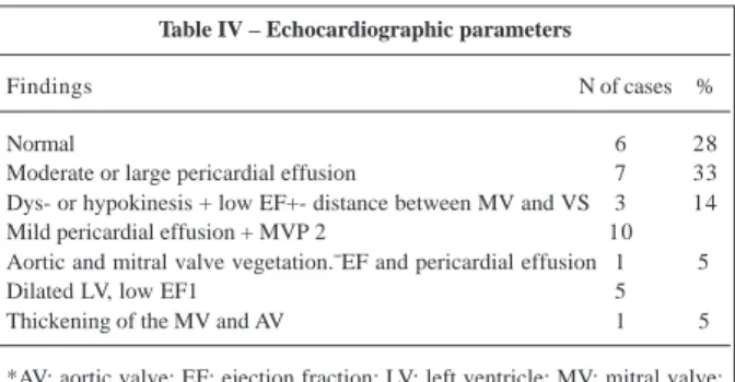
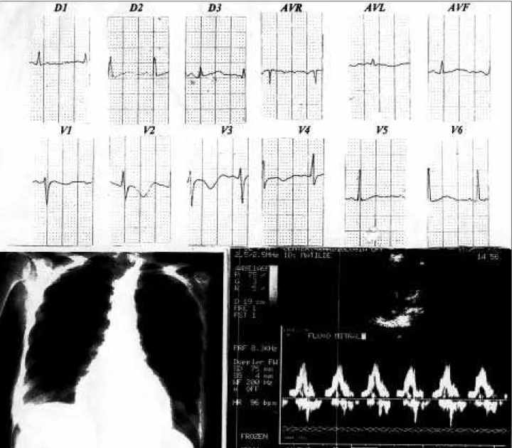
Fig. 2 - Case 14 – Six months after (close to time of death): severe abnormalities of the ST-T segment, severe increase in the cardiac silhouette due to a large pericardial effusion confirmed by echocardiography.
complications. Recurrent diarrhea followed by weight loss was a frequent complaint. The absorbing surface in the in-testine of patients infected by the virus of the acquired immunodeficiency syndrome is jeopardized by the shorte-ning of the villi intestinales, as shown in biopsies, as well as by abnormalities in the enterocytes 9. Deficiency of
oligo-elements can occur due to malnutrition 10.
Other frequently found complications in this series, such as tuberculosis and toxoplasmosis, have also had an impact at the beginning of the epidemic in developed coun-tries, because therapies, which had almost been forgotten because the diseases had been eradicated, had to be revie-wed and restarted 11.
Several of our patients had pericarditis or myocarditis. Cardiac involvement can happen secondary to a great varie-ty of causal agents 12. The virus of the acquired
immunode-ficiency syndrome itself can be the causal agent, because
the virus has been described in the myocardium by several authors 5,6. Other viral agents with cardiotropism, such as
cytomegalovirus, can be frequently found in myocarditis in children 13,14. The presence of the Epstein-Barr virus is a
pre-dictive factor for development of chronic cardiac failure 15.
We had two cases of severe myocarditis; one was caused by Toxoplasma gondii, which was the cause of death and is the protozoon most frequently involved in myocarditis 16.
Pericardial involvement was frequent in our series; some had cardiac tamponade and others had associated myocarditis. The described causes are infectious (viral,
Cryptococcus, Mycobacterium tuberculosis or Avium, Staphylococcus aureus) 16,17. Several of our patients had
tuberculosis, and one patient had cardiac tamponade cau-sed by Microsporida.
1. Jacob AJ, Boon NA. HIV cardiomyopathy. A dark cloud with a silver lining? Br Heart J l991; 66: 1-10.
2. Moskowitz L, Hensley GT, Chan JC. Immediate causes of death in acquired immu-nodeficiency Syndrome. Arch Pathol Lab Med 1989; 109: 735-9.
3. Baroldi G, Corallo S, Moroni M, et al. Focal lymphocytic myocarditis in AIDS. A correlative morphological and clinical study in consecutive 26 fatal cases. J Am Coll Cardiol 1988; 12: 463-9.
4. Herdy GVH, Ramos R, Bazin AR, et al. Correlação clínico-patológica de 50 casos de SIDA. Estudo retrospectivo. Arq Bras Cardiol 1994; 62: 95-8.
5. Lipshultz S, Fox. C, Perez–Atayde A. Identification of HIV-1 RNA and DNA in the heart of a child with cardiovascular abnormalities and congenital AIDS. Am J Cardiol 1990; 66: 246-50.
6. Grody W, Cheng L, Pang M, Lewis W. Direct infection of heart by HIV Abstract-.Circulation 1989; 80(supII): II-665.
7. Akras F. HIV and opportunistic infections which makes the heart vulnerable? Br J Clin Prat 1993; 47: 232-8.
8. CDC-Current Trends. Classification system for human T lymphotropic virus. Morbidity Mortality Weekly report(MMWR), 1986; 35: 334-9.
9. Soares RLS. Aspectos clínicos da síndrome de má-absorção em pacientes infecta-dos pelo HIV. Valor d-xilose como marcador de alteração funcional da mucosa jeju-nal. An Acad Nac Med 1996; 156: 79-82.
10. Dworkin BM, Antonechia PP, Smith F, et al. Reduced cardiac selenium content in AIDS.J Parenter Nutr 1989; 13: 644-7.
11. Cotton D. The impact of AIDS in the medical care system. JAMA 1988; 156: 79-82. 12. Herdy- GVH, Carvalho R, Vasconcelos MC. AIDS e coração. In: Celmo Celeno Porto – Doenças do Coração. Cap.206. Rio de Janeiro: Guanabara Koogan, 1998: 982-5.
References
13. Herdy GVH, Lopes VGS, Ramos RG. Correlação clínico patológica de 12 casos de SIDA em crianças. Arq Bras Ped 1996; 3: 133-7.
14. Kostianosky M, Orenstein JM, Schaff Z, et al. CMV observed in AIDS. Arch Pa-thol Lab Med 1987; 11: 218-23.
15. Luginbuhl LM, Orav EJ, Mc Intosh K. Cardiac morbidity and related mortality in children with HIV-1 infection. JAMA 1993; 169: 2869-75.
16. Kaul S, Fishbein MC, Siegel RJ. Cardiac manifestations of AIDS. 1991 update. Am Heart J 1991; 122; 535-9.
17. Araujo DV, Albanesi FM, Menezes MEC, et al. Pericardite tuberculosa como ma-nifestação inicial da SIDA. Arq Bras Cardiol 1995; 65: 497-500.
18. Hakas J, Generalovich T. Spontaneons regression of cardiomyopathy in a patient with AIDS.Chest 1991; 99: 770-2.
19. Deyton L, Walker R, Kovacs J, et al. Reversible cardiac dysfunction associated with interferon alfa.N Eng J Med l989; 321: 1246-9.
20. Dias FS. A disfunção cardiovascular no choque séptico e seu tratamento. Arq Bras Cardiol l993; 60: 43-9.
21. Girardin E, Gran E, Dayer JM, et al. The J5 study group, Lambert. Tumor necrose factor and interleukin in the serum of children with severe infectious purpura. N Engl J Med 1988; 319: 397-400.
22. Ognibene FP, Rosemberg AS, Lotze MT, et al. Interleukin-2 administration cau-ses reversible hemodynamic changes and left ventricular disfunction similar to those seen in septic shock.Chest 1988; 94: 750-4.
23. De Meules JE, Pigula FA, Mueller M, et al. Tumor necrosis factor and cardiac func-tion J Trauma 1992; 32: 686-92.
24. Bryant D, Becker L, Richardson J, et al. Cardiac failure in transgenic mice with myo-cardial expression of tumor necrosis factor-alpha. Circulation 1998; 97: 1375-81.
endocarditis, which has been the most frequently reported form of endocarditis in these patients 4,16, did not occur in our series.
Several cases of dilated cardiomyopathy with severe involvement of the ventricular function were present in our series. Some of these patients showed improvement in sys-tolic function and a decrease in the dimensions of the heart chambers during their hospital stay. Several reports have been published of improvement in cardiac function after adequate control of associated infection was obtained in patients with acquired immunodeficiency syndrome 18,19.
During severe infections, some patients develop con-gestive heart failure and echocardiographic parameters of dilated cardiomyopathy 20. It is known that, during these
infections (viral or bacterial), an increase in the blood levels of interleukins occurs, which relates to the severity of the disease 21. Interleukin 2 is produced by lymphocytes T, and
it increases the toxicity of killer cells that promote the synthesis of the tumor necrosis factor. Both interleukin 2 and the tumor necrosis factor can decrease the ejection fraction of the left ventricle 22,23. Bryant et al 24 recently used
alpha tumor necrosis factor and showed severe decrease in cardiac function with biventricular enlargement and decrease of the ejection fraction in transgenic mice.
Therefore, in our patients with congestive heart failure, with low systolic function indexes by echocardiogram and no significant myocardial abnormalities on necropsy examina-tion, the above mediators probably affected the heart during an episode of a severe infection, with subsequent recovery of systolic function with adequate control of the infection.