ANA RITA PEREIRA AZEVEDO
DEVELOPMENT OF MONOCLONAL ANTIBODIES FOR BLADDER
CANCER BASED ON GLYCOBIOMARKERS: IDENTIFICATION OF
RELEVANT
GLYCOANTIGENS
AND
SYNTHESIS
METHODOLOGIES THEREOF
Tese de Candidatura ao grau de Doutor em
Patologia e Genética Molecular submetida ao
Instituto de Ciências Biomédicas Abel Salazar da
Universidade do Porto.
Orientador: José Alexandre Ribeiro de Castro
Ferreira
Categoria: Investigador Auxiliar
Afiliação: Instituto Português de Oncologia do
Porto.
Co-orientador: Lúcio José de Lara Santos
Categoria: Professor Afiliado e Coordenador
Afiliação: Instituto de Ciências Biomédicas Abel
Salazar da Universidade do Porto e Instituto
Português de Oncologia do Porto.
Co-orientadora: Áurea Rosa Nunes Pereira Lima
Categoria: Professora Auxiliar
Afiliação: Instituto Universitário de Ciências da
Saúde - Cooperativa de Ensino Superior, Politécnico
e Universitário.
Aos meus pais, irmão, e a toda minha família e amigos que, com muito carinho e apoio, não mediram esforços para que eu chegasse até esta etapa da minha vida! ♥ E claro, a mim…
“Por vezes sentimos que aquilo que fazemos não é senão uma gota de água no mar. Mas o mar seria menor se lhe faltasse uma gota.” Madre Teresa de Calcutá
v
NOTA PRELIMINAR
Declaro que este trabalho é integralmente da minha autoria, estando devidamente referenciadas e identificadas as fontes de informação e as obras consultadas. Não contém, por isso, qualquer tipo de plágio de textos publicados ou trabalhos académicos, qualquer que seja o meio dessas publicações.
Os trabalhos de investigação conducentes a esta tese foram realizados no Grupo de Patologia e Terapêutica Experimental do Centro de Investigação do Instituto Português de Oncologia do Porto, EPE.
Declaro ainda que o presente trabalho foi financiado pela Fundação para a Ciência e a Tecnologia, pela atribuição de uma bolsa individual de doutoramento, com a referência SFRH/BD/105355/2014. A Fundação para a Ciência e a Tecnologia é co-financiada pelo Fundo Europeu de Desenvolvimento Regional da União Europeia, através do Programa Operacional Fatores de Competividade do Quadro de Referência Estratégico Nacional. Reconheço também o financiamento, pela Fundação para a Ciência e a Tecnologia, do Centro de Investigação do Instituto Português de Oncologia do Porto (PEst-OE/SAU/UI0776/201; IPOP-29-2014; CI-IPOP-58-2015). Por último, delaro que o presente trabalho teve ainda o suporte financeiro do Instituto de Ciências Biomédicas Abel Salazar da Universidade do Porto.
vi
Em obediência ao disposto no nº 2 do Artigo 8º do Decreto-Lei nº 388/70, o autor declara que participou na conceção e na execução do trabalho experimental, bem como na interpretação dos resultados e na redação dos trabalhos a seguir referenciados, estejam estes já publicados ou em fase de publicação e que integram a tese aqui apresentada:
a.
Rita Azevedo, José Alexandre Ferreira, Andreia Peixoto, Manuel Neves, Nuno Sousa, Aurea Lima, Lucio Lara Santos. Emerging antibody-based therapeutic strategies for bladder cancer: A systematic review. Journal ofControlled Release (Impact Factor in 2014: 7.70, Q1). 2015; 214: 40-61.
doi:10.1016/j.jconrel.2015.07.0022015.
b.
Rita Azevedo, Andreia Peixoto, Aurea Lima, José Alexandre Ferreira, Lucio Lara Santos. Emerging Antibody-Based Therapeutic Strategies For Bladder Cancer: A Systematic Three-Year Update (2015-2018). 2019. (Submitted)c.
Rita Azevedo*, Andreia Peixoto*, Cristiana Gaiteiro, Elisabete Fernandes,Manuel Neves, Luís Lima, Lúcio Lara Santos, José Alexandre Ferreira. Over forty years of bladder cancer glycobiology: Where do glycans stand facing precision oncology? Oncotarget (Impact Factor in 2016: 5.17, Q1). 2017. doi:10.18632/oncotarget.19433.
d.
Rita Azevedo, André M. N. Silva, Celso A. Reis, Lúcio Lara Santos, José Alexandre Ferreira. In silico approaches for unveiling novel glycobiomarkers in cancer. Journal of Proteomics. (Impact Factor in 2016: 3.91, Q1). 2018. 171:95-106. doi:10.1016/j.jprot.2017.08.004. Publication by invitatione.
Rita Azevedo*, Janine Soares*, Cristiana Gaiteiro, Andreia Peixoto, LuísLima, Dylan Ferreira, Marta Relvas-Santos, Elisabete Fernandes, Ana Tavares, Sofia Cotton, Ana Luísa Daniel-da-Silva, Lúcio Lara Santos, Rui Vitorino, Francisco Amado, José Alexandre Ferreira. Glycan affinity magnetic nanoplatforms for urinary glycobiomarkers discovery in bladder cancer.
Talanta (Impact Factor in 2016: 4.16, Q1). 2018. 184:347-355.
vii
f.
Rita Azevedo, Cristiana Gaiteiro, Andreia Peixoto, Marta Relvas-Santos, Luis Lima, José Alexandre Ferreira, Lucio Lara Santos. CD44 glycoprotein in cancer: A molecular conundrum hampering clinical applications. Clinical Proteomics (Impact Factor in 2016: 3.27, Q1). 2018. 15:22. doi: 10.1186/s12014-018-9198-9.g.
Sofia Cotton*, Rita Azevedo*, Cristiana Gaiteiro, Dylan Ferreira, Luís Lima, Andreia Peixoto, Elisabete Fernandes, Manuel Neves, Diogo Neves, Teresina Amaro, Ricardo Cruz, Ana Tavares, Maria Rangel, André M. N. Silva, Lúcio Lara Santos, José Alexandre Ferreira. Targeted O‐glycoproteomics explored increased sialylation and identified MUC16 as a poor prognosis biomarker in advanced stage bladder tumours. Molecular Oncology. (Impact Factor in 2016: 5.37, Q1). 2017. doi:10.1002/1878-0261.12035.h.
Rita Azevedo*, Dylan Ferreira*, Andreia Peixoto*, Marta Relvas-Santos, Cristiana Gaiteiro, Janine Soares, Elisabete Fernandes, Rui Freitas, Sofia Cotton, Luís Lima, Ana Tavares, Carlos Palmeira, Gabriela Martins, Áurea Lima, André M. N. Silva, Lúcio Lara Santos, José Alexandre Ferreira. Glycomics and Glycoproteomics identify GLUT1 and HOMER3 as top-ranked potentially targetable biomarkers in bladder cancer. 2019. (In preparation)i.
Rita Azevedo*, Elisabete Fernandes*, Marta Relvas-Santos, Andreia Peixoto,Rui Freitas, Dylan Ferreira, Lúcio Lara Santos, André M. N. Silva, José Alexandre Ferreira. Single-pot enzymatic synthesis of short-chain MUC16 O-sialoglycopeptides and protein glycoconjugates for bioanalytical and biomedical applications. 2019. (In preparation, subjected to patenting and
of condidential nature)
viii
Para além dos trabalhos científicos anteriormente mecionados e que integram esta tese, durante o período de doutoramento, o autor da presente tese participou nos seguintes artigos complementares:
o Andreia Peixoto, Marta Relvas-Santos, Rita Azevedo, Lucio L. Santos, José Alexandre Ferreira. Protein glycosylation and tumour microenvironment alterations driving cancer hallmarks. Frontiers in Oncology (Impact Factor in 2018: 4.42, Q1). 2019. 9:380. doi: 10.3389/fonc.2019.00380.
o Manuel Neves*, Rita Azevedo*, Luís Lima, Marta I. Oliveira, Andreia Peixoto, Dylan Ferreira, Janine Soares, Elisabete Fernandes, Cristiana Gaiteiro, Carlos Palmeira, Sofia Cotton, Stefan Mereiter, Diana Campos, Luís Pedro Afonso, Ricardo Ribeiro, Avelino Fraga, Ana Tavares, Hélder Mansinho, Eurico Monteiro, Paula Videira, Paulo P. Freitas, Celso A. Reis, Lúcio Lara Santos, Lorena Dieguez, José Alexandre Ferreira. Exploring sialyl-Tn expression in microfluidic-isolated circulating tumour cells: a novel biomarker and an analytical tool for precision oncology applications. New Biotechnology (Impact Factor in 2017: 3.73, Q1). 2018. 49:77-78. doi: 10.1016/j.nbt.2018.09.004.
o Rita Azevedo, Janine Soares, Andreia Peixoto, Sofia Cotton, Luís Lima, Lúcio Lara Santos, José Alexandre Ferreira. Circulating Tumour Cells in Bladder Cancer: Emerging Technologies and Clinical Implications Foreseeing Precision Oncology. Urologic Oncology: Seminars and Original
Investigations. (Impact Factor in 2016: 3.77, Q1). 2018. 36(5):221-236.
doi:10.1016/j.urolonc.2018.02.004.
o Milene A Carrascal, Mariana Silva, José Alexandre Ferreira, Rita Azevedo, Dylan Ferreira, André M. N. Silva, Dário Ligeiro, Lúcio Lara Santos, Robert Sackstein, Paula A. Videira. A functional glycoproteomics approach identifies CD13 as a novel E-selectin ligand in breast cancer. Biochim Biophys Acta (Impact Factor in 2016: 4.70, Q1). 2018. S0304-4165(18)30145-4. doi: 10.1016/j.bbagen.2018.05.013.
o Luís Lima*, Manuel Neves*, Marta I. Oliveira, Lorena Dieguez, Rui Freitas, Rita Azevedo, Cristiana Gaiteiro, Janine Soares, Dylan Ferreira, Andreia
ix
Peixoto, Elisabete Fernandes, Diana Montezuma, Ana Tavares, Ricardo Ribeiro, Ana Castro, Manuel Oliveira, Avelino Fraga, Celso A. Reis, Lúcio Lara Santos, José Alexandre Ferreira. Sialyl-Tn identifies muscle-invasive bladder cancer basal and luminal subtypes facing decreased survival, being expressed by circulating tumor cells and metastases. Urologic Oncology:
Seminars and Original Investigations. (Impact Factor in 2016: 3.77, Q1).
2017. 35(12):675.e1-675.e8. doi:10.1016/j.urolonc.2017.08.012.
o Andreia Peixoto*, Elisabete Fernandes*, Cristiana Gaiteiro*, Luís Lima, Rita Azevedo, Janine Soares, Sofia Cotton, Beatriz Parreira, Manuel Neves, Teresina Amaro, Ana Tavares, Filipe Teixeira, Carlos Palmeira, Maria Rangel, André M. N. Silva, Celso A. Reis, Lúcio Lara Santos, Maria José Oliveira, José Alexandre Ferreira. Hypoxia enhances the malignant nature of bladder cancer cells and concomitantly antagonizes protein O-glycosylation extension. Oncotarget (Impact Factor in 2016: 5.17, Q1). 2016. doi:10.18632/oncotarget.11257. Publication by invitation
xi
PREFÁCIO DO AUTOR
Toda a minha vida segui o pensamento que tudo se consegue, por mais longo e difícil que seja o caminho a percorrer para atingir os meus objetivos. E “sorte” é apenas o culminar de muito esforço, trabalho e dedicação! Esta é a plataforma que tem suportado e fundamentado todas as etapas da minha vida profissional, logo a partir do momento que os meus pais me ofereceram “ferramentas” para a iniciar.
Apesar das grandes dificuldades que senti em escolher uma área que iria no futuro dedicar o meu tempo e a minha vida, pois sempre me senti dividida entre o mundo das artes, da tecnologia e da saúde, o meu “acordar” para o mundo da investigação foi-se revelando cada vez mais à medida que me licenciava. Foi a partir desse momento que finalmente percebera que, ao dedicar-me à investigação, poderia ser e fazer um pouco de todas as outras áreas pelas quais me sentia dividida: fazer investigação de qualidade na área da saúde, com toda a arte e perícia que envolvem a experimentação e as ilustrações científicas, apimentada com toda a (bio)tecnologia envolvida nestes processos.
Todo este “novo” mundo foi-se revelando, à medida que passava por várias fases da minha vida científica, e por várias pessoas envolvidas na investigação. O meu percurso científico começou na farmacogenética e artrite reumatoide, onde tive o prazer de aprender com a Professora Doutora Áurea Lima (professora auxiliar do Instituto Universitário de Ciências da Saúde - Cooperativa de Ensino Superior, Politécnico e Universitário e investigadora no Grupo de Oncologia Molecular e Patologia Viral do Centro de Investigação do Instituto Português de Oncologia do Porto, coordenado pelo Professor Doutor Rui Medeiros). Mas, rapidamente, o meu impulso pela tecnologia fez-me mudar o rumo da farmacogenética para a área da “mHealth”, onde pude explorar a potencialidade da tecnologia “smartphone” para a gestão de doentes com artrite reumatoide, com todo o apoio do Professor Doutor João Fonseca da Faculdade de Medicina da Universidade do Porto e da Professora Doutora Áurea Lima. No entanto, o “bichinho” para explorar a área da oncologia foi cada vez estando mais presente, muito influenciado pelo facto de (com)viver ba grande casa que é o Instituto Português de Oncologia do Porto. Assim, logo que surgiu a oportunidade de trabalhar naquela área, aproveitei-a de imediato, começando o percurso desafiante do doutoramento. Soube que, ao tomar esta decisão, tudo seria diferente, deixaria de “nadar na minha zona de conforto ao pé da margem do rio e passaria a nadar em mar aberto a desafios e oportunidades,
xii
mas também perto de grandes tubarões”. O início foi turbulento, recheado de dificuldades em entender os princípios básicos da glicosilação, em como os açúcares (glicanos) poderiam ser importantes no cancro, mais especificamente, nos tumores da bexiga. Felizmente, arrastei neste meu barco alguém da minha inteira confiança e que hoje é uma verdadeira amiga – a Áurea Lima – que acumulava alguma experiência em oncologia. Ainda mais confortável fiquei, quando percebi que as pessoas que me acolheram, nomeadamente, o Professor Doutor José Alexandre Ferreira, o Professor Doutor Lúcio Lara Santos, o Professor Doutor Luís Lima e o restante Grupo de Patologia e Terapêutica Experimental do Instituto Português de Oncologia do Porto, estavam dispostos a remar na mesma direção que a minha. Todos eles contribuíram, significativamente, para o meu “input” profissional e pessoal. Já não sou a mesma pessoa que era há quatro anos atrás! Foi graças à grande excelência e capacidades excecionais de todas estas pessoas que hoje sou quem sou!
Este é mais um marco da minha vida, de grande importância, que culminará com a defesa pública de todo o trabalho apresentado nesta tese. Tantas coisas aprendi e vivi, tanto cresci, quantas memórias, sorrisos e lágrimas guardo de coração cheio… olhando para trás, nada aconteceu por acaso, tudo estava preparado para este mesmo momento, aquele em que suspiro de orgulho e emoção… E a minha mente grita: “Agora já sei nadar em mar aberto com grandes tubarões, mas ainda tenho tanto para nadar, tenho todo um oceano infinito de conhecimento e experiências”!
xiii
AGRADECIMENTOS
Foram várias as pessoas que ao longo deste percurso de quatro anos, direta ou indiretamente, contribuíram significativamente para a minha concretização profissional e pessoal. Deixo aqui o meu agradecimento e sentimento de gratidão a todas elas, pelo carinho, amizade, apoio e cumplicidade. Todos vocês fizeram-me crescer tanto, que fico sempre com um sentifizeram-mento de dívida extrema a todos que me fizeram sentir em “casa”. No entanto, não poderia deixar de dedicar um agradecimento especial a cada uma delas… Sem que a ordem pelo qual vou ditando as pessoas que marcaram este meu percurso de doutoramento influencie o nível de importância ou o impacto que tiveram na minha vida, um MUITO OBRIGADA…
Ao meu orientador Professor Doutor José Alexandre Ferreira, por estar sempre presente mesmo quando fisicamente ausente, pelo esforço e dedicação diários para garantir o suporte das minhas atividades científicas. A quem um Muito Obrigada nunca será suficiente para agradecer todo o empenho e paciência que teve durante este percurso. Obrigada por todos os ensinamentos, por toda a ajuda, por todos os sensatos conselhos científicos e não científicos, por toda a confiança depositada em mim e no meu trabalho. Certamente, graças à sua orientação, cresci enquanto investigadora e enquanto pessoa. Agradeço a sua amizade e toda a disponibilidade e prontidão com que respondeu às minhas perguntas, dúvidas, inquietudes e incertezas. Obrigada pela sua racionalidade, por ter tentado ensinar-me a ser mais paciente e a acreditar mais em mim. Do fundo do ensinar-meu coração, muito obrigada por não me deixar cair.
Ao meu co-orientador Professor Doutor Lúcio Lara Santos, pela oportunidade de fazer parte do seu grupo de investigação que coordena e pela confiança depositada. É um grande exemplo para mim, como profissional e como pessoa, e é com grande orgulho que digo que fiz parte da grande família que é o Grupo de Patologia e Terapêutica Experimental (a.k.a. equipa GPTE!) do Centro de Investigação do Instituto Português de Oncologia do Porto.
À minha co-orientadora Professora Doutora Áurea Lima, pela amizade e carinho, por me ter proporcionado a oportunidade de escolha para o doutoramento e pela grande contribuição na minha formação, pois sempre acreditou em mim e naquilo que eu poderia ser. Agradeço todas as portas e janelas que me abriu, e
xiv
toda a companhia e amparo em ambientes fora da minha zona de conforto. Parecem não haver palavras suficientes para descrever o quanto lhe agradeço por me ter feito crescer consigo tanto profissionalmente como pessoalmente… e por toda a disponibilidade com que respondeu às minhas incertezas e preocupações. Hoje somos verdadeiras amigas e a amizade significa o que nós sabemos.
Ao Professor Doutor Luís Lima, pela amizade, pela orientação e impacto que teve em todo este percurso da minha vida. Agradeço a calma e a paciência com que lidou com os meus momentos de ansiedade, incerteza e angústia; por complementar a falta de paciência do meu orientador, e pela capacidade de resolução rápida e lógica de todos os tipos de “bichos de cabeça”. O grupo não seria igual se não houvesse a dupla Luís/Alexandre, porque vocês complementam-se e são o suporte um do outro. E, é com grande orgulho que digo que o Luís é meu orientador mesmo que este título não esteja escrito oficiosamente, da mesma forma que é orientador de cada elemento da equipa GPTE! Sem si não seria igual… À equipa GPTE, meus colegas e amigos, como me sinto em dívida extrema a todos vocês, que me fizeram sentir em “casa”. Vou tentar fazer um rápido resumo… Querida “mãe” Elisabete, obrigada por todas as bases laboratoriais que me deste. Esse teu lado multifacetado facilitou-me muito a integrar-me novamente no laboratório e permitiu-me apreender uma panóplia de novas técnicas laboratoriais novas e a saber gerir corretamente um laboratório partilhado, juntamente com todas as questões de logística associadas. Andreia, obrigada pela tua exigência e ensino em como “comportar-me” corretamente num laboratório partilhado… vou sempre lembrar-me da tua grande transformação para uma mulher mais independente, descontraída e carinhosa. À Janine, pela grande simpatia e por me ter ajudado a dar os “primeiros passos” no grupo… marcas-me pela tua grande força de vontade… tu estravasas felicidade e boa disposição. À Cris, que tanto me ensinaste e ajudaste a crescer tanto como investigadora e pessoa… a tua felicidade, as tuas cantorias e as conversas “calientes” no laboratório ficarão sempre presentes na minha memória. À Sofi, pela amizade próxima que manténs comigo e por seres tão direta com os teus conselhos. És uma força da natureza e uma inspiração para mim… és fantástica! Ao Dylan, por seres tão querido, prestável e alegre… vou sempre lembrar-me dos teus falcetes “saídos do nada” no laboratório e da tua cumplicidade com a Sofia e a Cris… sem ti as conversas “calientes” não seriam a mesma coisa. À Marta, que me “aturou” nos
xv
primeiros tempos nas longas esperas da “SpeedVac”, as tuas manifestações de felicidade sempre que chegava material para ti nunca vão ser esquecidos… vou sempre lembrar-me das tuas manifestações com os comerciais, és única! Ao Rui, por seres tão querido e prestável, tens imenso potencial nesta área… Ainda bem que chegaste a esta “casa” para completares o coração da Janine. Aos restantes membros do grupo, especificamente à Ana Tavares, ao Manuel Neves e ao Diogo Neves, por toda a contribuição neste trabalho.
Ao Professor Doutor Manuel Teixeira, diretor do Centro de Investigação do do Instituto Português de Oncologia do Porto, pela oportunidade facultada ao aceitar-me como membro integrante do Centro de Investigação que coordena. É enorme a gratidão que tenho por ter antecipadamente visto o meu potencial e qualidade como investigadora, e de que conseguiria corresponder aos elevados padrões de qualidade existentes nos investigadores que integram este centro de investigação.
Ao Professor Doutor André Silva, da Universidade de Ciências da Universidade do Porto, por todo o tempo disponibilizado e auxílio na avaliação das nossas intermináveis amostras no espectrómetro de massa. Sem si, este projeto não teria sido possível! Obrigada pelo todo o ensinamento sobre espectrometria de massa e por toda a acessibilidade e disponibilidade em relação aos dados gerados das experiências.
Ao Professor Doutor Manuel A. Coimbra, da Universidade de Aveiro, por prontamente ter aceite que eu aprendesse no seu laboratório as experiências de acetilação e permetilação de O-glicanos. Obrigada a toda a sua equipa de investigação por me demonstrarem os “cantos à casa”. Um agradecimento especial à Professora Doutora Cláudia Nunes, por todo o tempo e ajuda dedicados a esta parte do trabalho.
Aos Professores Doutores Hassan Bousbaa, Vítor Seabra e Áurea Lima, pela disponibilidade e por ter proporcionado todas as condições para a utilização de equipamentos no Instituto de Investigação e Formação Avançada em Ciências e Tecnologias da Saúde da Cooperativa de Ensino Superior, Politécnico e Universitário. À Dra. Virgínia Gonçalves, membro do Instituto de Investigação e
xvi
Formação Avançada em Ciências e Tecnologias da Saúde, pelo carinho e por todo o apoio prestado nas questões de logística de laboratório.
À Professora Doutora Maria de Fátima Gärtner, pelo exemplo de rigor, confiança e otimismo, com que sempre me brindou. Obrigada por ter aceite a minha candidatura ao Programa Doutoral em Patologia e Genética Molecular do Instituto de Ciências Biomédicas Abel Salazar da Universidade do Porto/Faculdade de Medicina da Universidade do Porto e pela prontidão na resposta a todas as minhas dúvidas relativas ao doutoramento e a questões de logística. Obrigada à Dra. Ana Paula de Lima Pereira do Secretariado do Gabinete de Pós-Graduação/Doutoramentos do Instituto de Ciências Biomédicas Abel Salazar da Universidade do Porto pela ajuda na resolução de todas as minhas questões relativas ao doutoramento. E claro não me posso esquecer de agradeçer ao Sr. Rui Sousa do armazém do Instituto de Ciências Biomédicas Abel Salazar da Universidade do Porto pela simpatia e prontidão na agilização de processos em toda a logística das compras.
Às minhas colegas e amigas Teresa, Paula, Joana e Fátima do Programa Doutoral em Patologia e Genética Molecular do Instituto de Ciências Biomédicas Abel Salazar da Universidade do Porto/Faculdade de Medicina da Universidade do Porto. Muito obrigada por tornarem a experiência do doutoramento mais agradável ainda e pelos lanches ao final do dia. Vocês já sabem, mas digo na mesma – são pessoas fantásticas! Adoro-vos!
À Sandra e à Carla, minhas amigaças desde o meu início, no Instituto Português de Oncologia do Porto, obrigada pelo incentivo e por todo o apoio. São muito especiais para mim. Obrigada aos restantes membros do Grupo de Oncologia Molecular e Patologia Viral.
À Sra. Marta, a nossa técnica de laboratório no Centro de Investigação do Centro de Investigação do Instituto Português de Oncologia do Porto, obrigada pelo seu auxílio nas “pequenas coisas”, como a lavagem de material e entrega de requisições, que por si só ocupam tempo inestimável aos investigadores… Sem si não poderíamos dedicar-nos a tempo inteiro ao que “realmente interessa” no nosso percurso de investigação.
xvii
Ao Nuno Gonçalves, ao Sr. José Manuel Silva, ao Miguel Azevedo, ao Sr. Eduardo, ao Sr. José Estácio, ao Sr. Armando, à Sra. Armanda Santos e ao Sr. Salvador pela disponibilidade e ajuda na resolução de todas as questões de logística das compras e do arranjo de equipamentos no Centro de Investigação do Instituto Português de Oncologia do Porto.
Aos meus pais, que tanto se esforçaram para me darem “todas as ferramentas” para chegar onde estou hoje. Espero que estejam orgulhosos de mim. Sem vocês não teria conseguido o que consegui. Obrigada pelo apoio incondicional e pela paciência que tiveram comigo nos dias mais difíceis… Obrigada pela grande ajuda e preocupação quando entrei numa fase de “breakdown” psicológico, que me incapacitou durante cerca de um ano, bem perto da meta de conclusão deste doutoramento. Todo o meu esforço poderia ter sido perdido se vocês não me tivessem ajudado a ultrapassar aquela fase da melhor forma, com toda a ajuda médica que carecia. Ao meu querido irmão, pela preocupação que sempre teve com a sua “maninha”. Espero que o meu percurso académico sirva de modelo para conseguires atingir todos os teus objetivos. Às minhas avós e restante família, que tanto me ajudaram neste precurso, e me mantiveram sã psicologicamente. Vocês são o meu orgulho. Adoro-vos, do fundo do meu coração. E claro, a todos os meus amigos!
xix
RESUMO
A gestão de doentes com cancro de bexiga encontra dificuldades significativas associadas a uma elevada taxa de recidiva, rápida progressão e disseminação da doença, aliadas à resposta modesta à quimioterapia convencional. Além disso, o atraso na introdução de novas terapêuticas dirigidas, comparativamente a outros tipos de tumores sólidos, tem vindo a gestão destes doentes. Várias evidências sustentam que os tumores de bexiga comportam um reportório distinto de glicanos e glicoproteínas, proporcionando uma oportunidade única para a intervenção clínica. Neste sentido, o objetivo principal deste trabalho incide no estabelecimento das bases para a análise glicoproteómica do cancro de bexiga, com ênfase aos O-glicanos sialilados de cadeia curta (em resíduos de serina e treonina), visando assinaturas moleculares com potencial terapêutico. Mais ainda, estabelecer métodos de síntese quimioenzimática de glicopéptidos clinicamente relevantes, bem como de glicoconjugados com potencial imunogénico.
O trabalho aqui apreentado tem ínicio com o desenvolvimento de um tutorial
in silico, guiando a descoberta de biomarcadores de cancro, contornando as
dificuldades associadas à identificação de glicoproteínas por protocolos convencionais de proteómica. O conhecimento daí gerado fundamentou a exploração do glicoproteoma em distintas amostras biológicas (urina, modelos celulares e tumores) visando biomarcadores clinicamente relevantes.
Um sistema inovador, baseado em nanoplataformas funcionalizadas com lectinas, foi usado pela primeira vez para a descoberta não invasiva de biomarcadores em urina. Foram identificadas assinaturas glicoproteómicas distintas para todos os estadios da doença, que devem ser consideradas no futuro aquando do desenvolvimento de novos métodos de deteção não-invasivos. Nomeadamente, o marcador de células tumorais estaminais de bexiga CD44 foi consistentemente identificado em tumores humanos, estando ainda significativamente expresso na urina de doentes com cancro de bexiga musculo-invasivo, refletindo o seu aumento em tumores. Contudo, a elevada expressão de CD44 em tecidos normais, a existência de uma vasta gama de isoformas estruturalmente semelhantes, o seu caráter altamente glicosilado e a falta de consenso na nomenclatura desta proteína constituem, atualmente, constrangimentos significativos ao desenvolvimento de terapias dirigidas.
Numa segunda parte, este trabalho baseou-se na identificação de biomarcadores de prognóstico dos doentes com cancro de bexiga, além do
xx
desenvolvimento de novos alvos terapêuticos em tumores de bexiga e modelos celulares relevantes. A presença de O-glicanos de cadeia curta, geralmente sobre-expressos (antigénio T e a sua forma sialilada ST) ou sobre-expressos de novo em tumores humanos (antigénio Tn e a sua forma sialilada STn), foi determinada em tumores de bexiga quimioresistentes. As formas sialiladas do antigénio Tn e T, encontraram-se sobre-expressas em tumores de bexiga comparativamente às formas neutras, estando associadas a lesões de alto grau e invasão muscular. Em particular, o antigénio STn encontra-se significativamente aumentado em lesões mais agressivas e em metástases. Desta forma, uma estratégia glicoproteómica focada em glicoproteínas STn+ resultou na identificação de vários biomarcadores de cancro, incluindo o CD44 e a MUC16. Foram concentrados esforços no biomarcador MUC16, identificado pela primeira vez em cancro de bexiga, tendo-se concluído que a glicoforma STn-MUC16 associa-se com uma resposta modesta à quimioterapia baseada em cisplatina, enquanto que a MUC16 e o STn não têm valor preditivo por si só. Estas observações reforçam a importância de glicoformas específicas na valorização do valor biomarcador de glicoproteínas.
Posteriormente, procedeu-se à caracterização do O-glicoma de três dos mais estudados modelos celulares tumorais de bexiga (5637, T24, HT1376) por espectrometria de massa. O antigénio ST foi identificados como o O-glicano mais abundante nas três linhas celulares, corroborando as observações em tumores humanos. O estudo glicoproteómico destes modelos levou à identificação de mais de 1300 glicoproteínas potencialmente sialiladas ao nível do antigénio T, muitas das quais previamente encontradas em tumores de bexiga e urina, incluindo a MUC16 e o CD44. Uma estratificação das glicoproteínas identificadas de acordo com o seu potencial para terapia guiada em cancro de bexiga foi realizada recorrendo a ferramentas bioinformáticas. Desta forma, as glicoproteínas GLUT1 e HOMER3 emergiram no topo da classificação, refletindo a sua ausência em tecidos normais e uretélio saudável. Além disso, ambas as glicoproteínas encontram-se sobre-representadas com o aumento da severidade das lesões, sendo detetadas em metástases e associadas com mau prognóstico. Estudos estruturais detalhados de espectrometria de massa confirmaram a existência das glicoformas GLUT1-ST/STn e HOMER3-GLUT1-ST/STn em tumores de bexiga, fornecendo a informação molecular para a configuração de terapias guiadas. Por fim, a glicoproteína MUC16 classificou-se nos primeiros 10% da tabela de potenciais alvos, enquanto o CD44 foi severamente penalizado pela sua presença na maior parte dos tecidos humanos.
xxi
A última parte do trabalho dedicou-se ao estabelecimento de métodos de síntese quimioenzimática e purificação de glicopéptidos com diferentes O-glicanos de cadeia curta (antigénios Tn, STn, T, T mono e di-sialilado), glicosilados em diferentes extensões, tendo em vista o desenvolvimento de anticorpos e vacinas. Em suma, o presente estudo define uma estratégia para a descoberta de biomarcadores em cancro de bexiga, explorando o potencial de ferramentas bioinformáticas, glicómica e glicoproteómica. Identificaram-se glicoformas MUC16-STn como biomarcadoras de respostas modestas/diminuídas à quimioterapia baseada em cisplatina, bem como glicoformas GLUT1-ST/STn e HOMER3-ST/STn como potenciais alvos terapêuticos em cancro de bexiga. Ademais, estabelecem-se as fundações para a sínteestabelecem-se e purificação de glicoepítopos. O racional estrutural e clínico que suporta o desenvolvimento de aplicações teragnósticas (deteção e terapia) baseadas em glicanos encontra-se agora definido. Estudos futuros deverão focar-se nas implicações funcionais destas descobertas no desenvolvimento e progressão do cancro de bexiga, tendo em vista intervenções clínicas mais eficazes.
xxiii
ABSTRACT
The management of bladder cancer patients presents significant hurdles due to high recurrence rates, rapid progression, dissemination, and poor response to chemotherapy. In addition, there has been a substantial lag in the introduction of effective targeted therapeutics compared to other solid tumours, which has frustrated hopes of significant improvements in bladder cancer treatment. Several evidences support that bladder tumours express a unique repertoire of glycans and glycoproteins, providing an opportunity for clinical intervention. Therefore, the main objective of this work was to establish the foundations for a comprehensive interrogation of the bladder cancer glycoproteome, with emphasis on sialylated short-chain O-glycans (modifications of Ser and Thr residues), aiming at potentially targetable molecular signatures. Finally, it focused on setting up chemoenzymatic synthesis methods for glycopeptides of clinical interest and potentially immunogenic glycoconjugates. The work started by providing an in silico step-by-step tutorial for biomarker discovery in cancer, circumventing difficulties associated with the identification of glycoproteins by conventional proteomics workflows. This critical know-how set the foundations for interrogating the glycoproteome of distinct biological milieus (urine, cell models and bladder tumours) for clinically relevant biomarkers. An innovative lectin-affinity nanoenrichment proteomics workflow was first used for glycobiomarker discovery using urine as a starting point. Distinct glycoproteomics signatures were identified for all stages of the disease and should be considered in the development of future non-invasive detection methods. Namely, bladder cancer stem-cell marker CD44 was consistently found in patient’s samples and was significantly increased in urine of muscle-invasive bladder cancer patients, reflecting its increase in tumours. Nevertheless, this work discussed that the high expression of CD44 in normal tissues associated with a wide array of structurally similar isoforms, dense glycosylation and inadvertent lack of nomenclature consensus posed a significant challenge for targeted therapeutics at this stage. The second part of the work devoted to the identification of prognosis biomarkers and novel therapeutic targets in bladder tumours and relevant cell models. Chemoresistant bladder tumours were first screened for short-chain O-glycans generally overexpressed (T and sialylated T antigens-ST) or expressed de novo in human tumours (Tn and sialyl-Tn-STn). Sialoglycans (STn and ST) were predominant over neutral glycoforms (Tn and T antigens), being associated with high-grade lesions and muscle invasion in bladder
xxiv
tumours. In particular, the STn antigen was significantly increased in more aggressive lesions as well as in the metastasis. Targeted glycoproteomics focusing on STn-expressing glycoproteins resulted in the identification of several key cancer-associated biomarkers, including CD44 and MUC16. Particular focus was set on MUC16, a relevant cancer biomarker, identified for the first time in bladder cancer. MUC16-STn glycoforms were associated with decreased response to cisplatin-based therapeutics whereas MUC16 and STn evaluated independently were not. Such observations reinforced the importance of targeting specific protein glycoforms when envisaging the improvement of glycoproteins biomarker value. Efforts were then set on characterizing the glycome of three of the most widely studied bladder cancer cell models (5637, T24, HT1376) by mass spectrometry. ST antigens were identified as the most abundant O-glycoforms, in agreement with observations in human tumours. Targeted glycoproteomics led to the identification of over 1300 protein potentially carrying these sialylated glycans, many previously found in bladder tumours and urine samples, such as MUC16 and CD44. A bioinformatics-assisted scoring method was then established to rank the identified glycoproteins based on their potential relevance for targeting bladder cancer while minimizing off-target effects. GLUT1 and HOMER3 emerged as top-ranked glycoproteins, reflecting its lack of expression in the healthy urothelium and other healthy tissues. Moreover, both GLUT1 and HOMER3 were increased with the severity of the lesions, being also detected in the metastasis and associated with poor prognosis. More detailed structural studies by mass spectrometry confirmed the existence of GLUT1-ST/STn and HOMER3-ST/STn glycoforms in bladder tumours and provided the molecular foundations for designing targeted therapeutics. Finally, MUC16 was ranked on the top 10% of the biomarker listtarget score and CD44 was severely penalized due to its presence in most human organs. The last part of the work was devoted to the establishment of methods for the chemoenzymatic synthesis and purification of glycopeptides carrying different short-chain O-glycans (Tn, STn, T, mono and di-sialylated T antigens) with different degrees of glycosylation, envisaging the development of antibodies and cancer vaccines. In summary, the present study has set a roadmap for glycobiomarker discovery in bladder cancer, exploring the full potential of bioinformatics and state-of-the-art high throughput glycomics and glycoproteomics. It has identified MUC16-STn glycoforms as biomarkers of decreased response to chemotherapy and GLUT1, HOMER3 and also MUC16 carrying short-chain O-glycans as potentially targetable glycobiomarkers in bladder cancer, setting the structural basis for
xxv
targeted therapeutics. Moreover, it has set the foundations for glycoepitopes synthesis and purification. The necessary structural and clinical rationale now exists for supporting the development of novel glycan-based theragnostics applications (detection, therapy). Future studies should now focus on disclosing the functional implications of these findings for bladder cancer progression and development envisaging more effective clinical interventions.
xxvii
GRAPHICAL ABSTRACT
GRAP
H
ICAL
A
BS
TR
AC
T
xxix
TABLE OF CONTENTS
NOTA PRELIMINAR ... v PREFÁCIO DO AUTOR ... xi AGRADECIMENTOS ... xiii RESUMO ... xix ABSTRACT ... xxiii GRAPHICAL ABSTRACT... xxvii TABLE OF CONTENTS ... xxix LIST OF FIGURES ... xxxi LIST OF TABLES ... xxxvii ABBREVIATIONS & ACRONYMS ... xxxix INTRODUCTION ... 1 Overview Of Bladder Cancer, Clinical Management Challenges And Glycan-Based Opportunities ... 3 Emerging Antibody-Based Therapeutic Strategies For Bladder Cancer: A Systematic Review ... 11 Emerging Antibody-Based Therapeutic Strategies For Bladder Cancer: A Systematic Three-Year Update (2015-2018) ... 71 Over Forty Years Of Bladder Cancer Glycobiology: Where Do Glycans Stand Facing Precision Oncology? ... 107 AIMS & STUDY OUTLINE ... 185 CHAPTER IGLYCOBIOMARKERS DISCOVERY IN BLADDER CANCER ... 195
Section I
In Silico Approaches For Unveiling Novel Glycobiomarkers In Cancer ... 197 Section II
Glycan Affinity Magnetic Nanoplatforms For Urinary Glycobiomarkers Discovery In Bladder Cancer ... 229
Section III
CD44 Glycoprotein In Cancer: A Molecular Conundrum Hampering Clinical Applications ... 263
Section IV
Targeted O-Glycoproteomics Explored Increased Sialylation And Identified MUC16 As A Poor Prognosis Biomarker In Advanced Stage Bladder Tumours ... 273
xxx Section V
Glycomics and Glycoproteomics identify GLUT1 and HOMER3 as top-ranked potentially targetable biomarkers in bladder cancer ... 319 CHAPTER II
SYNTHESIS OF GLYCOANTIGENS ... 363 Single-pot enzymatic synthesis of short-chain MUC16 O-sialoglycopeptides and protein glycoconjugates for bioanalytical and biomedical applications ... 365 CONCLUDING REMARKS & FUTURE PERSPECTIVES ... 393
xxxi
LIST OF FIGURES
INTRODUCTIONArticle a
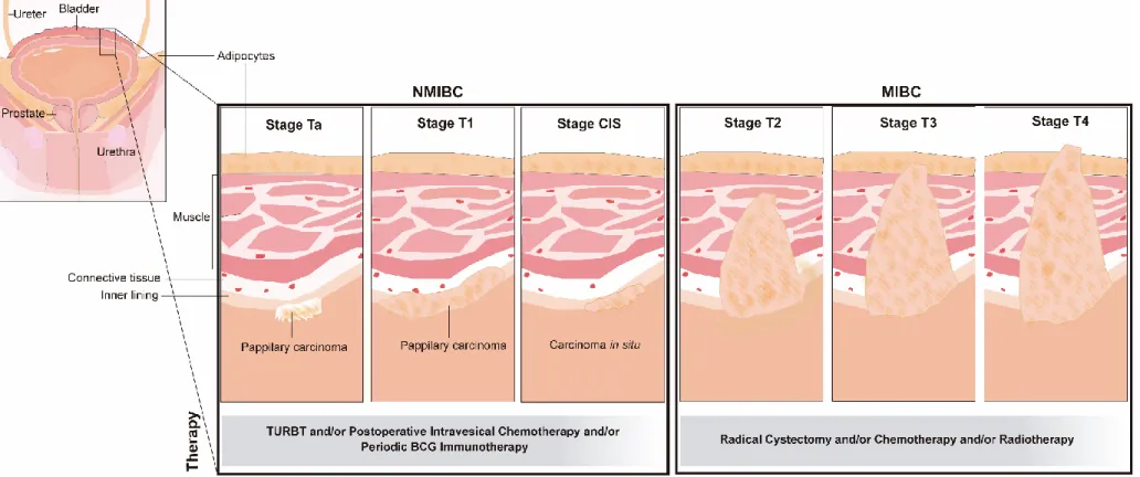
Figure 1. Bladder cancer stages and recommended therapy……… 16
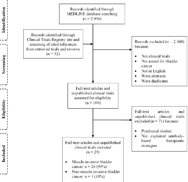
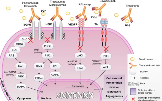
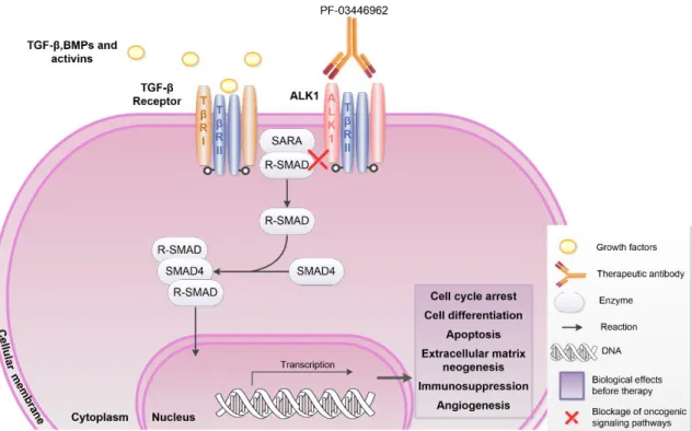
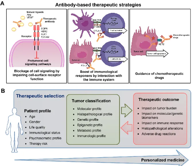
Figure 2. Flow diagram evidencing the different stages of study selection…… 18 Figure 3. Antibody-mediated inhibition of cellular signaling pathways by blocking of growth factors binding to tyrosine kinase receptors……… 19 Figure 4. Antibody-mediated inhibition of SMAD-mediated pathway by blocking
of growth factor binding to serine/threonine kinase receptors……… 20
Figure 5. Antibody-mediated inhibition of epithelial cell adhesion molecule
pathway……….. 31
Figure 6. Distinct mechanisms of programmed cell death protein 1 and cytotoxic T lymphocyte-associated antigen 4 on immunosuppression and
antibody-based immunotherapy………. 36
Figure 7. Overview on antibody-based therapeutic strategies for bladder cancer and a tentative approach to individualize cancer treatment and improved the
outcome……….. 44
Article b
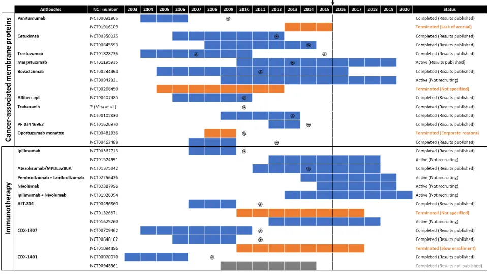
Figure 1. Timeline and status of the 29 clinical trials concerning
antibody-based therapies in bladder cancer conducted before to 2015………... 74 Figure 2. Flow diagram of studies selection for systematic review………. 76 Figure 3. Antibody-mediated inhibition and/or drug delivery of bladder cancer cells using cancer-associated membrane proteins and immunotherapy through
checkpoint-antibody-inhibitors……….. 88
Article c
Figure 1. Schematic representation of bladder cancer stage and grade………… 109 Figure 2. Schematic representation of protein-associated glycan structures.... 113 Figure 3. Schematic representation of short-chained O-linked glycan
structures……….………. 119
Figure 4. Schematic representation of the main glycomolecules with biological
relevance in bladder cancer……….. 128
Figure 5. Schematic representation of the main biologically relevant glycosphingolipids and glycosylphosphatidylinositon-anchored proteins in
xxxii
Figure 6. Schematic representation of the glycomolecule-mediated
metastization model and diagnostic value of glycans………. 141
Figure 7. Schematic representation of literature associations between (altered) expression of glycans and glycoconjugates and bladder tumour stage, grade
and invasion/metastasis, and patients’ diagnosis and
prognosis……… 153
CHAPTER I. GLYCOBIOMARKERS DISCOVERY IN BLADDER CANCER
Article d (Section I)
Figure 1. In silico roadmap for mining O-glycoproteomics datasets for relevant cancer biomarkers holding potential for targeted therapeutics……… 202 Figure 2. A. Panther main screen with selected functional classification viewed in pie chart. B. Cellular component GO term viewed in a pie chart at Panther
results screen……… 206
Figure 3. Results screen of retrieve ID/mapping tool of UniProtKB additionally
presenting topological domain of queried proteins………. 208
Figure 4. Results screen of retrieve ID/mapping tool of UniProtKB only showing
protein sequence………. 210
Figure 5. NetOGlyc results screen that lists putative O-glycosylated proteins… 211 Figure 6. A. Panther main screen with selected functional classification viewed in gene list. B. GO terms viewed in a gene list at Panther’s results
screen……… 213
Figure 7. Molecular function, biological process and protein class GO terms
viewed in a pie chart at Panther results screen………. 214
Figure 8. Oncomine results screen that provides the expression of each protein in our database (assumed by gene expression) in bladder cancer compared to normal tissue and tap our list to possible glycobiomarkers in bladder
cancer……… 217
Figure 9. Network presenting the most significant biological functions of our possible glycomarlers in bladder cancer clustered by disease stages…………. 221 Figure 10. Candidate STn-expressing membrane glycoproteins in bladder cancer comprehensively distributed by Oncomine’s rank of expression and
xxxiii Article e (Section II)
Figure 1. Protein content and total sialic acids of urines from controls and bladder cancer patients with low-grade NMIBC, high-grade NMIBC and MIBC…. 239 Figure 2. Urinary glycoproteins identified using MNP@ConA, MNP@WGA and MNP@SNA lectins in controls and low-grade NMIBC, high-grade NMIBC and
MIBC……… 246
Figure 3. Urine glycoproteins identified in controls and low-grade NMIBC, high-grade NMIBC and MIBC using MNP@ConA, MNP@WGA and MNP@SNA
lectins………. 247
Figure 4. Slot-blot analysis of urine samples from fifteen controls and
thirty-one bladder cancer patients using the ConA, WGA and SNA lectins……… 248 Figure 5. Graphical representation of cancer-specific urine glycobiomarkers with affinity to MNP@ConA, MNP@WGA and MNP@SNA lectin nanoprobes distributed according to their expression in bladder cancer………. 250 Figure 6. Graphical representation of cancer-specific urine glycobiomarkers distributed according to their overexpression in bladder cancer by Oncomine… 251 Figure 7. Network presenting the most significant biological functions of the
candidate glycobiomarkers in bladder cancer………... 253
Figure 8. Validation of CD44 expression in urine samples and immunohistochemistry of thirty-one bladder cancer patients………. 254
Article f (Section III)
Figure 1. Schematic representation of experimentally confirmed human CD44
pre-mRNA and respective isoforms………... 268
Article g (Section IV)
Figure 1. Schematic representation protein O-GalNac glycosylation biosynthesis evidencing the cancer-associated short-chain glycans explored in
this study……… 278
Figure 2. Schematic representation of the analytical strategy for S6T and S3T
evaluation by immunohistochemistry………... 282
Figure 3. Analytical workflow for A) whole proteome analysis starting from FFPE tissues and B) identification of STn expressing glycoproteins in bladder
tumours……….. 285
Figure 4. Immunohistochemistry for sialyl-Tn (STn) antigen evidencing A) expression in cells longing and invading the basal layer in high-grade NMIBC and B) extensive staining including in cells invading the muscle layer in MIBC…. 293
xxxiv
Figure 5. Immunohistochemistry for sialylated T antigens (ST: corresponding to mono and di-sialylated T glycoforms; S3T and S6T) for low and high grade superficial papillary muscle invasive bladder tumours……… 294 Figure 6. Proteins isolated from FFPE muscle-invasive bladder tumours distributed according to cellular localization (A), molecular (B) and cell
functions (C) based on gene ontology analysis……….. 298
Figure 7. Distribution of candidate STn-expressing glycoproteins in muscle-invasive bladder tumours (detailed in Table 3) comprehensively integrated according to cellular localization (A), molecular (B) and cell functions (C) based on gene ontology analysis by Panther bioinformatics tool……….. 300 Figure 8. Candidate STn-expressing glycoproteins in muscle-invasive bladder tumours comprehensively distributed according to its association with the
severity of the lesions………..… 301
Figure 9. A) Western blot for glycoproteins expressing the STn antigen in advanced bladder tumours. B) Identification of STn glycoforms in CD44 and ITGB1 glycoproteins isolated from advanced bladder tumours by immunoprecipitation. C) Immunohistochemistry and PLA for CD44, ITGB1 and
STn in bladder tumours……..……… 303
Figure 10. A) Exemplificative annotated nanoLC-ESI-LTQ-Orbitrap-CID-MS/MS spectra for a MUC16 glycopeptide substituted with a HexNAc residue evidencing the specific glycosite; B) Co-localization of MUC16 and STn in bladder tumours by immunohistochemistry; C) Expression of MUC16
STn-glycoforms in bladder tumours based on PLA analysis……….… 305
Figure 11. Annotated nanoLC-ESI-LTQ-Orbitrap-CID-MS/MS spectra for a MUC16 glycopeptide substituted with a HexNAc and HexNAc-Hex residues evidencing the specific glycosites (highlighted in the assignment table bellow). 306 Figure 12. A) Associations of MUC16 with the stage; B) grade of the disease and C) decreased overall survival in MIBC patients subjected to cisplatin-based
chemotherapy………. 308
Figure 13. Association between MUC16 classification by
immunohistochemistry in FFPE cancer tissues (IHC; negative vs positive) and
MUC16 expression……… 309
Article h (Section V)
Figure 1. Illustration of bioinformatics-assisted protocol for glycobiomarkers identification using cell models……….…………... 330
xxxv
Figure 2. Glycome repertoire for 5637, T24 and HT1376 cell lines in bladder
cancer………. 333
Figure 3. Expression of T and ST antigens by FACS……….……… 334 Figure 4. A) Sub-cellular location, B) molecular functions and C) biological processes associated to the identified glycoproteins……….. 336 Figure 5. Identification of targetable glycoproteins in bladder cancer supported
by bioinformatics analysis……… 341
Figure 6. Glycoproteins expression matrix ordered based on its expression levels in bladder tumours and healthy tissues according to the Human Protein
Atlas……….. 343
Figure 7. A) Expression of GLUT1 (red), HOMER3 (red), T (green) and ST (green) antigens in 5637, T24 and HT1376 cell lines. B) Western blot for GLUT1/HOMER3 and ST antigens in IP for both glycoproteins in cell line T24….. 345 Figure 8. Expression of A) ST antigens, B) GLUT1, C) HOMER3 in the healthy urothelium as well as T1, T1, T2, T3 and T4 bladder tumours and metastasis……….
348 Figure 9. Illustrative panel of the number of ST vs STn positive cases according
to the severity of the disease………... 349
Figure 10. Overall survival (OS) for bladder cancer patients with tumours exhibiting A) GLUT1 overexpression, B) HOMER3 at the plasma membrane
(memHOMER3) and C) memHOMER3 in the MIBC subgroup……….. 351
Figure 11. A) Co-localization of STn and ST, GLUT1 and HOMER3 positive areas. B) GLUT1 and C) HOMER3 glycopeptides presenting STn and ST antigens identified by nanoLC-EThcD-MS/MS. D) Expression of STn, GLUT1 and HOMER3 in healthy tissues (thyroid, liver, gallbladder, testis, lung, stomach, pancreas,
colon/small intestine/appendix………. 353
Figure 12. GLycosites identified by nanoLC-EThcD-MS/MS for A) GLUT1 and B)
HOMER3 having the canonical forms as reference……… 354
CHAPTER II. SYNTHESIS OF GLYCOANTIGENS
Article i
Figure 1. C18 reverse phase nanoLC-ESI-MS profiles for reaction products resulting from the glycosylation of 20 mer MUC16-VRT peptide by
xxxvi
Figure 2. C18 reverse phase nanoLC-ESI-MS profiles for the main reaction products resulting from single-pot enzymatic synthesis of MUC16-STn
glycopeptides before and after TiO2 enrichment……….. 376
Figure 3. C18 reverse phase nanoLC-ESI-MS profiles for the main reaction products resulting from single-pot enzymatic synthesis of MUC16-S3T glycopeptides before and after TiO2 enrichment………... 379 Figure 4. C18 reverse phase nanoLC-ESI-MS profiles for the main reaction products resulting from single-pot enzymatic synthesis of MUC16-S6T
glycopeptides before and after TiO2 enrichment………. 382
Figure 5. Monitoring reaction products of MUC16-Tn/STn biosynthesis by
xxxvii
LIST OF TABLES
INTRODUCTIONArticle a
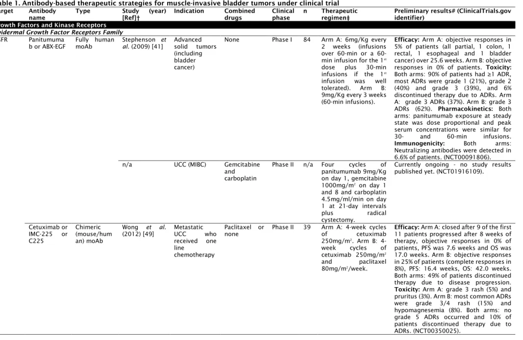
Table 1. Antibody-based therapeutic strategies for muscle-invasive bladder
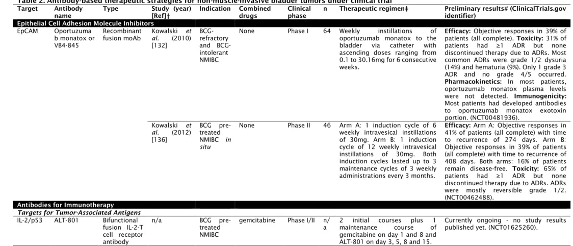
tumors under clinical trial………... 45 Table 2. Antibody-based therapeutic strategies for non-muscle-invasive bladder
tumors under clinical trial………. 52
Article b
Table 1. Antibody-based therapeutic strategies for bladder cancer under clinical
trial………. 91
Article c
Table 1. Biological and clinical significance of altered glycans and related
biosynthetic enzymes in bladder cancer………... 142
CHAPTER I. GLYCOBIOMARKERS DISCOVERY IN BLADDER CANCER
Article e (Section II)
Table 1. Extracellular membrane-bound or secreted glycoproteins with affinity to ConA, WGA or SNA lectins showing N- or O-glycosylation that were identified
in urine samples from controls……… 240
Table 2. Cancer-specific extracellular membrane-bound or secreted glycoproteins with affinity to ConA, WGA or SNA lectins showing N- or O-glycosylation that were identified in urine samples from bladder cancer
patients………. 242
Article f (Section III)
Table 1. Proposed CD44 nomenclature for experimentally observed isoforms, its correspondence with UniProt and NCBI databases and predicted N- and
O-glycosylation sites………. 269
Article g (Section IV)
Table 1. Expression of short-chain O-GalNAc glycans in bladder tumours of
different clinicopathological natures determined by
xxxviii Article h (Section V)
Table 1. Target Score for upregulated glycoproteins in MIBC distributed
according to the cell lines………... 337
CHAPTER II. SYNTHESIS OF GLYCOANTIGENS
Article i
Table 1. Products from the single-pot enzymatic synthesis of MUC16-STn glycopeptides analyzed by C18 reverse phase nanoLC-ESI-MS before and after
TiO2 enrichment………. 375
Table 2. Products from the single-pot enzymatic synthesis of MUC16-S3T glycopeptides analyzed by C18 reverse phase nanoLC-ESI-MS before and after
TiO2 enrichment………. 378
Table 3. Products from the single-pot enzymatic synthesis of MUC16-S6T glycopeptides analyzed by C18 reverse phase nanoLC-ESI-MS before and after
xxxix
ABBREVIATIONS & ACRONYMS
ADAM17: a disintegrin and metalloproteinase domain 17 ADCC: antibody-dependent cell-mediated cytotoxicity ADR: adverse drug reaction
ALK1: activin receptor-like kinase 1 Ang: angiopoietin
BC: bladder cancer
BCG: bacillus Calmette-Guérin BMP: bone morphogenetic protein BSA: bovine serum albumin
C1GalT1: core 1 synthase or N-Acetylgalactosamine 3-Beta-Galactosyltransferase 1 C1GalT1C1: C1GalT1 chaperone 1
CAMK: calmodulin-dependent protein kinase CD44: cluster of differentiation 44
CDC: complement-dependent cytotoxicity cDNA: complementary deoxyribonucleic acid CEA: carcinoembryonic antigen
CID: collision induced dissociation CIS: carcinoma in situ
CMP-NeuAc: cytidine-5′-monophospho-N-acetylneuraminic acid CSS: cancer-specific survival
CTLA-4: cytotoxic T lymphocyte-associated antigen 4 DAG: diacylglycerol
DAGR: Database of Anti-Glycan Reagents DHB: 2,5-dihydroxybenzoic acid
DMF: dimethylformamide DNA: deoxyribonucleic acid dST: disialylated T
DTT: 1,4-dithiothreitol ECM: extracellular matrix
EDTA: ethylenediamine tetraacetic acid EGF: epidermal growth factor
EGFR: epidermal growth factor receptor EGTA: ethylene glycol tetraacetic acid EMT: epithelial-to-mesenchymal transition
xl
EpEX: EpCAM extracellular domain EpICD: EpCAM intracellular domain ER: endoplasmic reticulum
ESI: electrospray ionisation
FACS: fluorescence-activated cell sorting FBS: fetal bovine serum
Fc: fragment crystallizable portion FDA: Food and Drug Administration
FFPE: formalin-fixed, paraffin embedded tissue section FHL2: four and a half lim domain protein 2
FOLFOX-4: oxaliplatin, leucovorin and 5-fluorouracil FUT-I: alpha1,2-fucosyltransferase 1
FUT-VI: alpha1,3-fucosyltransferase VI GA: Golgi apparatus
Gal: galactose
GalNAc: N-acetylgalactosamine GC: gemcitabine/cisplatin GlcNAc: N-acetylglucosamine
GLUT1: glucose transporter 1 or solute carrier family 2, facilitated glucose transporter member 1 (SLC2A1)
GM-CSF: granulocyte-macrophage colony-stimulating factor GO: gene ontology
GPAA1: GPI anchor attachment 1
GRB2: growth factor receptor bound protein-2 HAS: hyaluronic acid synthases
HCELL: hematopoietic cell E-/L-selectin ligand hCG-β: human chorionic gonadotropin beta-chain
HEPES: 4-(2-hydroxyethyl)-1-piperazineethanesulfonic acid HER2: human epidermal growth factor receptor 2
HRP: horseradish peroxidase
ICC/IF: immunocytochemistry/immmunofluorescence IGF-IR: insulin-like growth factor receptor I
IL: interleukin
IP3: inositol 1,4,5-triphosphate ITGA6: alpha6beta4 integrin ITGAV: αv integrin receptors
xli
ITGB1: integrin beta 1 JAK1: janus kinase 1
LEF-1: lymphoid enhancer-binding factor 1
LTQ-Orbitrap XL: hybrid linear ion trap-orbitrap mass spectrometer MAPK: mitogen-activated protein kinase
MBS: 3-maleimidobenzoic acid N-hydroxysuccinimide ester MgCl2: magnesium chloride
MHC: major histocompatibility complex MIBC: muscle invasive bladder cancer moAb: monoclonal antibody
MnCl2: manganese(II) chloride MR: mannose receptors
mRNA: messenger ribonucleic acid MS/MS: tandem mass spectrometry MS: mass spectrometry
MUC1: mucin 1 MUC16: mucin 16
MVAC: methotrexate, vinblastine, cisplatin and doxorubicin nanoLC: nano liquid chromatography
NMIBC: non-muscle invasive bladder cancer NMP22: nuclear matrix protein 22
O-GalNAc: O-N-acetylgalactosamine OS: overall survival
OSTase: oligosaccharide transferase complex PCR: polymerase chain reaction
PD-1: programmed cell death protein 1 PD-L1: programmed cell death ligand-1 PFA: paraformaldehyde
PFS: progression-free survival
PGAP1: post-GPI attachment to proteins 1 PI3K: phosphatidylinositol-3-kinase
PIGF: placental growth factor
PIP2: phosphatidylinositol 4,5-bisphosphate PLA: in situ proximity ligation assay
PLCG: phospholipase C gamma PNA: peanut agglutinin
xlii
poly-ICLC: polyinosinic-polycytidylic acid
ppGalNAc-Ts: UDP-GalNAc:polypeptide N-acetylgalactosaminyl transferases PRISMA: Preferred Reporting Items for Systematic Reviews and Meta-Analyses PRKC: protein kinase C
PSEN2: presenilin 2
PSM: peptide-to-spectrum match KCl: potassium chloride
Ref: reference
RHAMM: hyaluronan-mediated motility RhoGDI2: RhoGTP dissociation inhibitor 2 RIP: regulated intramembrane proteolysis RNA: ribonucleic acid
RNAseq: RNA sequencing
R-SMAD: receptor-regulated SMAD S3T: sialyl-3-T
S6T: sialyl-6-T
SARA: SMAD anchor for receptor activation SDS: sodium dodecyl sulphate
SDS-PAGE: sodium dodecyl sulphate-polyacrylamide gel electrophoresis SHC: src-homology and collagen homology
SLea: sialyl lewis a SLex: sialyl lewis x
SMAD: sma and mad related family SOS: son of sevenless
SPSS: Statistical Package for Social Sciences ST: sialyl-T
ST6GalNAc: α-N-acetylgalactosaminide α-2,6-sialyltransferase STAT: signal transducer and activator of transcription
STn: sialyl-Tn
STRAP: Software Tool for Researching Annotations of Proteins STRING: Search Tool for the Retrieval of Interacting Genes/Proteins TAA: tumor-associated antigen
TBS: Tris-buffered saline TCF: transcription factor. TCR: T cell receptor TFA: trifluoroacetic acid
xliii
TGF: transforming growth factor TLR: toll-like receptor
Tn: GalNAcα-O-Ser/Thr TUR: transurethral resection
TURBT: transurethral resection of the bladder tumour TβRI: transforming growth-factor beta type I receptor TβRII: transforming growth-factor beta type II receptor UCC: urothelial cell carcinoma
UDP: uridine diphosphate
VEGF: vascular endothelial growth factor
VEGFR: vascular endothelial growth factor receptor WHO: World Health Organization
The information provided in this section is based in the following publications: Rita Azevedo, José Alexandre Ferreira, Andreia Peixoto, Manuel Neves, Nuno Sousa, Aurea Lima, Lucio Lara Santos. Emerging antibody-based therapeutic strategies for bladder cancer: A systematic review. Journal of Controlled Release
(Impact Factor in 2014: 7.70, Q1). 2015; 214: 40-61.
doi:10.1016/j.jconrel.2015.07.0022015.
Rita Azevedo, Andreia Peixoto, Aurea Lima, José Alexandre Ferreira, Lucio Lara Santos. Emerging Antibody-Based Therapeutic Strategies For Bladder Cancer: A Systematic Three-Year Update (2015-2018). 2019. (Submitted)
Rita Azevedo*, Andreia Peixoto*, Cristiana Gaiteiro, Elisabete Fernandes, Manuel Neves, Luís Lima, Lúcio Lara Santos, José Alexandre Ferreira. Over forty years of bladder cancer glycobiology: Where do glycans stand facing precision oncology?
Oncotarget (Impact Factor in 2016: 5.17, Q1). 2017.
DEVELOPMENT OF MONOCLONAL ANTIBODIES FOR BLADDER CANCER BASED ON GLYCOBIOMARKERS:
IDENTIFICATION OF RELEVANT GLYCOANTIGENS AND SYNTHESIS METHODOLOGIES THEREOF 3
Overview Of Bladder Cancer, Clinical Management Challenges
And Glycan-Based Opportunities
Bladder Cancer: Detection And Main Therapeutic Challenges
Bladder cancer is the ninth most common malignancy, being in the top fifteen causes of death worldwide, showing three times higher incidence in men, especially after the age of the fifty [1, 2]. In Western world, cigarette smoking is the most relevant risk factor, accounting for approximately 50% of bladder cancer cases [2, 3]. Furthermore, occupational exposure to chemicals, genetic factors, and infection with Schistosoma haematobium, especially in rural areas of Africa and the Middle East, have also been described to increase the risk of bladder cancer development [4-6].
At diagnosis, approximately ninety percent of bladder cancers are urothelial cell carcinomas, 5% are squamous cell carcinomas, and less than 2% are adenocarcinomas [2]. Seventy percent of all newly diagnosed urothelial cell carcinomas are non-muscle invasive bladder tumours (NMIBC; Tis, Ta or T1) [2, 7, 8], and 30% comprise muscle-invasive tumours (MIBC; T2-T4), of which 10-15% are already metastatic [9]. The clinical diagnosis ensues careful evaluation of the patient’s background, including risk factors and family history assessment [2, 9], invasive bladder cytoscopic examination and urine cytology [10]. At presentation, over 80% of patients experience macroscopic urine haematuria, with urine examination generally done to exclude parasite infection [11]. Bladder cystoscopy and urine cytology also constitute surveillance tools for bladder cancer follow-up. The first provides information about tumour location, appearance, and size, while having suboptimal sensitivity for low-grade papillary tumours (Ta and T1) and high-grade Tis lesions [12]. In turn, urine cytology has reduced accuracy, especially for low-grade and low-stage tumours [10]. To overcome these limitations, several molecular non-invasive urine-based clinical tests were approved by Food and Drug Administration (FDA) agency. Namely, immunoassays to detect urinary proteins, including bladder cancer-associated antigens (BTA STAT®, BTA TRAK®), the nuclear matrix protein NMP22 immunocytofluorescence-based test (ImmunoCyt®/uCytt®), and the fluorescence in situ hybridization-based assay (UroVysion®) [10]. Still, the performance of these tests varies significantly since it is not always reproducible
DEVELOPMENT OF MONOCLONAL ANTIBODIES FOR BLADDER CANCER BASED ON GLYCOBIOMARKERS:
IDENTIFICATION OF RELEVANT GLYCOANTIGENS AND SYNTHESIS METHODOLOGIES THEREOF 4 in independent patient populations [13, 14]. Also, there are no serum-based tests currently available for bladder cancer detection and surveillance in clinical practice. Therefore, the search for novel secreted biomarkers are still required to improve the accuracy of available non-invasive detection tools and reduce the need for invasive tests.
Despite the knowledge on bladder cancer clinicopathological features, it has remained an “orphan disease” regarding the introduction of novel therapeutics [15, 16]. The mainstay treatment for NMIBC is endoscopic transurethral resection of the bladder tumour (TURBT) [2]. Further management is based upon risk factors, stage, grade, multimodality and recurrence history [2, 17]. Accordingly, patients can be advocated for surveillance, single-installation of intravesical chemotherapy (e.g. mitomycin C, epirubicin and doxorubicin), a course of intravesical chemotherapy or immunotherapy (bacillus Calmette-Guérin - BCG), or further surgery [2, 18, 19]. Despite the administration of standard therapy, 50-70% of NMIBC will recur, which constitutes the single highest recurrence rate amongst solid tumours [16, 18]. Moreover, roughly 10–20% of these tumours will progress to muscle-invasive disease (pT2–4), usually with metastasis and localized persistent disease, within 2 years if managed only by TURBT and intravesical therapy [20-22]. Specifically, patients with low grade Ta disease have a 15-year progression-free survival of 95% with no cancer-specific mortality [7]. In turn, patients with high-grade Ta tumours have a progression-free survival of 61% and a disease-specific survival of 74%, whereas patients with T1 disease have a progression-free survival of 44% and a disease-specific survival of 62%, supporting the notion that lamina propria invasion is a prognostic indicator of disease progression and reduced survival [5]. Consequently, high-grade NMIBC patients present strikingly high recurrence rate and progression to muscle-invasive disease [18], requiring frequent follow-up and repeated treatments, making the cost per patient from diagnosis to death the highest of all cancers [23, 24]. This encompasses a significant burden for both patients and health care systems, urging the early identification of BCG responders better served by alternative and often more aggressive therapeutics. Regarding MIBC treatment, the widely adopted therapeutic options includes radical cystectomy with pelvic lymphadenectomy and adjuvant or neoadjuvant chemotherapy (cisplatin-based combinations: methotrexate, vinblastine, cisplatin and doxorubicin - MVAC, or gemcitabine and cisplatin - GC) [9, 24]. Even with the administration of available therapy, the relapse rate after radical cystectomy is as
DEVELOPMENT OF MONOCLONAL ANTIBODIES FOR BLADDER CANCER BASED ON GLYCOBIOMARKERS:
IDENTIFICATION OF RELEVANT GLYCOANTIGENS AND SYNTHESIS METHODOLOGIES THEREOF 5 high as 50%, depending on the pathological stage of the primary tumour and the presence of loco-regional or distant metastasis [25]. Also, despite the introduction of cisplatin-based chemotherapy in recent therapeutic schemes has allowed improving survival in comparison to cystectomy alone (14 months vs 6 months), the five-year overall survival does not exceed 25% and many patients die prematurely from adverse drug reactions [17]. Therefore, the management of bladder cancer patients presents significant hurdles due to high recurrence rates, rapid progression, dissemination, and poor response to chemotherapy. Moreover, the substantial lag in the introduction of novel targeted therapeutics has frustrated hopes of significant improvements in bladder cancer treatment.
Currently, the success of bladder cancer management depends on early prognosis, but prognostication difficulties are often aggravated by the lack of efficient non-invasive follow-up strategies, early detection of occult micrometastasis, real-time monitoring of therapy response and metastatic risk assessment, which would enable timely and eventually life-saving interventions. Moreover, the high molecular heterogeneity of bladder tumours of apparently similar histology is responsible for significant variations in disease course, inconsistencies in therapeutic response and prognostic uncertainty [26].
In summary, the main clinical challenges for bladder cancer management include: i) lack of tools for non-invasive early diagnosis and follow-up; ii) difficulties in tumour classification and prognostication due to high molecular heterogeneity iii) lack of biomarkers to predict treatment response (BCG immunotherapy, chemotherapy), specially for NMIBC cases; iv) lack of molecular-based targeted therapeutics capable of delay or avoid recurrence and progression of bladder cancer. Facing these limitations, intense efforts are ongoing to integrate significant genomic and transcriptomic data into comprehensive molecular models for improving disease management beyond conventional histopathological evaluation. However, many of the adopted methodologies do not fully reflect the tumour molecular microheterogeneity or allow predictions of its evolution throughout the disease course. More importantly, there is an unmet need for context-specific biomarkers capable of guiding the development of targeted therapeutics.
Glycan-Based Opportunities Facing Bladder Cancer Clinical Challenges
Although genetic and epigenetic alterations are considered primary causes of cancer development, downstream phenotypic changes at the protein level are